. 2013 Jul 4;32(27):3246-53.
doi: 10.1038/onc.2012.351. Epub 2012 Aug 13.

Silencing of miR-148a in cancer-associated fibroblasts results in WNT10B-mediated stimulation of tumor cell motility

Affiliations

Silencing of miR-148a in cancer-associated fibroblasts results in WNT10B-mediated stimulation of tumor cell motility

O Aprelikova et al. Oncogene. .

Abstract

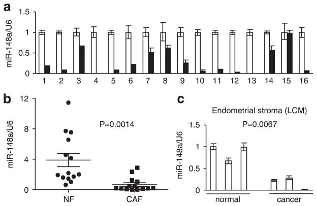
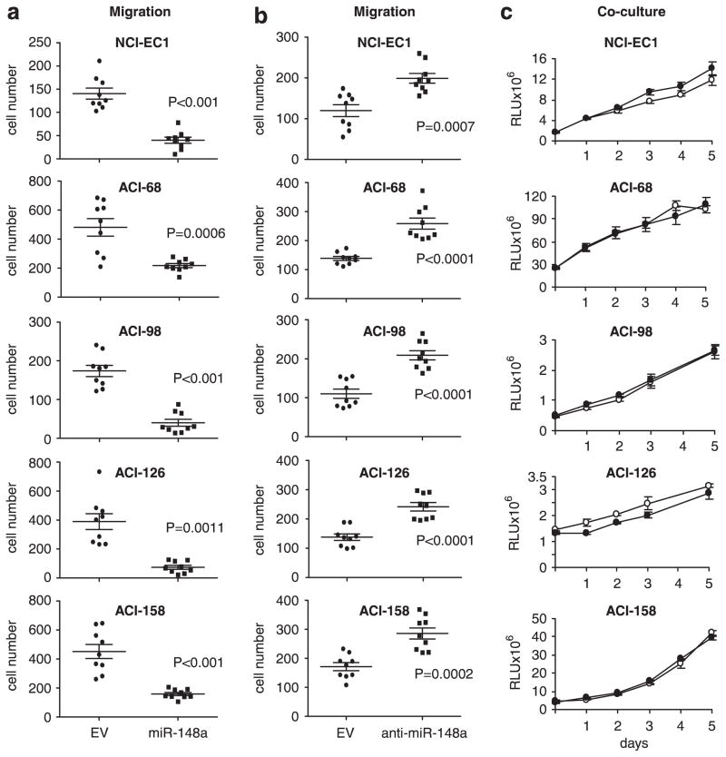
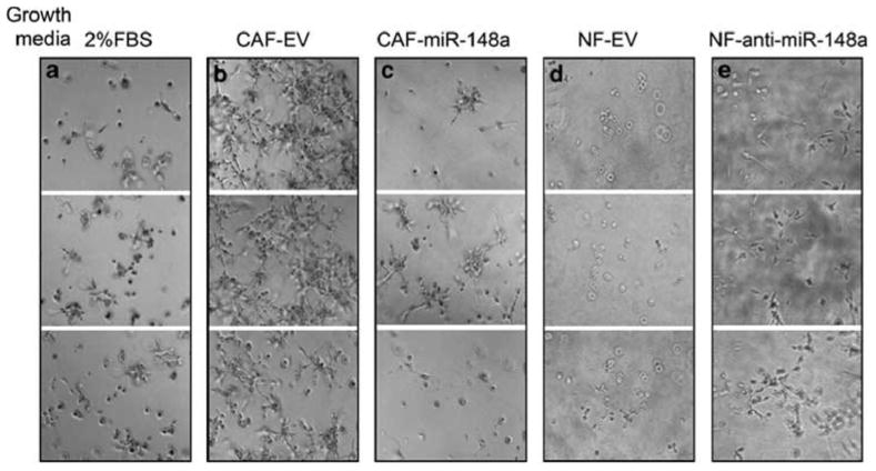
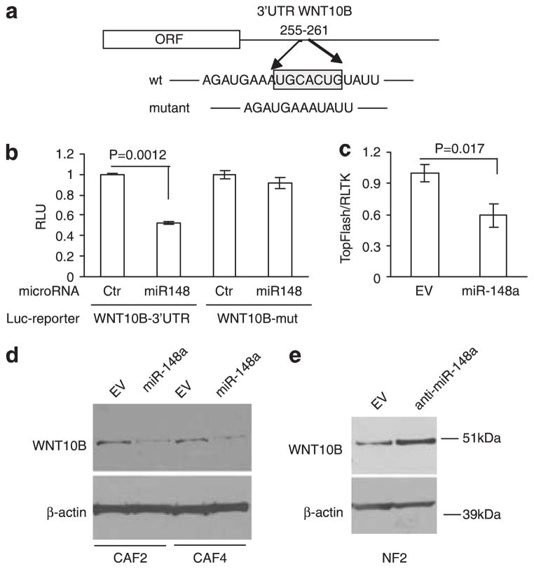
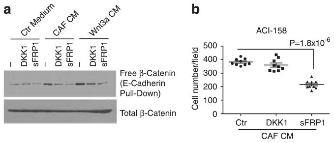
The tumor microenvironment has an important role in cancer progression. Here we show that miR-148a is downregulated in 15 out of 16 samples (94%) of cancer-associated fibroblasts (CAFs) compared with matched normal tissue fibroblasts (NFs) established from patients with endometrial cancer. Laser-capture microdissection of stromal cells from normal tissue and endometrial cancer confirmed this observation. Treatment of cells with 5-aza-deoxycytidine stimulated the expression of miR-148a in the majority of CAFs implicating DNA methylation in the regulation of miR-148a expression. Investigation of miR-148a function in fibroblasts demonstrated that conditioned media (CM) from CAFs overexpressing miR-148a significantly impaired the migration of five endometrial cancer cell lines without affecting their growth rates in co-culture experiments. Among predicted miR-148a target genes are two WNT family members, WNT1 and WNT10B. Activation of the WNT/β-catenin pathway in CAFs was confirmed by microarray analysis of gene expression and increased activity of the SuperTOPFlash luciferase reporter. We found elevated levels of WNT10B protein in CAFs and its level decreased when miR-148a was re-introduced by lentiviral infection. The 3'-UTR of WNT10B, cloned downstream of luciferase cDNA, suppressed luciferase activity when co-expressed with miR-148a indicating that WNT10B is a direct target of miR-148a. In contrast to the effect of miR-148a, WNT10B stimulated migration of endometrial cancer cell lines. Our findings have defined a molecular mechanism in the tumor microenvironment that is a novel target for cancer therapy.


Conflict of interest statement

CONFLICT OF INTEREST

The authors declare no conflict of interest.

Figures

Figure 1
Figure 1
Expression of miR-148a in endometrial cells, (a) qRT-PCR of miR-148a expression in CAFs relative to matched NFs from 16 patients. White bars: NFs; black bars: CAFs. Results are presented as mean ± s.e.m. of triplicate measurements, (b) Distribution of miR-148a expression values in NF and CAFs. Same as in (a), but all samples normalized to NF1. Analysis shows mean values with 95% confidence levels, (c) Expression of miR-148a in laser capture microdissected endometrial stromal cells from normal and cancer patients. Results are means ± s.e.m. of triplicate measurements.
Figure 2
Figure 2
Effect of miR-148a overexpression in fibroblasts on endometrial tumor cell motility and growth, (a) Transwell migration of five endometrial cancer cell lines towards CAFs expressing empty lentiviral vector (circles) or miR-148a (squares). Results are the means with 95% confidence interval of migrated cells/field. P-value was calculated using unpaired two-tailed t-test. All experiments were performed in triplicate, with three fields analyzed/replicate, and repeated at least two times, (b) Transwell migration of endometrial cancer cell lines towards NFs expressing empty lentiviral vector (circles) or anti-miR-148a (squares). The data are analyzed as in (a), (c) CAFs expressing empty lentiviral vector or miR-148a were co-cultured with luciferase-expressing endometrial cancer cell lines during 5-day time course. The luciferase activity was measured every day in three replicate experiments. White circles: CAF-EV; black circles: CAF-miR-148a. Results are means ± s.e.m.
Figure 3
Figure 3
ACI-158 endometrial cancer cells in 3D matrigel culture. Cells were grown in growth factor-reduced matrigel and DMEM containing 2% FBS (a), CM from CAFs expressing empty vector (b) or miR-148a (c), or NFs expressing vector control (d) or anti-miR-148a (e) cultured in DMEM/2% FBS. Media were replaced every 2 days and images were taken from three different fields at day 5. Experiments were repeated four times and a representative experiment is shown.
Figure 4
Figure 4
WNT pathway activation in CAFs. (a) Top ranking pathways enriched in CAFs versus NFs using SLEPR method. The analysis was run with KEGG pathway annotation with inclusion of fibroblast-specific WNT-responsive genes obtained from Klapholz-Brown et al. 103 permutations were performed. Permutated P-values: P-value of terms derived from permutated data; FDR-false discovery rates. FDR q values: FDR of terms derived from permutated data, (b) Heat map of WNT-responsive genes expressed in samples of CAFs and NFs. The rows of the heat map represent the WNT-responsive genes and the columns correspond to individual cell lines. The red color indicates enriched expression. (c) SuperTOPFIash reporter assay of CM from NFs vs CAFs. Paired samples from two patients were analyzed in luciferase reporter assay normalized either to Renilla-TK or SuperFopFlash (reporter with mutated TCF binding sites). Results are mean ± s.d. of triplicate measurements, (d) Western blot analysis of WNT10B in NFs and CAFs. Numbers below blots indicate WNT10B level in CAF normalized to β-actin and expressed relative to NF from the same patient, and miR-148a expression levels in CAFs relative to NF from Figure 1a.
Figure 5
Figure 5
WNT10B is a direct target of miR-148a in endometrial fibroblasts, (a) Schematic representation of WNT10B 3′UTR with putative miR-148a-binding site. The seed region is boxed and mutated reporter construct has seed region deleted, (b) Luciferase activity of HeLa cells co-transfected with reporter vector containing either wild-type or mutant WNT10B 3′-UTR and miR-148a or non-targeting control. Results are mean ± s.d. of triplicate measurements. (c) miR-148a suppresses SuperTopFlash reporter activity. CAFs stably expressing empty vector (EV) or lentiviral miR-148a construct were electroporated with the SuperTOPFIash and Renilla-TK. Luciferase activity was measured after 24 h. Results are mean ± s.d. of triplicate measurements, (d) Western blot analysis of WNT10B in CAF cell lysates after stable transfection with miR-148a construct or empty vector. Transfection and immunoblotting were performed with CAFs from two patients (CAF2 and CAF4). β-actin blot served as loading control, (e) Western blot analysis of WNT10B in NF cell lysates after stable transfection with anti-miR-148a construct or empty vector.
Figure 6
Figure 6
WNT10B stimulates migration of endometrial cancer cells. (a) Transwell migration assay of three endometrial cancer cell lines in response to CM from 293 T cells transfected with human WNT10B construct or empty vector control. Results are the means with 95% confidence interval of migrated cells/field. P-value was calculated using unpaired two-tailed t-test. (b) β-catenin stabilization assay following immunodepletion of WNT10B. ACI-158 cells were incubated for 3 h with untreated CAF CM (−), or CM treated with control IgG or anti-WNT10B. Wnt3a CM was used a positive control. Free (soluble) β-catenin obtained with a GST-Ecadherin pull-down protocol and β-catenin in whole cell lysates (total β-catenin) were detected by immunoblotting. (c) Transwell migration assay of ACI-158 cells towards CAF CM pre-treated with control IgG or anti-WNT10B. Assay was performed as described in (a), (d) Transwell migration assay of ACI-158 cells towards CM from CAF with stable expression of miR-148a and transient expression of WNT10B or empty vector, (e) Transwell migration assay of ACI-158 cells towards CM from NF with stable expression of anti-miR-148a immunodepleted with anti-WNT10B antibody or control IgG.
Figure 7
Figure 7
WNT10B stimulates tumor cell migration via non-canonical signaling, (a) β-catenin stabilization assay in ACI-158 cells treated with control or CAF CM or Wnt3a CM. Assay as described in (Figure 6b), with cells either pre-incubated with DKK1 (1 μg/ml) or sFRP1 (10 μg/ml) added to the CM. (b) Transwell migration assay of ACI-158 in the presence of DKK1 or sFRP1. Assay was performed as described in (Figure 6a) without addition of recombinant protein (Ctr) or with DKK1 (1 μg/ml) or sFRPI (10 μg/ml).

References

    1. Bissell MJ, Radisky D. Putting tumours in context. Nat Rev Cancer. 2001;1:46–54. - PMC - PubMed
    1. Polyak K, Haviv I, Campbell IG. Co-evolution of tumor cells and their micro-environment. Trends Genet. 2009;25:30–38. - PubMed
    1. Kalluri R, Zeisberg M. Fibroblasts in cancer. Nat Rev Cancer. 2006;6:392–401. - PubMed
    1. Aguirre-Ghiso JA. Models, mechanisms and clinical evidence for cancer dormancy. Nat Rev Cancer. 2007;7:834–846. - PMC - PubMed
    1. Kuperwasser C, Chavarria T, Wu M, Magrane G, Gray JW, Carey L, et al. Reconstruction of functionally normal and malignant human breast tissues in mice. Proc Natl Acad Sci USA. 2004;101:4966–4971. - PMC - PubMed

Publication types