. 2013 Feb;44(2):581-95.
doi: 10.1007/s00726-012-1376-x. Epub 2012 Aug 14.

Characterizing circular peptides in mixtures: sequence fragment assembly of cyclotides from a violet plant by MALDI-TOF/TOF mass spectrometry

Affiliations

Characterizing circular peptides in mixtures: sequence fragment assembly of cyclotides from a violet plant by MALDI-TOF/TOF mass spectrometry

Hossein Hashempour et al. Amino Acids. 2013 Feb.

Abstract

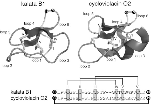
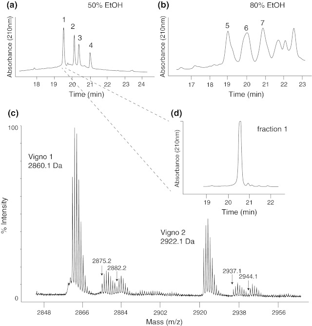
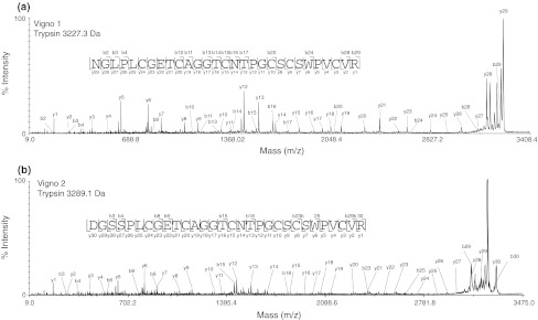
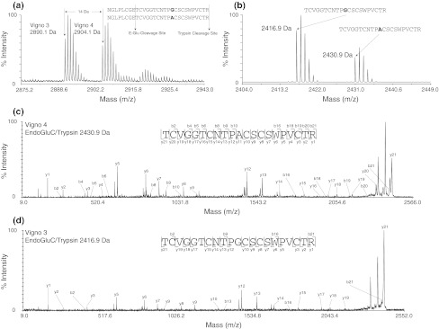
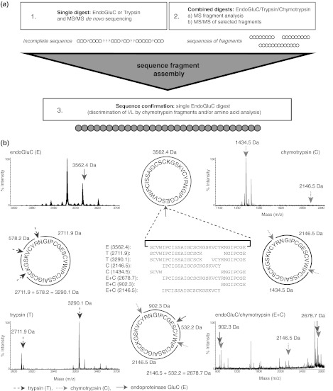
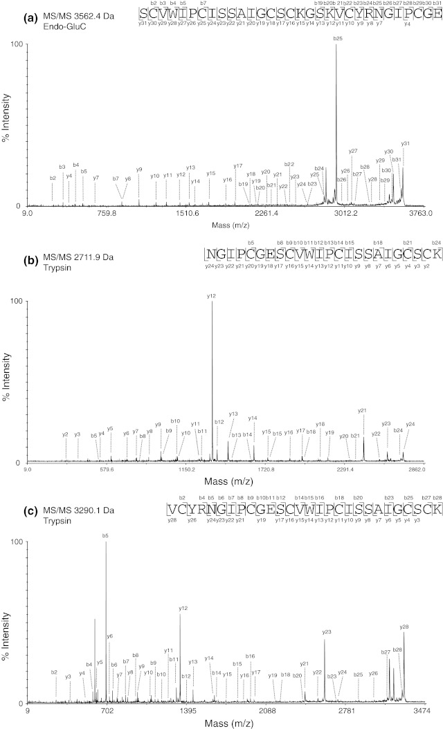
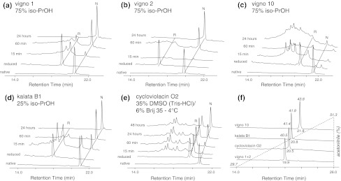
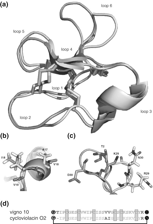
Cyclotides are a very abundant class of plant peptides that display significant sequence variability around a conserved cystine-knot motif and a head-to-tail cyclized backbone conferring them with remarkable stability. Their intrinsic bioactivities combined with tools of peptide engineering make cyclotides an interesting template for the design of novel agrochemicals and pharmaceuticals. However, laborious isolation and purification prior to de novo sequencing limits their discovery and hence their use as scaffolds for peptide-based drug development. Here we extend the knowledge about their sequence diversity by analysing the cyclotide content of a violet species native to Western Asia and the Caucasus region. Using an experimental approach, which was named sequence fragment assembly by MALDI-TOF/TOF, it was possible to characterize 13 cyclotides from Viola ignobilis, whereof ten (vigno 1-10) display previously unknown sequences. Amino acid sequencing of various enzymatic digests of cyclotides allowed the accurate assembly and alignment of smaller fragments to elucidate their primary structure, even when analysing mixtures containing multiple peptides. As a model to further dissect the combinatorial nature of the cyclotide scaffold, we employed in vitro oxidative refolding of representative vigno cyclotides and confirmed the high dependency of folding yield on the inter-cysteine loop sequences. Overall this work highlights the immense structural diversity and plasticity of the unique cyclotide framework. The presented approach for the sequence analysis of peptide mixtures facilitates and accelerates the discovery of novel plant cyclotides.


Figures

Fig. 1
Fig. 1
Ribbon structures of the cyclotides kalata B1 (left panel), a representative of the Möbius subfamily and cycloviolacin O2 (right panel) belonging to the bracelet subfamily are shown as cartoons. The unique cyclic cystine-knot (CCK) motif with three conserved disulfide bonds (yellow) and the cyclized backbone (black dots and connecting line) as well as typical secondary structure elements of α-helices (blue) and β-sheets (red) and their respective sequences are shown (PDB code: 1NB1 and 2KNM, respectively). The disulfide connectivity CI–IV, CII–V and CIII–VI has been indicated with black lines (color figure online)
Fig. 2
Fig. 2
HPLC fractionation of cyclotides from Viola ignobilis extract. Analytical HPLC traces of the 50 % (a) and 80 % (b) ethanolic solid-phase extracts yielding seven cyclotide-containing subfractions (labelled 17). (c) MALDI-TOF/TOF spectrum of fraction 1 indicating the masses of several cyclotides including the most abundant Möbius cyclotides vigno 1 and vigno 2, which are co-eluting (d) on analytical HPLC
Fig. 3
Fig. 3
MALDI-TOF/TOF sequencing of co-eluting vigno 1 and 2. MS/MS spectra of the precursor masses of 3,227.3 Da (a) and 3,289.1 Da (b) of a tryptic digest (reduced and S-carbamidomethylated) of fraction 1 (see Fig. 2) is shown. Observed C-terminal y- and N-terminal b-ions that allowed sequence characterization are labelled
Fig. 4
Fig. 4
MALDI-TOF/TOF identification of the co-eluting peptides vigno 3 and vigno 4. The difference of 14 Da can be observed in crude (a) and within the combined trypsin and endoproteinase GluC digest (b) of fraction 4. MS/MS sequencing of the endo-GluC/trypsin-digested precursors with 2430.9 Da (c) and 2416.9 Da (d), respectively, allowed unambiguous assignment of the sequences of these two peptides
Fig. 5
Fig. 5
Sequence fragment assembly approach for vigno 6. a An overview of the sequence fragment assembly workflow that has been used to elucidate cyclotide sequences in mixtures is presented. b The combination of single trypsin, chymotrypsin and endoproteinase GluC and a combination of chymotrypsin/endo-GluC digests of fractions containing multiple cyclotides together with the alignment of partial sequences and assembling of peptide-specific fragments allowed the discrimination and unambiguous assignment and elucidation of the cyclotide sequences. MS spectra of four digests using endo-GluC (upper left panel), chymotrypsin (upper right), trypsin (lower left) and a combination of endoproteinase GluC and chymotrypsin (lower right) are shown for vigno 6. The cleavage sites and resulting peptide fragments of the different enzymes are indicated by arrows (trypsin: blue/dashed line, chymotrypsin: green/dotted line, endo-GluC: red/straight line). The alignment of obtained MS sequence fragments (middle) together with MS/MS sequence data of selected precursors (see Fig. 6) allows the unambiguous sequence elucidation of the novel cyclotide vigno 6 (color figure online)
Fig. 6
Fig. 6
MS/MS sequencing of vigno 6. Three MS/MS spectra of (a, b) the precursors with the molecular weight of 2,711.9 and 3,290.1 Da, respectively, from a tryptic digest and (c) the linearized cyclotide precursor with a molecular weight of 3,265.4 Da from an endoproteinase GluC digest are shown. The sequences were obtained by assigning the y- and b-ions series
Fig. 7
Fig. 7
Refolding of cyclotides. RP-HPLC traces of native, reduced and refolded peptides under conditions leading to the highest yields of vigno 1 (a), vigno 2 (b), vigno 10 (c), kalata B1 (d) and cycloviolacin O2 (e) are offset aligned for clarity. The folding of vigno peptides and kalata B1 (ad) was performed in 0.1 M NH4HCO3 at 20 °C and cycloviolacin O2 (e) folding was carried out in Tris-buffer at 4 °C (see “Materials and methods” for further details). f Difference in hydrophobicity of all peptides used in this study is shown
Fig. 8
Fig. 8
Structural alignment of vigno 10 and cycloviolacin O2. The structures of cycloviolacin O2 (cyan PDB code: 2GJ0) and the homology model of vigno 10 (green) were aligned using PyMOL (root-mean-square deviation = 1.324 Å) and are shown in cartoon representation (a). The cyclotide loops and disulfide bonds (yellow) are indicated. Differences in the side-chain orientation of the distinct residues of both cyclotides of loop 3 (b) and loop 6 (c) are indicated in stick representation and amino acids are labelled in one-letter code, numbered according to their position in the cyclotide sequence (starting from G1, see d). Images have been prepared using PyMOL. (d) Sequence alignment of vigno 10 and cycloviolacin O2 with the three conserved disulfide bridges (shown in yellow) and the cyclized backbone (black dots and connecting line). Residues differing between those two cyclotides have been highlighted in red (color figure online)

References

    1. Aboye TL, Clark RJ, Burman R, Roig MB, Craik DJ, Göransson U. Interlocking disulfides in circular proteins: toward efficient oxidative folding of cyclotides. Antioxid Redox Signal. 2011;14(1):77–86. doi: 10.1089/ars.2010.3112. - DOI - PubMed
    1. Barbeta BL, Marshall AT, Gillon AD, Craik DJ, Anderson MA. Plant cyclotides disrupt epithelial cells in the midgut of lepidopteran larvae. Proc Natl Acad Sci USA. 2008;105(4):1221–1225. doi: 10.1073/pnas.0710338104. - DOI - PMC - PubMed
    1. Claeson P, Goransson U, Johansson S, Luijendijk T, Bohlin L. Fractionation protocol for the isolation of polypeptides from plant biomass. J Nat Prod. 1998;61(1):77–81. doi: 10.1021/np970342r. - DOI - PubMed
    1. Clark RJ, Daly NL, Craik DJ. Structural plasticity of the cyclic-cystine-knot framework: implications for biological activity and drug design. Biochem J. 2006;394(Pt 1):85–93. - PMC - PubMed
    1. Colgrave ML, Craik DJ. Thermal, chemical, and enzymatic stability of the cyclotide kalata B1: the importance of the cyclic cystine knot. Biochemistry. 2004;43(20):5965–5975. doi: 10.1021/bi049711q. - DOI - PubMed

Publication types

MeSH terms