. 2012 Nov 15;590(22):5845-60.
doi: 10.1113/jphysiol.2012.238113. Epub 2012 Aug 13.

Neurocardiac dysregulation and neurogenic arrhythmias in a transgenic mouse model of Huntington's disease

Affiliations

Neurocardiac dysregulation and neurogenic arrhythmias in a transgenic mouse model of Huntington's disease

Helen Kiriazis et al. J Physiol. .

Abstract

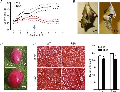
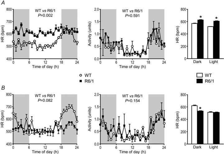
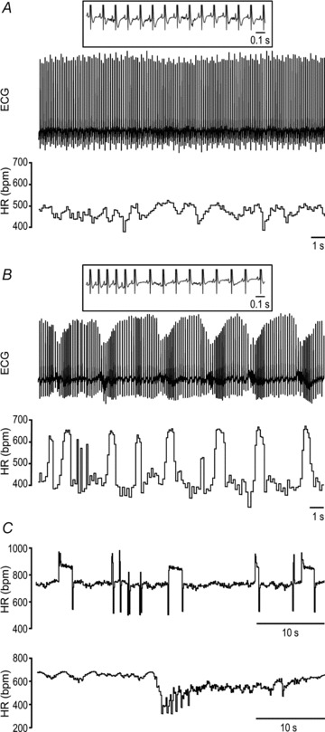
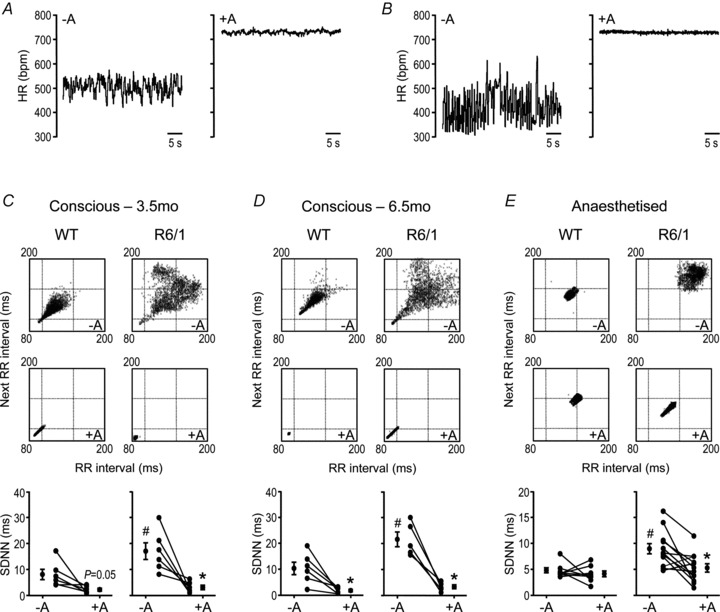
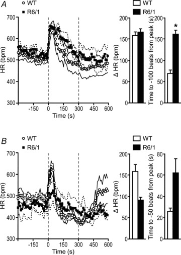
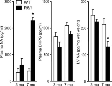
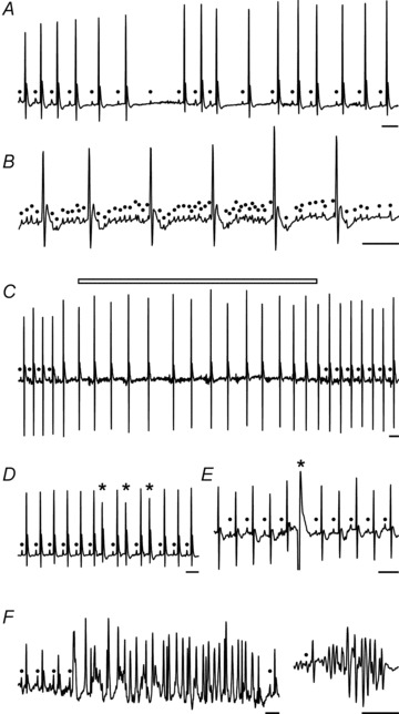
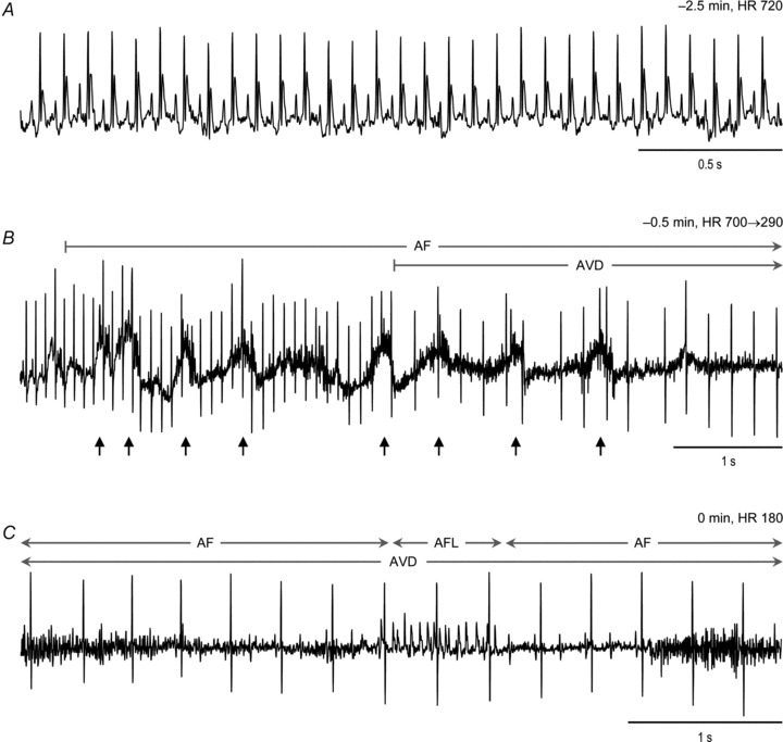
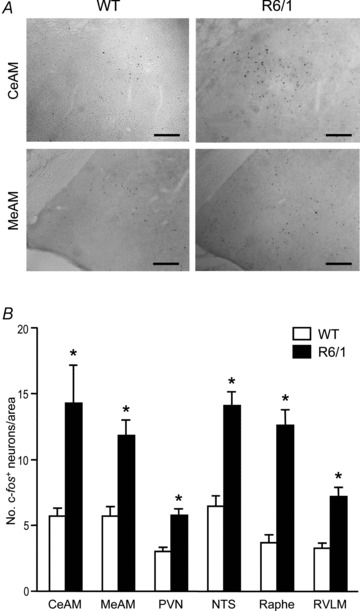
Huntington's disease (HD) is a heritable neurodegenerative disorder, with heart disease implicated as one major cause of death. While the responsible mechanism remains unknown, autonomic nervous system (ANS) dysfunction may play a role. We studied the cardiac phenotype in R6/1 transgenic mice at early (3 months old) and advanced (7 months old) stages of HD. While exhibiting a modest reduction in cardiomyocyte diameter, R6/1 mice had preserved baseline cardiac function. Conscious ECG telemetry revealed the absence of 24-h variation of heart rate (HR), and higher HR levels than wild-type littermates in young but not older R6/1 mice. Older R6/1 mice had increased plasma level of noradrenaline (NA), which was associated with reduced cardiac NA content. R6/1 mice also had unstable R-R intervals that were reversed following atropine treatment, suggesting parasympathetic nervous activation, and developed brady- and tachyarrhythmias, including paroxysmal atrial fibrillation and sudden death. c-Fos immunohistochemistry revealed greater numbers of active neurons in ANS-regulatory regions of R6/1 brains. Collectively, R6/1 mice exhibit profound ANS-cardiac dysfunction involving both sympathetic and parasympathetic limbs, that may be related to altered central autonomic pathways and lead to cardiac arrhythmias and sudden death.


Figures

Figure 1
Figure 1. Gross phenotype and cardiac histology of R6/1 mice at early HD (3 months old) and advanced HD (7 months old) stages
A, growth curves (mean ± SD) showing cessation of body weight gain in R6/1 mice prior to the age when they exhibit typical HD motor symptoms (arrow). B, when suspended by the tail, R6/1 mice showed a typical hindlimb clasping, whereas wild-type (WT) mice spread their four limbs. C, reduced heart size in male R6/1 mice at 7 months old. D, there were no overt histological abnormalities of the LV myocardium (×20, Masson's trichrome), except for a 10–15% reduction in cardiomyocyte (CM) diameter at both 3 and 7 months old. Data are means ± SEM, *P < 0.05 vs. age-matched WT (two-way ANOVA); n= 4 per group.
Figure 2
Figure 2. Changes in conscious heart rate (HR) and physical activity across 24 h in R6/1 and wild-type (WT) mice
Plots of hourly averaged conscious HRs across 24 h for mice at 3.5 (A) and 6.5 (B) months old show a variation with time of day in WT (n= 4–6/group, effect of time P < 0.001 both ages) but not in R6/1 mice (n= 5–6/group, effect of time P > 0.05 both ages). HR levels over the 24 h period were significantly higher in the 3.5-month-old R6/1 than age-matched WT mice (P= 0.002), but not in the older mice (P= 0.082). Notably, HR was lower in old compared with young R6/1 mice (P= 0.001). Activity was not significantly different between the groups at both ages (P= 0.591 and 0.154, respectively). Shaded areas represent lights-off periods of the day; 24 h data were analysed using ANOVA for repeated measures. Bar graphs illustrate the HR averaged over the 12 h light and 12 h dark periods, and show that compared with age-matched WT mice, young R6/1 mice have a higher HR throughout the day; whereas, older R6/1 mice have a lower HR during the dark phase (*P < 0.05, Student's unpaired t test).
Figure 3
Figure 3. Variation of cardiac rhythm in conscious mice
Telemetry ECG and/or ECG-derived heart rate (HR) from a typical wild-type (A) and R6/1 mouse at 3 months old (B and C), with the latter showing marked instability in HR. Insets show P waves coupled with QRS.
Figure 4
Figure 4. Heart rate (HR) variation and effect of the muscarinic antagonist atropine in conscious and isoflurane-anaesthetised mice
A and B, ECG-derived HR in representative wild-type (WT) (A) and R6/1 (B) mice immediately before and 10 min after atropine (1.2 mg kg−1 i.p.). C–E, Poincaré plots (upper, no atropine, and middle, with atropine) showing beat-to-beat HR variation in conscious (C, at 3.5 months old, and D, 6.5 months old) and isoflurane anaesthesia (E, at 4–5 months old) R6/1 and WT representative mice, with each R–R interval plotted against the preceding R–R interval, derived from 5 min recordings (∼2000–3500 beats). HR variation was attenuated by atropine. The findings are summarised in the graphs (bottom) showing standard deviation of normal R–R intervals (SDNN), with the mean ± SEM plotted either side of data from individual mice (circles + lines). At baseline (no atropine), SDNN was significantly higher for R6/1 versus its age-matched WT group (#P < 0.05, Student's unpaired t test). Following atropine, SDNN was attenuated compared with the corresponding pre-atropine values, and this was more clearly evident in R6/1 mice (*P < 0.05, Student's paired t test). There were no group differences in SDNN in the presence of atropine (P > 0.05, Student's unpaired t test). n= 5–12; –A, no atropine; +A, with atropine.
Figure 5
Figure 5. Heart rate (HR) response to the β-adrenergic agonist isoproterenol in conscious and isoflurane-anaesthetised mice
A and B, absolute HR and change in HR (Δ HR) following injection with isoproterenol (indicated by filled triangle, 4 μg kg−1, i.p.), in conscious 3.5- (A) and 6.5-month-old (B) R6/1 and wild-type (WT) mice (n= 5–6). Group differences were more prominent at 6.5 months old. *P < 0.05, ANOVA for repeated measures. C, representative traces from R6/1 and WT anaesthetised 4–5-month-old mice showing a blunted HR response to isoproterenol (filled triangle, 4 μg kg−1, i.p.) and the effect of pre-treatment with atropine (1.2 mg kg−1 i.p.). D, grouped data show that the blunted HR response to isoproterenol (filled triangle) in R6/1 mice was normalised by atropine pre-treatment (n= 3–5 per group). Symbols and faint lines represent means ± SEM. –A, no atropine; +A, with atropine.
Figure 6
Figure 6. Heart rate (HR) response to shake stress test
Animals that were 3.5 (A, n= 6–7) and 6.5 (B, n= 4) months old were subjected to the shake stress test for 5 min (indicated by the period between the dashed vertical lines), and HR was derived from telemetry ECG recordings. Plotted data are means ± SEM (the latter indicated by the accompanying faint lines). A, compared with wild-type (WT) mice, R6/1 mice at 3.5 months old showed a similar peak HR (P= 0.804, Student's unpaired t test) but a slower HR recovery to baseline (*P= 0.013, Student's unpaired t test). B, at 6.5 months old, although there was a tendency for a decrease in the peak HR level and a slower recovery towards baseline in response to the shake stress test in R6/1 compared with WT mice, these parameters were not different between the two groups (P= 0.112 and 0.134, respectively, Student's unpaired t test).
Figure 7
Figure 7. Plasma levels of noradrenaline (NA) and 3,4-dihydroxyphenylglycol (DHPG), and cardiac content of NA
Plasma NA was significantly increased and LV NA decreased in 7-month-old R6/1 compared with age-matched wild-type (WT) mice. These changes were not evident at 3 months old. There were no significant changes in plasma DHPG levels between R6/1 and WT mice at 3 or 7 months old. *P < 0.05 vs. age-matched WT and 3-month-old R6/1 mice (two-way ANOVA). n= 4–6 per group.
Figure 8
Figure 8. Different types of arrhythmias detected by telemetry ECG in conscious R6/1 mice
A, sinus arrhythmias and atrial–ventricular conduction blockade (note the absence of consistent change in P–R duration). B, atrial flutter. C, paroxysmal AF (open bar) evidenced by loss of P waves, unstable baseline and irregularity of R–R intervals. D, supra- ventricular ectopic beats. E, ventricular premature beat. F, episodes of ventricular tachycardia; closed circles indicates P wave; asterisk indicates ectopic beats; scale bars: 0.1 s. Group data shown in Table 3.
Figure 9
Figure 9. Telemetry ECG from a 7-month-old R6/1 mouse revealing development of arrhythmias and sudden death
A and B, ECG traces starting from 2.5 min prior to death show the time course from normal cardiac rhythm (A) to development of atrial fibrillation (AF) immediately followed by complete atrial–ventricular disassociation (AVD) (B). Small arrows indicate changes in ECG baseline likely due to hypoxia-driven gasps just prior to death. C, finally, sustained AF interrupted by a short episode of atrial flutter (AFL), together with a slow and regular ventricular rhythm as the terminal presentation of ECG. HR, heart rate.
Figure 10
Figure 10. Density of active neurons in various brain regions controlling autonomic nervous activity
A, images of brain sections showing active neurons (black dots) detected by immunohistochemistry for c-Fos. Photomicrographs of coronal sections through the central (CeAM) and medial (MeAM) nuclei of the amygdala in 5.5-month-old wild-type (WT) and R6/1 mouse brains. Scale bars: 100 μm. B, bar graph showing grouped data of active neurons in brain regions that are known autonomic nervous centres (n= 4/group). CeAM and MeAM, central and medial nuclei of the amygdala, respectively; PVN, paraventricular nucleus of the hypothalamus; NTS, nucleus tractus solitarii; Raphe, raphe pallidus; RVLM, rostral ventrolateral medulla. *P < 0.05 vs. WT (ANOVA).

Similar articles

Cited by

References

    1. Andrich J, Schmitz T, Saft C, Postert T, Kraus P, Epplen JT, Przuntek H, Agelink MW. Autonomic nervous system function in Huntington's disease. J Neurol Neurosurg Psychiatry. 2002;72:726–731. - PMC - PubMed
    1. Arora R, Ulphani JS, Villuendas R, Ng J, Harvey L, Thordson S, Inderyas F, Lu Y, Gordon D, Denes P, Greene R, Crawford S, Decker R, Morris A, Goldberger J, Kadish AH. Neural substrate for atrial fibrillation: implications for targeted parasympathetic blockade in the posterior left atrium. Am J Physiol Heart Circ Physiol. 2008;294:H134–H144. - PubMed
    1. Aziz NA, Anguelova GV, Marinus J, van Dijk JG, Roos RA. Autonomic symptoms in patients and pre-manifest mutation carriers of Huntington's disease. Eur J Neurol. 2010;17:1068–1074. - PubMed
    1. Aziz NA, Pijl H, Frolich M, Schroder-van der Elst JP, van der Bent C, Roelfsema F, Roos RA. Delayed onset of the diurnal melatonin rise in patients with Huntington's disease. J Neurol. 2009;256:1961–1965. - PMC - PubMed
    1. Bar KJ, Boettger MK, Andrich J, Epplen JT, Fischer F, Cordes J, Koschke M, Agelink MW. Cardiovagal modulation upon postural change is altered in Huntington's disease. Eur J Neurol. 2008;15:869–871. - PubMed

Publication types

MeSH terms

LinkOut - more resources