. 2012 Oct;32(20):4181-94.
doi: 10.1128/MCB.00483-12. Epub 2012 Aug 13.

A eukaryotic translation initiation factor 4E-binding protein promotes mRNA decapping and is required for PUF repression

Affiliations

A eukaryotic translation initiation factor 4E-binding protein promotes mRNA decapping and is required for PUF repression

Nathan H Blewett et al. Mol Cell Biol. 2012 Oct.

Abstract

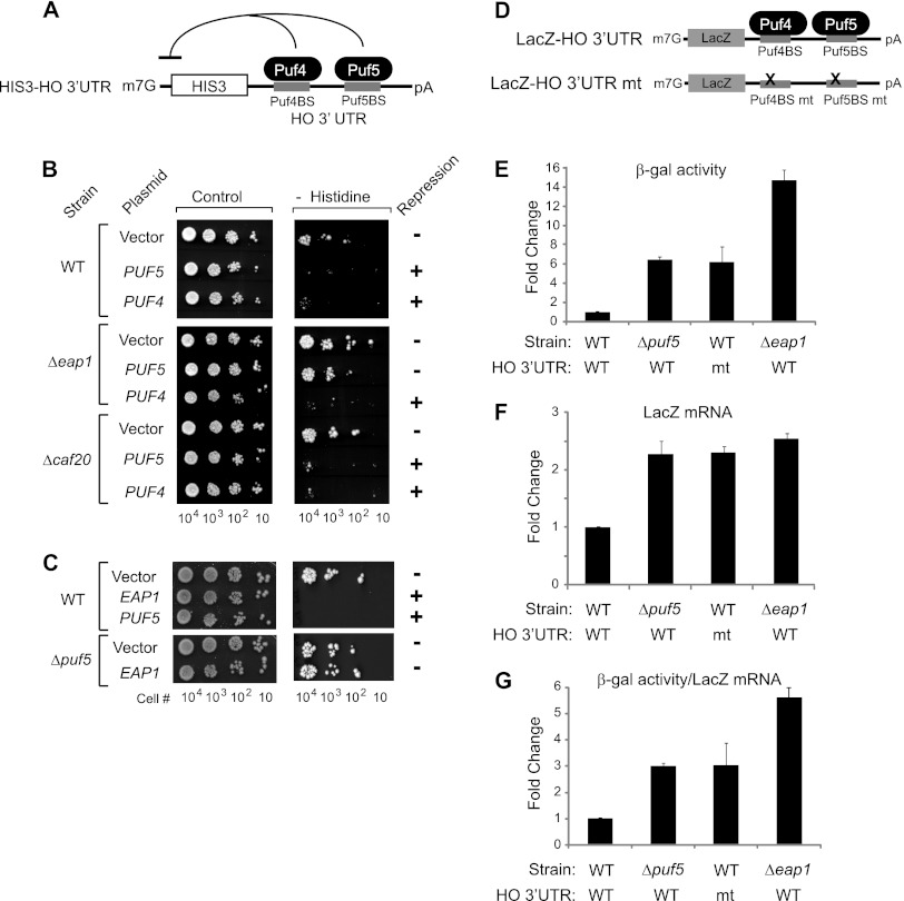
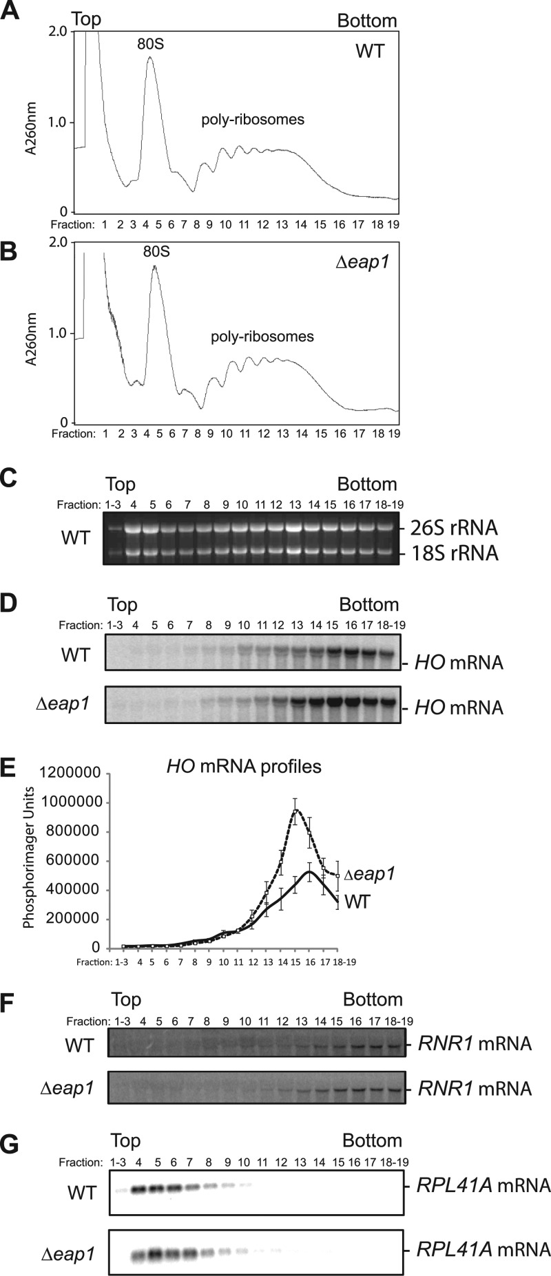
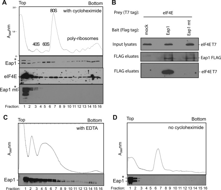
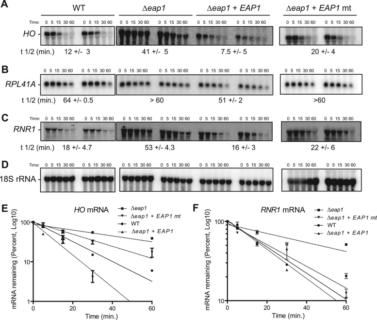
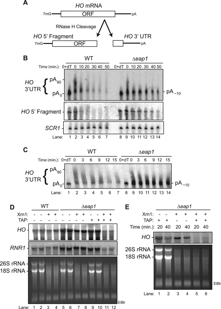
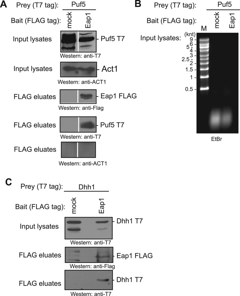
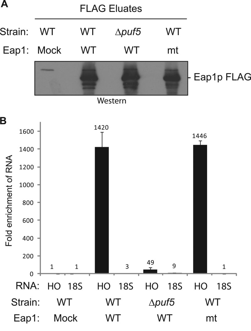
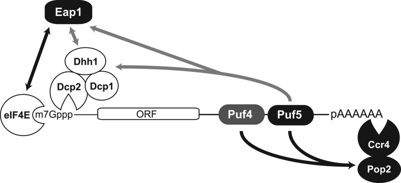
PUF proteins are eukaryotic RNA-binding proteins that repress specific mRNAs. The mechanisms and corepressors involved in PUF repression remain to be fully identified. Here, we investigated the mode of repression by Saccharomyces cerevisiae Puf5p and Puf4p and found that Puf5p specifically requires Eap1p to repress mRNAs, whereas Puf4p does not. Surprisingly, we observed that Eap1p, which is a member of the eukaryotic translation initiation factor 4E (eIF4E)-binding protein (4E-BP) class of translational inhibitors, does not inhibit the efficient polyribosome association of a Puf5p target mRNA. Rather, we found that Eap1p accelerates mRNA degradation by promoting decapping, and the ability of Eap1p to interact with eIF4E facilitates this activity. Deletion of EAP1 dramatically reduces decapping, resulting in accumulation of deadenylated, capped mRNA. In support of this phenotype, Eap1p associates both with Puf5p and the Dhh1p decapping factor. Furthermore, recruitment of Eap1p to downregulated mRNA is mediated by Puf5p. On the basis of these results, we propose that Puf5p promotes decapping by recruiting Eap1p and associated decapping factors to mRNAs. The implication of these findings is that a 4E-BP can repress protein expression by promoting specific mRNA degradation steps in addition to or in lieu of inhibiting translation initiation.


Figures

Fig 1
Fig 1
EAP1 is required for Puf5p-mediated repression. (A) The HIS3-HO 3′UTR reporter contains the HIS3 open reading frame (ORF) with the HO 3′ untranslated region (3′UTR) containing the Puf4p binding site (Puf4BS) and Puf5p binding site (PUF5BS). (B) Growth assays measure repression of HIS3-HO reporter in the wild-type (WT) strain and in eap1eap1) or caf20caf20) deletion strains transformed with plasmids expressing PUF4 or PUF5 or with control plasmid. The indicated numbers of cells (Cell #) were spotted onto medium with histidine (Control) or without histidine (− Histidine). Repression by each gene was scored by no growth on medium lacking histidine (+) or growth (−). (C) Growth assay for repression by PUF5 and EAP1 in wild-type and puf5 deletion (Δpuf5) cells. (D) Diagram of the LacZ-HO 3′UTR reporter mRNA. The LacZ HO 3′UTR mt reporter was created by mutating the Puf4p and Puf5p binding sites (Puf4BS mt and Puf5BS mt [mt stands for mutant]). (E) Graph of β-galactosidase (β-gal) activity from wild-type or mutant (mt) LacZ reporters measured from equal number of wild-type, Δpuf5, or Δeap1 cells. The fold change in relative light unit values is plotted relative to wild-type reporter in wild-type cells. (F) Graph of fold change in LacZ HO (WT or mt) mRNA levels, as measured by Northern blotting (see Fig. S1 in the supplemental material) and calculated relative to the wild-type reporter in wild-type cells. (G) Graph of fold change in the ratio of β-galactosidase activity in panel E to LacZ mRNA level in panel F. In each graph, mean values are plotted with standard errors (error bars) from multiple biological replicates.
Fig 2
Fig 2
Eap1p does not inhibit polyribosome association of a Puf5p-regulated mRNA. Ribosome profiles of sucrose density gradients from wild-type (WT) cells (A) and eap1 deletion (Δeap1) cells (B). UV absorbance at 260 nm (A260nm) was measured during collection of fractions to generate the chromatograms. “Top” and “Bottom” refer to the relative position in the gradient tube. Ribosome species are indicated within the chromatogram. (C) The ethidium bromide-stained gel shows the rRNA (26S and 18S rRNA) content of each fraction from WT cells. (D) Northern blots of HO mRNA in gradient fractions from WT and Δeap1 strains. (E) Quantitation of HO mRNA profile across gradient fractions from three biological replicate samples of wild-type and Δeap1 strains. Mean values are plotted with standard errors (error bars). (F and G) Northern blots of RNR1 (F) and RPL41A (G) mRNAs in gradient fractions from WT and Δeap1 strains.
Fig 3
Fig 3
Eap1p fractionates with polyribosomes. (A) (Top) Absorbance at 260 nm (A260nm) chromatogram of sucrose density gradient of sample from eap1 deletion strain expressing FLAG-tagged Eap1p with T7-tagged eIF4E. Western blots of wild-type Eap1p (Eap1), eIF4E, and Eap1p with Y109A and L114A mutations in the eIF4E binding motif (Eap1 mt) in gradient fractions are shown below the chromatogram. Cycloheximide was present in each sample. Western blot of Eap1 mt was overexposed 10-fold relative to wild-type Eap1. The asterisk indicates Eap1p with an electrophoretic mobility lower than that for the expected 70-kDa size. (B) Western blots of RNase-treated immunoprecipitations of FLAG-tagged, wild-type or mutant Eap1p from cells coexpressing T7-tagged eIF4E. Mock FLAG immunoprecipitation was from cells expressing eIF4E-T7 but not Eap1p-FLAG. (C) Chromatogram from cell extracts treated with EDTA to dissociate polyribosomes. FLAG-tagged Eap1p was detected by Western blotting. (D) Chromatogram from extracts incubated in the absence of cycloheximide resulted in ribosome runoff and accumulation of 80S ribosomes. Eap1p was detected by Western blotting.
Fig 4
Fig 4
Eap1p accelerates mRNA degradation. mRNA half-lives were measured by transcription shutoff with thiolutin. RNA samples were collected at the indicated time points (in minutes). Blots from duplicate samples are shown for each condition. The average mRNA half-lives are indicated below each sample. (A) Northern blot of HO mRNA from wild-type (WT), eap1 deletion (Δeap1), or eap1 deletion cells complemented with plasmids expressing wild-type Eap1p (Δeap1+EAP1) or eIF4E binding defective mutant Eap1p (Δeap1+EAP1 mt). (B) Northern blots of RPL41A mRNA from samples in panel A. (C) Northern blots of RNR1 mRNA from samples in panel A. (D) Northern blots of the stable 18S rRNA from samples in panel A. (E) Graph of HO mRNA half-lives in wild-type, Δeap1, Δeap1+EAP1, and Δeap1+EAP1 mt strains. (F) Graph of RNR1 mRNA half-lives in each strain.
Fig 5
Fig 5
Eap1p promotes decapping of HO mRNA. (A) HO mRNA was cleaved with RNase H and a DNA oligonucleotide to produce a 1,600-nucleotide 5′ fragment and a 253-nucleotide 3′ fragment with a poly(A) tail of up to 80 adenosines (pA80). (B) Northern blot analysis of HO mRNA decay in wild-type (WT) or eap1 deletion strain (Δeap1). RNA samples were collected over a time course (in minutes) following the addition of thiolutin and then cleaved as shown in panel A. The 0-min samples were also treated with RNase H and oligo(dT15) (designated 0+dT) to remove the poly(A) tail (pA0). (Top) Northern blot analysis of poly(A) tail length and decay rate of the HO 3′UTR with a specific probe to the 3′ fragment. The position of the oligo-adenylated HO intermediate (pA∼10) is indicated to the right of the blot. (Middle) Northern blot of the HO 5′ fragment. (Bottom) SCR1 RNA was detected by Northern blotting as a control for sample loading. (C) Analysis of HO mRNA decay and deadenylation over the initial 15 min of decay following the addition of thiolutin. (D) Duplicate RNA samples from panel B were treated with recombinant Xrn1 (+) or not treated with recombinant Xrn1 (−) to assess the status of the 5′ cap. For a control, where indicated, the samples were also treated with tobacco acid pyrophosphatase (TAP) to remove the 5′ cap. Northern blotting for HO and RNR1 mRNA are indicated on the left. Before transfer to membrane, the gel was stained with ethidium bromide (EtBr) (bottom blot) to visualize degradation of uncapped 18S and 26S rRNA. (E) Xrn1 sensitivity assay of HO mRNA decay intermediates from the 20- and 40-min time points in panel B.
Fig 6
Fig 6
Eap1p associates with Puf5p and Dhh1p. (A) T7-tagged Puf5p coimmunoprecipitates with FLAG-tagged Eap1p from cell extracts (input) treated with RNases A and One. Western blot detection of input lysates and FLAG peptide eluates from mock or Eap1-FLAG immunoprecipitations. (B) RNase-mediated destruction of RNA in extracts from panel A was confirmed by ethidium bromide (EtBr) staining of nucleic acids. The migration positions (in thousands of nucleotides or kilonucleotides [knt]) of RNA size markers (lane M) are shown to the left of the blot. (C) T7-tagged Dhh1p coimmunoprecipitates with Eap1-FLAG from RNase-treated cell lysates (input).
Fig 7
Fig 7
Eap1p associates with HO mRNA, dependent on Puf5p. (A) FLAG-tagged wild-type Eap1p (WT) or eIF4E binding defect mutant Eap1p (mt) were expressed and immunopurified from wild-type or Δpuf5 cells. For a negative control, mock immunoprecipitation was performed on cells that did not express tagged Eap1p. The blot is an anti-FLAG Western blot of FLAG peptide eluates. (B) Graph of fold enrichment of HO mRNA purified from eluted samples in panel A, detected using qRT-PCR. Fold enrichment was calculated relative to mock immunoprecipitation, normalized to input levels. As a negative control, 18S rRNA was also measured. Note that conditions used in this assay did not preserve polyribosomes.
Fig 8
Fig 8
Model of posttranscriptional regulation of HO mRNA by PUFs and Eap1p. Puf4p and Puf5p bind to their respective sites in the 3′UTR of HO mRNA. Both proteins recruit the Pop2p-Ccr4p deadenylase complex to accelerate deadenylation. Puf5p also recruits Eap1p and decapping factors Dhh1p and decapping enzyme Dcp1-Dcp2 to enhance decapping. Eap1p interacts with 5′ cap-bound eIF4E to facilitate mRNA decay. Direct protein interactions are shown by black lines. Gray lines indicate protein associations; direct protein-protein contacts remain unknown.

Similar articles

Cited by

References

    1. Altmann M, Schmitz N, Berset C, Trachsel H. 1997. A novel inhibitor of cap-dependent translation initiation in yeast: p20 competes with eIF4G for binding to eIF4E. EMBO J. 16:1114–1121 - PMC - PubMed
    1. Arava Y, et al. 2003. Genome-wide analysis of mRNA translation profiles in Saccharomyces cerevisiae. Proc. Natl. Acad. Sci. U. S. A. 100:3889–3894 - PMC - PubMed
    1. Asaoka-Taguchi M, Yamada M, Nakamura A, Hanyu K, Kobayashi S. 1999. Maternal Pumilio acts together with Nanos in germline development in Drosophila embryos. Nat. Cell Biol. 1:431–437 - PubMed
    1. Beelman CA, et al. 1996. An essential component of the decapping enzyme required for normal rates of mRNA turnover. Nature 382:642–646 - PubMed
    1. Blewett N, Coller J, Goldstrohm A. 2011. A quantitative assay for measuring mRNA decapping by splinted ligation reverse transcription polymerase chain reaction: qSL-RT-PCR. RNA 17:535–543 - PMC - PubMed

Publication types

MeSH terms