. 2012 Nov;80(11):3786-94.
doi: 10.1128/IAI.00647-12. Epub 2012 Aug 13.

Suppression of Clostridium difficile in the gastrointestinal tracts of germfree mice inoculated with a murine isolate from the family Lachnospiraceae

Affiliations

Suppression of Clostridium difficile in the gastrointestinal tracts of germfree mice inoculated with a murine isolate from the family Lachnospiraceae

Angela E Reeves et al. Infect Immun. 2012 Nov.

Abstract

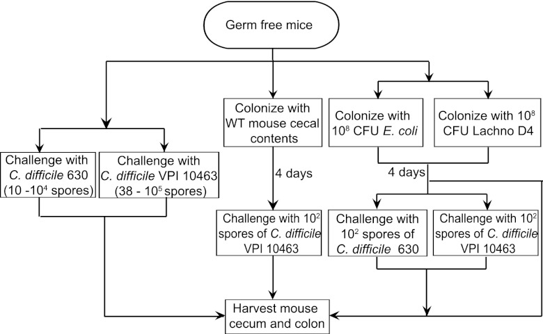
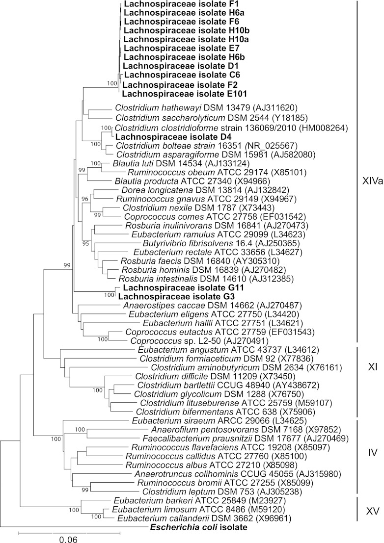
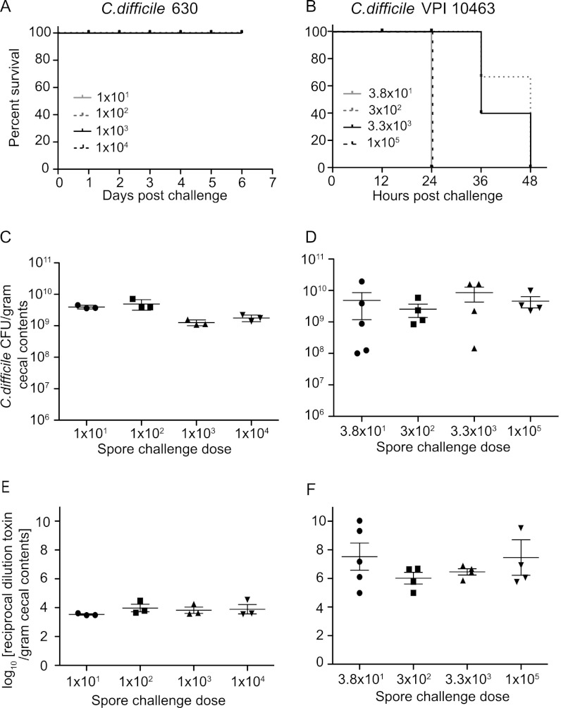
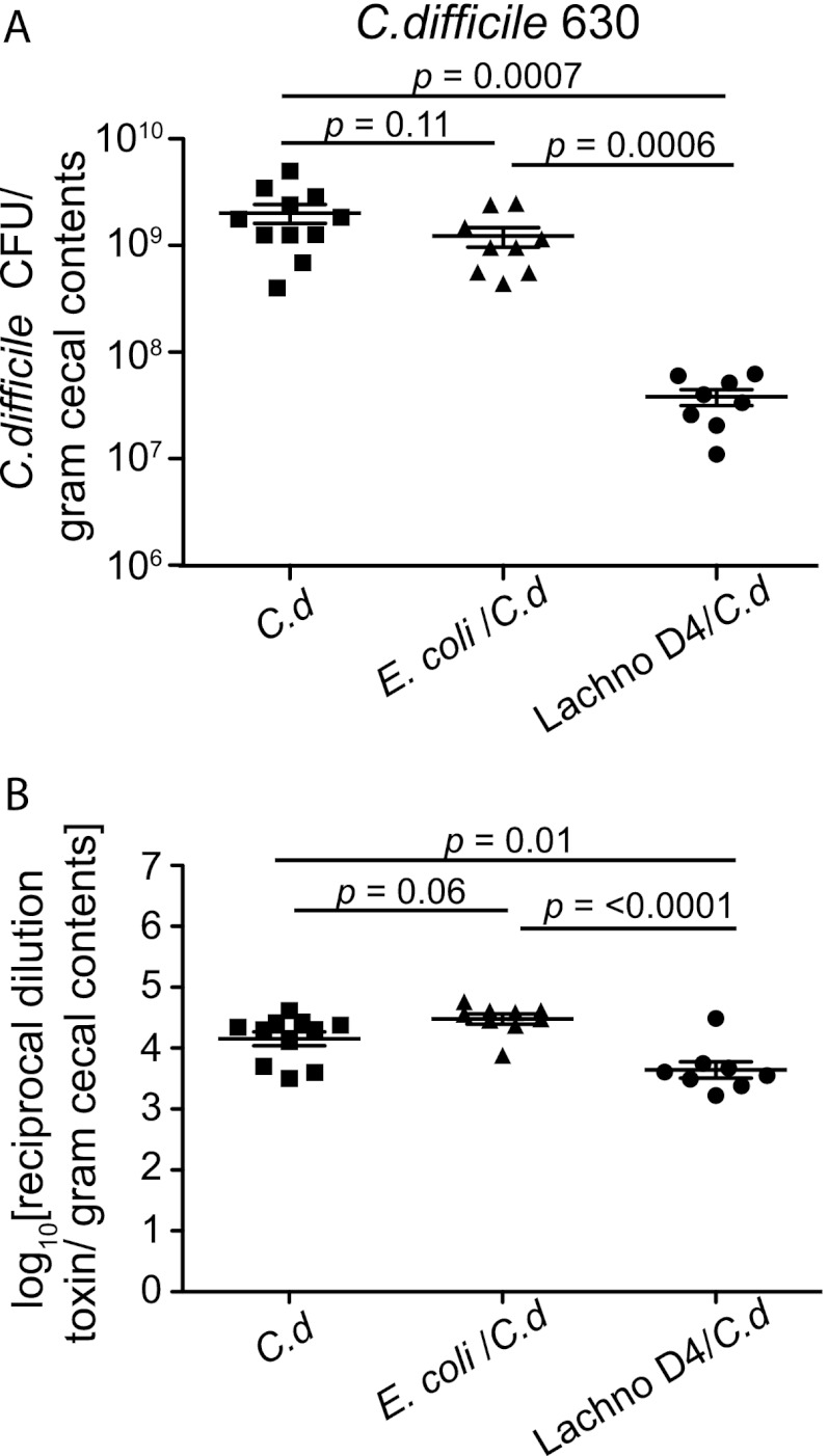
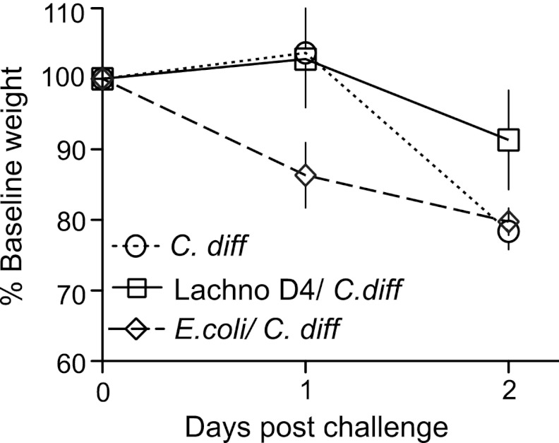
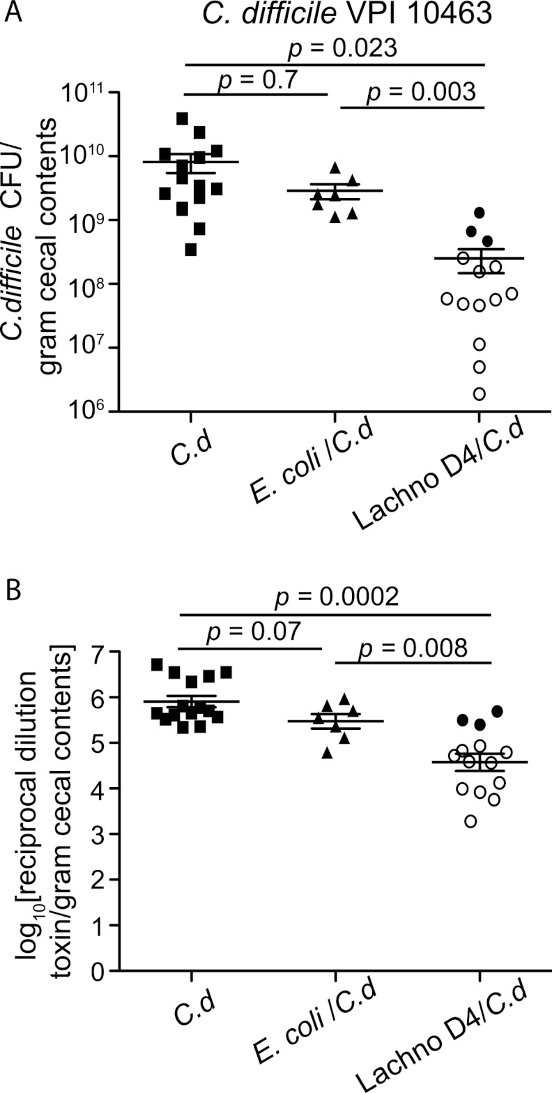
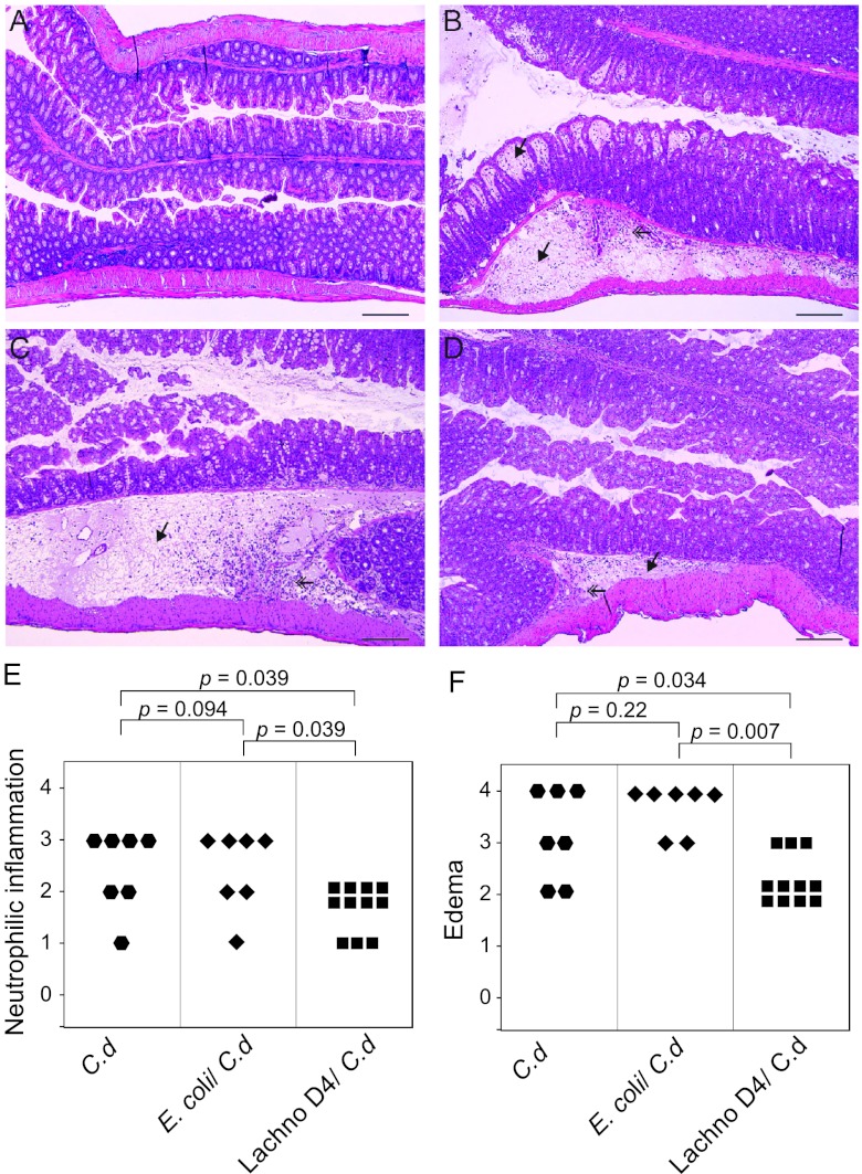
The indigenous microbial community of the gastrointestinal (GI) tract determines susceptibility to Clostridium difficile colonization and disease. Previous studies have demonstrated that antibiotic-treated mice challenged with C. difficile either developed rapidly lethal C. difficile infection or were stably colonized with mild disease. The GI microbial community of animals with mild disease was dominated by members of the bacterial family Lachnospiraceae, while the gut community in moribund animals had a predominance of Escherichia coli. We investigated the roles of murine Lachnospiraceae and E. coli strains in colonization resistance against C. difficile in germfree mice. Murine Lachnospiraceae and E. coli isolates were cultured from wild-type mice. The ability of each of these isolates to interfere with C. difficile colonization was tested by precolonizing germfree mice with these bacteria 4 days prior to experimental C. difficile challenge. Mice precolonized with a murine Lachnospiraceae isolate, but not those colonized with E. coli, had significantly decreased C. difficile colonization, lower intestinal cytotoxin levels and exhibited less severe clinical signs and colonic histopathology. Infection of germfree mice or mice precolonized with E. coli with C. difficile strain VPI 10463 was uniformly fatal by 48 h, but only 20% mortality was seen at 2 days in mice precolonized with the Lachnospiraceae isolate prior to challenge with VPI 10463. These findings confirm that a single component of the GI microbiota, a murine Lachnospiraceae isolate, could partially restore colonization resistance against C. difficile. Further study of the members within the Lachnospiraceae family could lead to a better understanding of mechanisms of colonization resistance against C. difficile and novel therapeutic approaches for the treatment and prevention of C. difficile infection.


Figures

Fig 1
Fig 1
Schematic for examining the effects of Lachnospiraceae D4 and E. coli on C. difficile colonization in germfree mice. Mice were infected with titrating doses of C. difficile 630 spores (10, 102, 103, and 104; n = 3 per dose) or C. difficile VPI 10463 spores (38, 3 × 10², 3.3 × 10³, and 1 × 10⁵; n = 4 per dose) and monitored for colonization. Animals were colonized with cecal contents from a wild-type (WT) mouse for 4 days prior to C. difficile VPI 10463 challenge (n = 5). Additionally, mice were either precolonized with E. coli and then challenged with C. difficile 630 (n = 9) or C. difficile VPI 10463 (n = 7) spores or precolonized with Lachnospiraceae D4 and then challenged with C. difficile 630 (n = 8) or C. difficile VPI 10463 (n = 14) spores. Mice monocolonized with C. difficile 630 (n = 11) or C. difficile VPI 10463 (n = 15) were used as controls. Other groups of mice were colonized with either E. coli (n = 4) or Lachnospiraceae D4 (n = 4) for 4 days and harvested. The cecum and colon from each animal were harvested for bacterial and cytotoxin quantification.
Fig 2
Fig 2
Phylogenetic tree showing clostridial clusters of low-moles-percent G+C Gram-positive bacteria based on 16S rRNA sequence. The tree was constructed using the neighbor-joining method, with the murine E. coli isolate used as the outgroup. Newly isolated murine Lachnospiraceae strains are shown in boldface. Accession numbers for sequences are given in parentheses. Bootstrap values greater than 95 (per 500 replicates) are shown at branch points. The scale bar represents genetic distance, and clostridial clusters are indicated by Roman numerals.
Fig 3
Fig 3
C. difficile infection in germfree mice. (A and B) Kaplan-Meier survival plots for mice infected with titrating doses of C. difficile 630 spores (1 × 101, 1 × 10², 1 × 10³, and 1 × 10⁴) (A) and C. difficile VPI 10463 spores (3.8 × 101, 3 × 10², 3.3 × 10³, and 1 × 10⁵) (B). (C and D) Quantification of C. difficile was determined by culturing cecal contents at the time of necropsy: at day 6 from mice infected with C. difficile 630 for each challenge dose (n = 3) (C) or at days 1 or 2 for mice infected with C. difficile VPI 10463 for each challenge dose (n = 4) (D). (E and F) Vero cell tissue culture was used to determine the log10 reciprocal cytotoxin dilution per gram of cecal contents from mice used for panels C and D infected with C. difficile 630 (E) or C. difficile VPI 10463 (F) for each challenge dose. Points on each graph represent individual animals. Error bars represent standard deviations.
Fig 4
Fig 4
Decreased levels of C. difficile 630 and cytotoxin in Lachnospiraceae D4-precolonized mice. (A) Quantification of C. difficile was determined by culturing cecal contents at the time of necropsy (day 6) from mice infected with C. difficile only (n = 11) or precolonized with either E. coli (n = 9) or Lachnospiraceae D4 (n = 8) and then infected with C. difficile. Each point represents the C. difficile level from an individual animal. Mice precolonized with Lachnospiraceae D4 had significantly decreased levels of C. difficile compared to those in C. difficile-infected controls or E. coli-precolonized mice. Error bars represent standard deviations. Comparisons between groups were performed using the nonparametric Kruskal-Wallis test. (B) Vero cell tissue culture was used to determine the log10 reciprocal cytotoxin dilution per gram of cecal contents from mice used for panel A infected with C. difficile only or precolonized with either E. coli or Lachnospiraceae D4 and then infected with C. difficile. Error bars represent standard deviations. Comparisons between groups were performed using the nonparametric Kruskal-Wallis test. C.d, C. difficile; Lachno D4, Lachnospiraceae D4.
Fig 5
Fig 5
Weight loss in C. difficile-infected mice. Weight loss curves for C. difficile-infected mice (n = 15), mice precolonized with Lachnospiraceae D4 and infected with C. difficile (n = 14), and mice precolonized with E. coli and infected with C. difficile (n = 7) are shown. Lachnospiraceae D4-precolonized mice lost less weight than C. difficile-infected control or E. coli-precolonized mice. Weight loss percentage is based on the starting weight on day 0. Error bars represent the standard deviations of the weights for animals within each group. Lachno D4, Lachnospiraceae D4, C. diff, C. difficile.
Fig 6
Fig 6
Decreased C. difficile VPI 10643 colonization and cytotoxin levels in Lachnospiraceae D4-precolonized mice. (A) Quantification of C. difficile was determined by culturing cecal contents at the time of necropsy (day 2) from mice infected with C. difficile only (n = 15) or precolonized with E. coli (n = 7) or Lachnospiraceae D4 (n = 14) and then infected with C. difficile. The levels of C. difficile colonization were decreased in Lachnospiraceae D4-precolonized mice compared to those in C. difficile-infected controls or E. coli-precolonized mice. Each point represents the C. difficile level from an individual animal. The open symbols represent animals that had improved CDI signs and did not lose significant weight, while the closed symbols represent animals that were moribund or dead at the time of necropsy. (B) Vero cell tissue culture was used to determine the log10 reciprocal cytotoxin dilution per gram of cecal contents from mice used for panel A infected with C. difficile only or precolonized with E. coli or Lachnospiraceae D4 and then infected with C. difficile. Error bars represent standard deviations. Comparisons between groups were performed using the nonparametric Kruskal-Wallis test. C.d, C. difficile; Lachno D4, Lachnospiraceae D4.
Fig 7
Fig 7
Germfree animals precolonized with Lachnospiraceae D4 have decreased colonic histopathology after challenge with C. difficile strain VPI 10463. (A) Colon of an uninfected germfree mouse. (B) Colon of a C. difficile-infected mouse showing severe edema in the submucosa and mucosal lamina propria (arrow) accompanied by neutrophilic inflammation. (C) Colon from a mouse precolonized with E. coli and infected with C. difficile showing submucosal edema (arrow) and neutrophilic inflammation similar to those for a C. difficile-infected mouse. (D) Colon of a mouse precolonized with Lachnospiraceae D4 and infected with C. difficile showing moderate neutrophilic mucosal inflammation and minimal submucosal edema (arrow). All micrographs are of H&E-stained tissue at an original magnification of ×100. Bars represent 100 μm. (E and F) Categorical scores of neutrophilic inflammation (E) and edema (F) in C. difficile-infected controls, C. difficile-infected, E. coli-precolonized mice, and C. difficile-infected, Lachnospiraceae D4-precolonized mice. Comparisons between groups were performed using the nonparametric Kruskal-Wallis test. C.d, C. difficile; Lachno D4, Lachnospiraceae D4.

References

    1. Barcenilla A, et al. 2000. Phylogenetic relationships of butyrate-producing bacteria from the human gut. Appl. Environ. Microbiol. 66:1654–1661 - PMC - PubMed
    1. Bartlett JG. 2010. Clostridium difficile: progress and challenges. Ann. N. Y. Acad. Sci. 1213:62–69 - PubMed
    1. Bartlett JG, Onderdonk AB, Cisneros RL, Kasper DL. 1977. Clindamycin-associated colitis due to a toxin-producing species of Clostridium in hamsters. J. Infect. Dis. 136:701–705 - PubMed
    1. Britton RA, Young VB. 2012. Interaction between the intestinal microbiota and host in Clostridium difficile colonization resistance. Trends Microbiol. 20:313–319 - PMC - PubMed
    1. Chen X, et al. 2008. A mouse model of Clostridium difficile-associated disease. Gastroenterology 135:1984–1992 - PubMed

Publication types

MeSH terms

Substances

LinkOut - more resources