Unusual intracranial spread of anaplastic oligodendroglioma
- PMID: 22891021
- PMCID: PMC4543360
- DOI: 10.1136/bcr-2012-006354
Unusual intracranial spread of anaplastic oligodendroglioma
Abstract
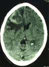
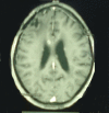
Oligodendrogliomas are the third most common type of glioma. Oligodendrogliomas are typically indolent tumours, yet the majority of them are incurable and most patients will ultimately die. Anaplastic oligodendrogliomas are important to recognise from other primary brain tumours, since they have unique molecular, histological and clinical features. Oligodendroglioma, especially the anaplastic type, has a tendency to intracranial and extracranial metastasis. In this paper, we present an unusual manifestation of anaplastic oligodendroglioma.
Conflict of interest statement
Figures


Similar articles
-
B cell non-Hodgkin cerebral lymphoma associated with an anaplastic oligodendroglioma.Clin Neuropathol. 2002 Nov-Dec;21(6):243-7. Clin Neuropathol. 2002. PMID: 12489671
-
Treatment of oligodendroglioma: an update.Neuro Oncol. 1999 Jan;1(1):61-8. Neuro Oncol. 1999. PMID: 11550303 Free PMC article. Review.
-
The treatment of cerebral oligodendrogliomas with particular reference to features indicating malignancy: report of seventy-seven cases.Neurosurg Rev. 1998;21(2-3):138-46. doi: 10.1007/BF02389319. Neurosurg Rev. 1998. PMID: 9795948
-
Extracranial metastases of anaplastic oligodendroglioma.J Clin Neurosci. 2011 Jan;18(1):136-8. doi: 10.1016/j.jocn.2010.05.028. J Clin Neurosci. 2011. PMID: 20888774
-
Combined surgery, radiation, and chemotherapy for oligodendroglial gliomatosis cerebri.Br J Neurosurg. 2004 Jun;18(3):306-10. doi: 10.1080/02688690410001732823. Br J Neurosurg. 2004. PMID: 15327240 Review.
Cited by
-
Extracranial metastasis of anaplastic oligoastrocytoma.Cancer Biol Med. 2018 Aug;15(3):311-313. doi: 10.20892/j.issn.2095-3941.2017.0181. Cancer Biol Med. 2018. PMID: 30197798 Free PMC article.
References
-
- Engelhard H, Stelea A, Mundt A. Oligodendroglioma and anaplastic oligodendroglioma: clinical features, treatment, and prognosis. Surg Neurol 2003;60:443–56. - PubMed
-
- Ozisik P, Isikay I, Oruckaptan H, et al. Unusual massive spinal metastasis of an intracranial oligodendroglioma. Turkish Neurosurg 2008;18:276–80. - PubMed
-
- Uzuka T, Kakita A, Inenaga C, et al. Frontal anaplastic oligodendroglioma showing multi-organ metastases after a long clinical course. Neural Med Chir (Tokyo) 2007;47:174–7. - PubMed
-
- Voldby B. Disseminated, muein-produeing oligodendroglioma report of two cases. Acta Neurochir 1974;30:299–307. - PubMed
Publication types
MeSH terms
LinkOut - more resources
Full Text Sources
Medical
