

An fMRI study on cortical responses during active self-touch and passive touch from others
- PMID: 22891054
- PMCID: PMC3412995
- DOI: 10.3389/fnbeh.2012.00051
An fMRI study on cortical responses during active self-touch and passive touch from others
Abstract
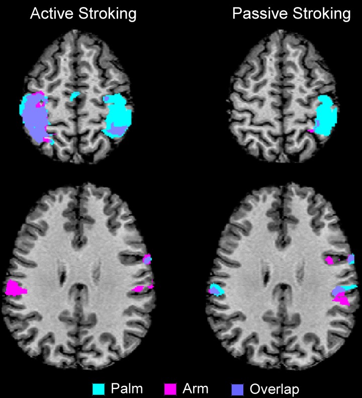
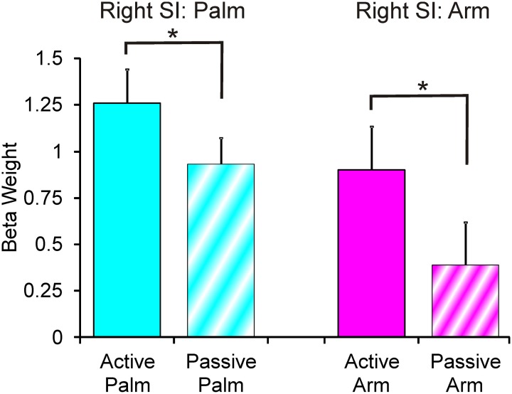
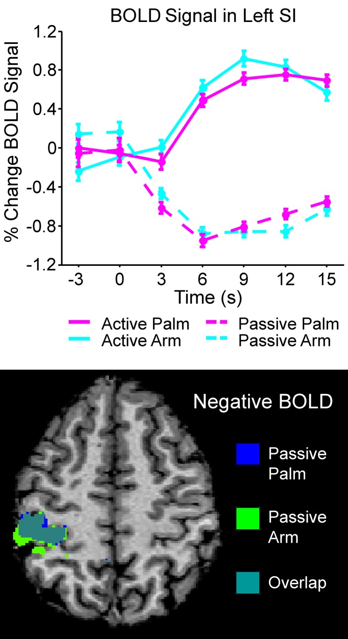
Active, self-touch and the passive touch from an external source engage comparable afferent mechanoreceptors on the touched skin site. However, touch directed to glabrous skin compared to hairy skin will activate different types of afferent mechanoreceptors. Despite perceptual similarities between touch to different body sites, it is likely that the touch information is processed differently. In the present study, we used functional magnetic resonance imaging (fMRI) to elucidate the cortical differences in the neural signal of touch representations during active, self-touch and passive touch from another, to both glabrous (palm) and hairy (arm) skin, where a soft brush was used as the stimulus. There were two active touch conditions, where the participant used the brush in their right hand to stroke either their left palm or arm. There were two similar passive, touch conditions where the experimenter used an identical brush to stroke the same palm and arm areas on the participant. Touch on the left palm elicited a large, significant, positive blood-oxygenation level dependence (BOLD) signal in right sensorimotor areas. Less extensive activity was found for touch to the arm. Separate somatotopical palm and arm representations were found in Brodmann area (BA) 3 of the right primary somatosensory cortex (SI) and in both these areas, active stroking gave significantly higher signals than passive stroking. Active, self-touch elicited a positive BOLD signal in a network of sensorimotor cortical areas in the left hemisphere, compared to the resting baseline. In contrast, during passive touch, a significant negative BOLD signal was found in the left SI. Thus, each of the four conditions had a unique cortical signature despite similarities in afferent signaling or evoked perception. It is hypothesized that attentional mechanisms play a role in the modulation of the touch signal in the right SI, accounting for the differences found between active and passive touch.
Keywords: glabrous; hairy; motor; sensorimotor; skin; somatosensory; stroking.
Figures
References
-
- Ageranioti-Bélanger S. A., Chapman C. E. (1992). Discharge properties of neurones in the hand area of primary somatosensory cortex in monkeys in relation to the performance of an active tactile discrimination task. II. Area 2 as compared to areas 3b and 1. Exp. Brain Res. 91, 207–228 - PubMed
-
- Bickley L., Szilagyi P. (2007). Bates' Guide to Physical Examination and History Taking, 9th Edn Philadelphia, PA: Lippincott Williams and Wilkins
LinkOut - more resources
Full Text Sources