

Regulation of glucose metabolism in T cells: new insight into the role of Phosphoinositide 3-kinases
- PMID: 22891069
- PMCID: PMC3413010
- DOI: 10.3389/fimmu.2012.00247
Regulation of glucose metabolism in T cells: new insight into the role of Phosphoinositide 3-kinases
Abstract
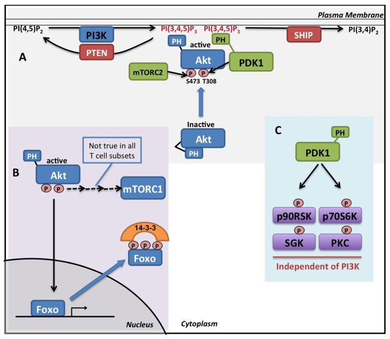
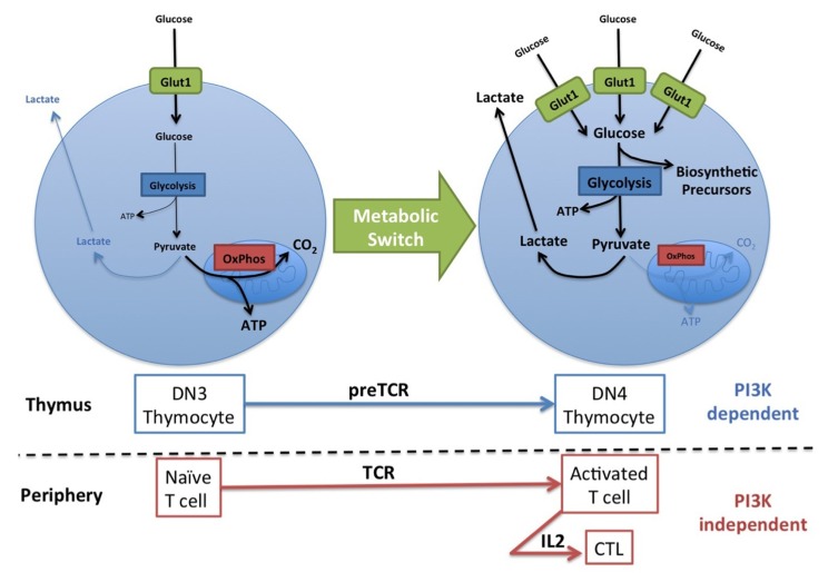
Naïve T cells are relatively quiescent cells that only require energy to prevent atrophy and for survival and migration. However, in response to developmental or extrinsic cues T cells can engage in rapid growth and robust proliferation, produce of a range of effector molecules and migrate through peripheral tissues. To meet the significantly increased metabolic demands of these activities, T cells switch from primarily metabolizing glucose to carbon dioxide through oxidative phosphorylation to utilizing glycolysis to convert glucose to lactate (termed aerobic glycolysis). This metabolic switch allows glucose to be used as a source of carbon to generate biosynthetic precursors for the production of protein, DNA, and phospholipids, and is crucial for T cells to meet metabolic demands. Phosphoinositide 3-kinases (PI3K) are a family of inositol lipid kinases linked with a broad range of cellular functions in T lymphocytes that include cell growth, proliferation, metabolism, differentiation, survival, and migration. Initial research described a critical role for PI3K signaling through Akt (also called protein kinase B) for the increased glucose uptake and glycolysis that accompanies T cell activation. This review article relates this original research with more recent data and discusses the evidence for and against a role for PI3K in regulating the metabolic switch to aerobic glycolysis in T cells.
Keywords: Akt; Glucose metabolism; PDK1; PI3K; T lymphocyte; aerobic glycolysis; c-Myc.
Figures
References
-
- Alessi D. R., Cohen P. (1998). Mechanism of activation and function of protein kinase B. Curr. Opin. Genet. Dev. 8 55–62 - PubMed
-
- Bayascas J. R., Wullschleger S., Sakamoto K., Garcia-Martinez J. M., Clacher C., Komander D., van Aalten D. M. F., Boini K. M., Lang F., Lipina C., Logie L., Sutherland C., Chudek J. A., van Diepen J. A., Voshol P. J., Lucocq J. M., Alessi D. R. (2008). Mutation of the PDK1 PH domain inhibits protein kinase B/Akt, leading to small size and insulin resistance. Mol. Cell. Biol. 28 3258–3272 - PMC - PubMed
LinkOut - more resources
Full Text Sources
Miscellaneous