. 2012 Aug 8:3:175.
doi: 10.3389/fpls.2012.00175. eCollection 2012.

Tyrosine Phosphorylation of the BRI1 Receptor Kinase Occurs via a Post-Translational Modification and is Activated by the Juxtamembrane Domain

Affiliations

Tyrosine Phosphorylation of the BRI1 Receptor Kinase Occurs via a Post-Translational Modification and is Activated by the Juxtamembrane Domain

Man-Ho Oh et al. Front Plant Sci. .

Abstract

In metazoans, receptor kinases control many essential processes related to growth and development and response to the environment. The receptor kinases in plants and animals are structurally similar but evolutionarily distinct and thus while most animal receptor kinases are tyrosine kinases the plant receptor kinases are classified as serine/threonine kinases. One of the best studied plant receptor kinases is Brassinosteroid Insensitive 1 (BRI1), which functions in brassinosteroid signaling. Consistent with its classification, BRI1 was shown in early studies to autophosphorylate in vitro exclusively on serine and threonine residues and subsequently numerous specific phosphoserine and phosphothreonine sites were identified. However, several sites of tyrosine autophosphorylation have recently been identified establishing that BRI1 is a dual-specificity kinase. This raises the paradox that BRI1 contains phosphotyrosine but was only observed to autophosphorylate on serine and threonine sites. In the present study, we demonstrate that autophosphorylation on threonine and tyrosine (and presumably serine) residues is a post-translational modification, ruling out a co-translational mechanism that could explain the paradox. Moreover, we show that in general, autophosphorylation of the recombinant protein appears to be hierarchical and proceeds in the order: phosphoserine > phosphothreonine > phosphotyrosine. This may explain why tyrosine autophosphorylation was not observed in some studies. Finally, we also show that the juxtamembrane domain of BRI1 is an activator of the kinase domain, and that kinase specificity (serine/threonine versus tyrosine) can be affected by residues outside of the kinase domain. This may have implications for identification of signature motifs that distinguish serine/threonine kinases from dual-specificity kinases.

Keywords: autophosphorylation; brassinosteroid signaling; hierarchical phosphorylation; juxtamembrane domain; signal transduction.


Figures

Figure 1
Figure 1
Time course of Flag-BRI1 production in E. coli. (A) Total recovery of recombinant protein from bacterial cell cultured for up to 16 h after addition of (?) mM IPTG at 22°C. (B) Immunoblot analysis of purified Flag-BRI1 protein harvested from cells at different stages of induction, using a variety of generic and modification-specific antibodies. Staining with ProQ Diamond phosphoprotein stain reported phosphorylation at all serine, threonine, and tyrosine sites, and immunoblotting with anti-Flag antibodies and staining with Coomassie Brilliant Blue (CBB) demonstrated equal protein loading (1.5 μg per lane) and the electrophoretic mobility shift accompanying autophosphorylation. (C) Peptide kinase activity of BRI1 purified at different stages of induction. The SP11 peptide (sequence: GRJRRIASVEJJKK, where J is norleucine) was used and reaction mixtures were incubated for 20 min at room temperature. (D) Time courses of SP11 peptide phosphorylation catalyzed by BRI1-Flag purified at different stages of induction.
Figure 2
Figure 2
Quantification of Flag-BRI1 autophosphorylation during induction in E. coli. (A) Densitometric analysis of immunoblot data in Figure 1 showing the increase in ProQ Diamond staining, and cross-reaction with anti-phosphothreonine or anti-phosphotyrosine antibodies, normalized for Flag-BRI1 protein based on detection by CBB staining. The dashed line in the left panel is the value obtained for the kinase-inactive BRI1 (K911E) directed mutant, which is completely unphosphorylated but reacts weakly with ProQ Diamond stain and provides a baseline for interpretation of the autophosphorylation of active Flag-BRI1. (B) Densitometry analysis of phosphorylation of specific sites based on immunoblotting with custom antibodies as in Figure 1. Residues monitored that are contained within the JM domain and KD are plotted separately as indicated.
Figure 3
Figure 3
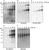
Time course of transphosphorylation of E. coli proteins during expression of Flag-BRI1 cytoplasmic domain. At each time point, Flag-BRI1 protein was immunopurified and the remaining supernatant (containing E. coli proteins and some residual Flag-BRI1 protein) was resolved by SDS-PAGE followed by staining or immunoblot analysis. (A) ProQ Diamond phosphoprotein stain. (B) Immunoblotting (IB) with anti-phosphotyrosine (anti-pY) or (C) anti-phosphotyrosine-956 (anti-pY956), or (D) anti-phosphothreonine (anti-pT) antibodies. (E) Staining with Coomassie Brilliant Blue (CBB).
Figure 4
Figure 4
Separation of E. coli proteins by 2-DE followed by immunoblot analysis with anti-phosphotyrosine antibodies. Samples were collected at (A) 0 h and (B) 16 h following addition of IPTG to induce Flag-BRI1 production. From each sample, the majority of the Flag-BRI1 protein was removed by immunopurification, and the remaining supernatant (300 μg protein) was subjected to 2-DE analysis.
Figure 5
Figure 5
Not all receptor kinases can transphosphorylate E. coli proteins during induction. (A) Alignment of subdomain VIb for the seven RD-type and two non-RD-type (SRF9 and FLS2) receptor kinases used in the present study. The RD dipeptide motif, when present, is underlined. (B) Autophosphorylation analysis of the nine receptor kinases, all expressed as fusion proteins with an N-terminal Flag tag, as analyzed by staining with ProQ Diamond phosphoprotein stain. (C) Transphosphorylation of E. coli proteins after 16 h of expression of the indicated receptor kinase. For each sample, the Flag-tagged receptor kinase was imunopurified and the remaining supernatant was subjected to SDS-PAGE, transferred to PVDF, and stained with ProQ Diamond.
Figure 6
Figure 6
Schematic representation of the primary structure of Flag-BRI1 cytoplasmic domain showing the serial truncations made through the JM domain. The positions of confirmed and possible phosphorylation sites are indicated with solid or dashed symbols, respectively.
Figure 7
Figure 7
The JM domain is an activator of the BRI1 kinase domain. Serial deletions of the JM domain were constructed to produce truncation mutants starting with the indicated residue. (A) Analysis of autophosphorylation of the JM truncation mutants by staining with ProQ Diamond and immunoblotting with generic and custom antibodies. (B) Peptide kinase activity of the JM truncation mutants measured using the SP11 synthetic peptide. Values are means of three determinations ± SEM. (C) Correlation between peptide kinase activity (from B) and relative phosphorylation of the threonine-872 site, determined by densitometric analysis of the immunoblots in A. (D) Correlation between peptide kinase activity and relative phosphorylation status of the serine-891 site.
Figure 8
Figure 8
Identification of specific JM domain residues that are essential for autophosphorylation of BRI1 on tyrosine residues. Two critical regions identified by truncation analysis were examined by alanine scanning of individual residues in the full-length Flag-BRI1 cytoplasmic domain. The recombinant proteins were expressed and autophosphorylation was analyzed by ProQ Diamond staining and immunoblotting with generic phosphotyrosine and phosphothreonine antibodies.

Similar articles

Cited by

References

    1. Clouse S. D. (2011). Brassinosteroid signal transduction: from receptor kinase activation to transcriptional networks regulating plant development. Plant Cell 23, 1219–123010.1105/tpc.111.084475 - DOI - PMC - PubMed
    1. Cole A., Frame S., Cohen P. (2004). Further evidence that the tyrosine phosphorylation of glycogen synthase-3 (GSK3) in mammalian cells is an autophosphorylation event. Biochem. J. 377, 249–25510.1042/BJ20031259 - DOI - PMC - PubMed
    1. Cowan S. W., Schirmer T., Rummel G., Steiert M., Ghosh R., Pauptit R. A., Jansonius J. N., Rosenbusch J. P. (1992). Crystal structures explain functional properties of two E. coli porins. Nature 358, 727–73310.1038/358727a0 - DOI - PubMed
    1. Cui J., Qasim S., Davidson A. L. (2010). Uncoupling substrate transport from ATP hydrolysis in the Escherichia coli maltose transporter. J. Biol. Chem. 285, 39986–3999310.1074/jbc.M110.147819 - DOI - PMC - PubMed
    1. Ehsan H., Ray W. K., Phinney B., Wang X., Huber S. C., Clouse S. D. (2005). Interaction of Arabidopsis brassinosteroid-insensitive 1 receptor kinase with a homolog of mammalian TFG-β receptor interacting protein. Plant J. 43, 251–26110.1111/j.1365-313X.2005.02448.x - DOI - PubMed

LinkOut - more resources