

Design of nanovectors for therapy and imaging of cardiovascular diseases
- PMID: 22891105
- PMCID: PMC3405780
- DOI: 10.14797/mdcj-8-1-13
Design of nanovectors for therapy and imaging of cardiovascular diseases
Abstract
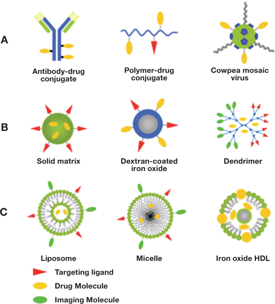
Cardiovascular diseases are widely prevalent in western societies, and their associated costs number in the billions of dollars and affect millions of patients each year. Nanovectors targeted to tissues involved in cardiovascular diseases offer great opportunities to improve cardiovascular treatment through their imaging and drug delivery capabilities. Vascular-targeted imaging particles may permit the early identification of atherosclerosis, discriminate between stable and vulnerable atherosclerotic plaques, or guide surgeons as they work on fragile vasculature. Tailored therapeutic nanoparticles may provide safer, more efficient and effective intervention through localization and release of encapsulated therapeutics. Nanovector design involves numerous considerations such as fabrication material, particle size, and surface-modification with ligands for targeting and increasing blood circulation times. Complex blood rheology may affect the efficiency with which dissimilarsized particles target ligand receptors associated with disease. Additionally, the intended use of a nanovector is a critical factor in its design as some materials with poor drug-loading qualities or release kinetics may be suitable for imaging purposes only. Overall, vectors targeted to the vasculature will need to be efficient in avoiding blood clearance, honing to the target location, and binding at the desired site.
Keywords: atheroma; atherosclerosis; contrast agent; liposomes; micelles; molecular imaging; nanovectors; phospholipids; stroke; vascular targeting.
Similar articles
-
The choice of targets and ligands for site-specific delivery of nanomedicine to atherosclerosis.Cardiovasc Res. 2020 Nov 1;116(13):2055-2068. doi: 10.1093/cvr/cvaa047. Cardiovasc Res. 2020. PMID: 32077918 Review.
-
High-resolution CT vascular imaging using blood pool contrast agents.Methodist Debakey Cardiovasc J. 2012 Jan;8(1):18-22. doi: 10.14797/mdcj-8-1-18. Methodist Debakey Cardiovasc J. 2012. PMID: 22891106 Free PMC article. Review.
-
Dynamic and cellular interactions of nanoparticles in vascular-targeted drug delivery.Mol Membr Biol. 2010 Oct;27(7):312-27. doi: 10.3109/09687688.2010.522117. Mol Membr Biol. 2010. PMID: 21028938 Review.
-
Liposomes and inorganic nanoparticles for drug delivery and cancer imaging.Ther Deliv. 2012 May;3(5):645-56. doi: 10.4155/tde.12.38. Ther Deliv. 2012. PMID: 22834408 Review.
-
Nanomedicine in cardiology: Precision drug delivery for enhanced patient outcomes.Life Sci. 2024 Dec 1;358:123199. doi: 10.1016/j.lfs.2024.123199. Epub 2024 Oct 31. Life Sci. 2024. PMID: 39488265 Review.
Cited by
-
IgA and IgM protein primarily drive plasma corona-induced adhesion reduction of PLGA nanoparticles in human blood flow.Bioeng Transl Med. 2017 May 22;2(2):180-190. doi: 10.1002/btm2.10064. eCollection 2017 Jun. Bioeng Transl Med. 2017. PMID: 28932819 Free PMC article.
-
Optimization of Chitosan-Decorated Solid Lipid Nanoparticles for Improved Flurbiprofen Transdermal Delivery.ACS Omega. 2023 May 21;8(22):19302-19310. doi: 10.1021/acsomega.2c08135. eCollection 2023 Jun 6. ACS Omega. 2023. PMID: 37305303 Free PMC article.
-
Abdominal aortic aneurysms targeted by functionalized polysaccharide microparticles: a new tool for SPECT imaging.Theranostics. 2014 Mar 11;4(6):592-603. doi: 10.7150/thno.7757. eCollection 2014. Theranostics. 2014. PMID: 24723981 Free PMC article.
-
Controlled and Targeted Drug Delivery Using Smart Nanovectors.Int J Drug Discov Pharm. 2023 Mar;2(1):84-90. doi: 10.53941/ijddp.0201010. Epub 2023 Mar 20. Int J Drug Discov Pharm. 2023. PMID: 39524321 Free PMC article.
-
In situ re-endothelialization via multifunctional nanoscaffolds.ACS Nano. 2014 Oct 28;8(10):10826-36. doi: 10.1021/nn504636n. Epub 2014 Sep 15. ACS Nano. 2014. PMID: 25222570 Free PMC article.
References
-
- Lloyd-Jones D, Adams RJ, Brown TM, Carnethon M, Dai S, De Simone G, et al. Heart disease and stroke statistics — 2010 update: a report from the American Heart Association. Circulation. 2010 Feb 23;121(7):e46–e215.. - PubMed
-
- Ambrose JA, Winters SL, Arora RR, Haft JI, Goldstein J, Rentrop KP, et al. Coronary angiographic morphology in myocardial infarction: a link between the pathogenesis of unstable angina and myocardial infarction. J Am Coll Cardiol. 1985;6(6):1233–8.. - PubMed
-
- Ambrose JA, Tannenbaum MA, Alexopoulos D, Hjemdahl-Monsen CE, Leavy J, Weiss M, et al. Angiographic progression of coronary artery disease and the development of myocardial infarction. J Am Coll Cardiol. 1988 Jul;12(1):56–62.. - PubMed
-
- Hackett D, Davies G, Maseri A. Pre-existing coronary stenoses in patients with first myocardial infarction are not necessarily severe. Eur Heart J. 1988 Dec;9(12):1317–23.. - PubMed
-
- Giroud D, Li JM, Urban P, Meier B, Rutishauer W. Relation of the site of acute myocardial infarction to the most severe coronary arterial stenosis at prior angiography. Am J Cardiol. 1992 Mar 15;69(8):729–32.. - PubMed
Publication types
MeSH terms
Substances
LinkOut - more resources
Full Text Sources
Medical
