. 2012 Oct;160(2):653-66.
doi: 10.1104/pp.112.202309. Epub 2012 Aug 13.

Structural characterization of Arabidopsis leaf arabinogalactan polysaccharides

Affiliations

Structural characterization of Arabidopsis leaf arabinogalactan polysaccharides

Theodora Tryfona et al. Plant Physiol. 2012 Oct.

Abstract

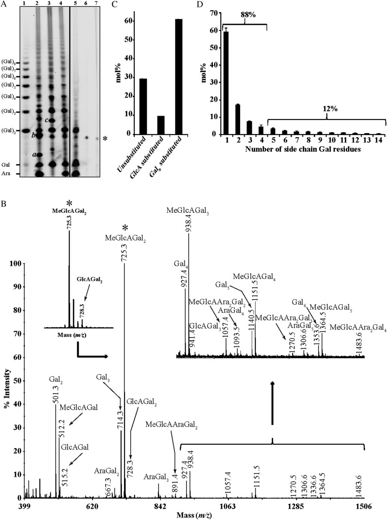
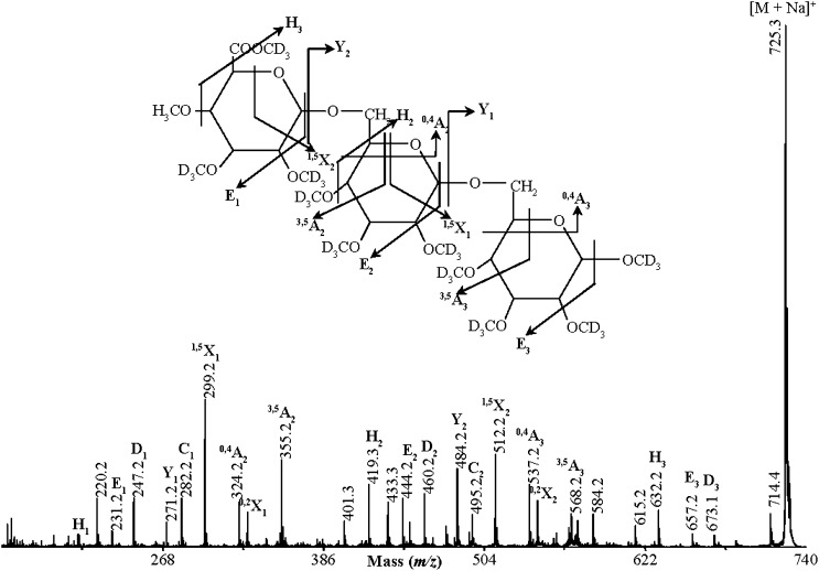
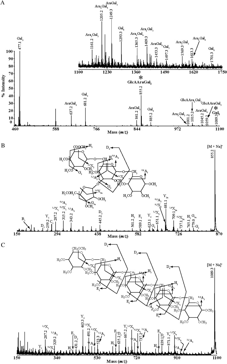
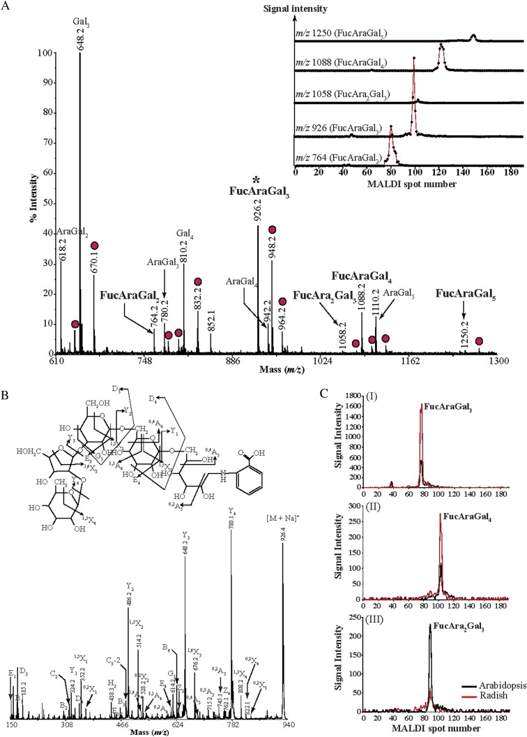
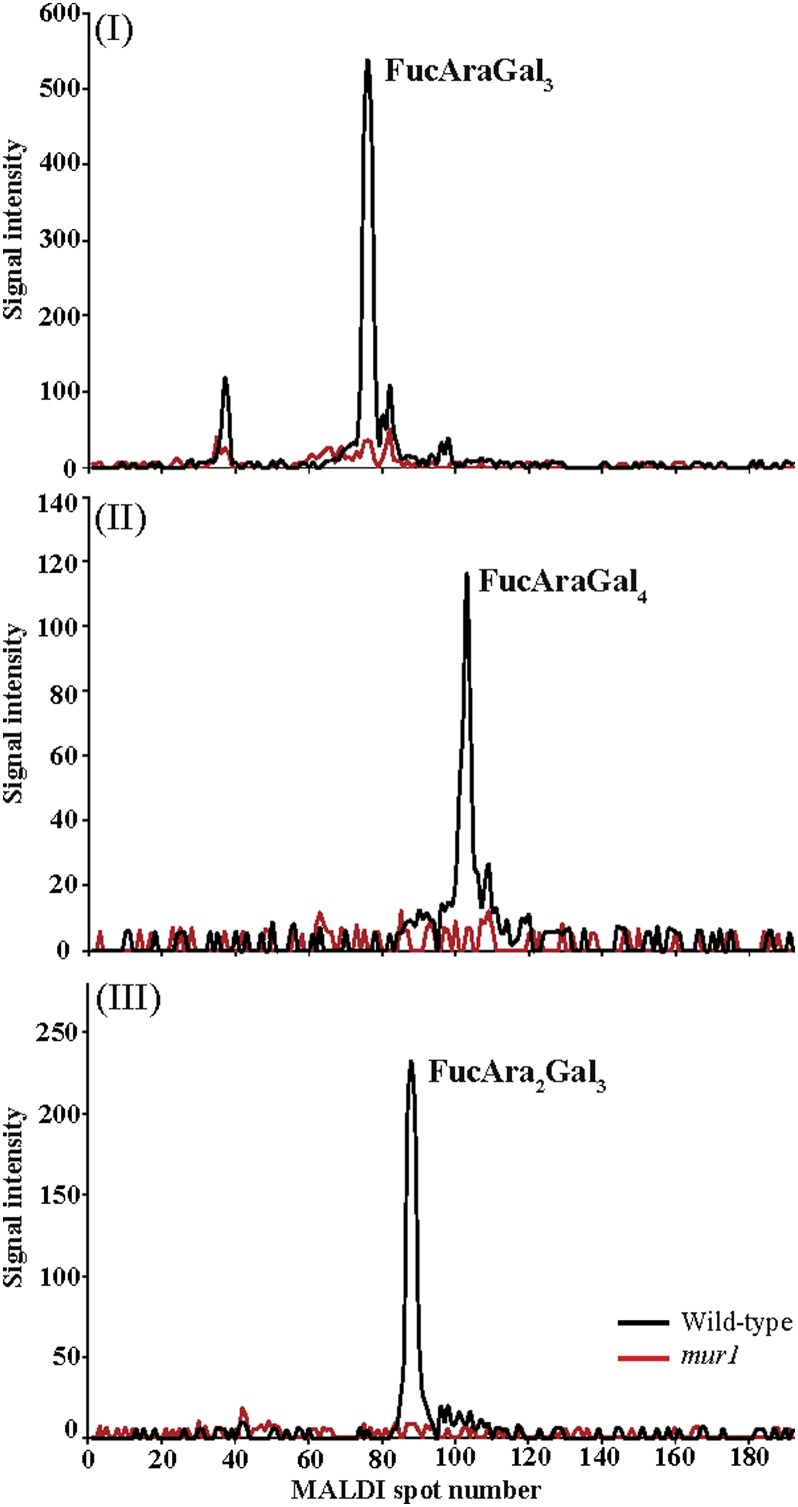
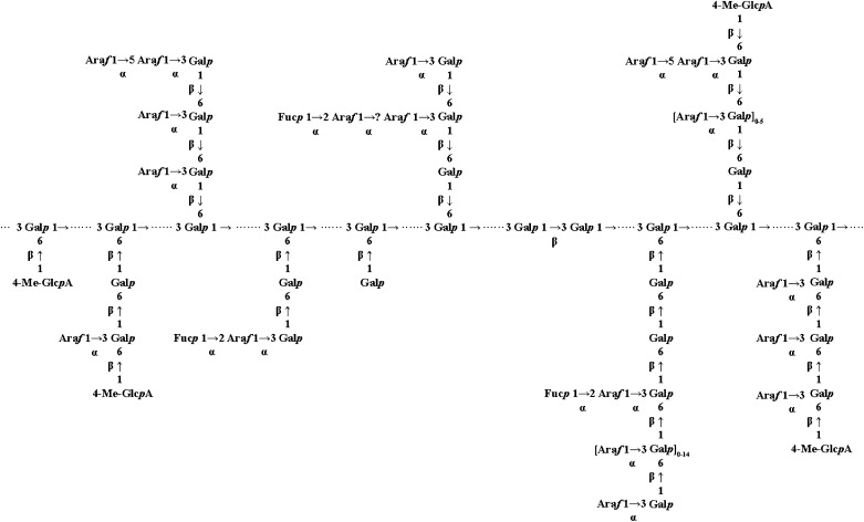
Proteins decorated with arabinogalactan (AG) have important roles in cell wall structure and plant development, yet the structure and biosynthesis of this polysaccharide are poorly understood. To facilitate the analysis of biosynthetic mutants, water-extractable arabinogalactan proteins (AGPs) were isolated from the leaves of Arabidopsis (Arabidopsis thaliana) plants and the structure of the AG carbohydrate component was studied. Enzymes able to hydrolyze specifically AG were utilized to release AG oligosaccharides. The released oligosaccharides were characterized by high-energy matrix-assisted laser desorption ionization-collision-induced dissociation mass spectrometry and polysaccharide analysis by carbohydrate gel electrophoresis. The Arabidopsis AG is composed of a β-(1→3)-galactan backbone with β-(1→6)-d-galactan side chains. The β-(1→6)-galactan side chains vary in length from one to over 20 galactosyl residues, and they are partly substituted with single α-(1→3)-l-arabinofuranosyl residues. Additionally, a substantial proportion of the β-(1→6)-galactan side chain oligosaccharides are substituted at the nonreducing termini with single 4-O-methyl-glucuronosyl residues via β-(1→6)-linkages. The β-(1→6)-galactan side chains are occasionally substituted with α-l-fucosyl. In the fucose-deficient murus1 mutant, AGPs lack these fucose modifications. This work demonstrates that Arabidopsis mutants in AGP structure can be identified and characterized. The detailed structural elucidation of the AG polysaccharides from the leaves of Arabidopsis is essential for insights into the structure-function relationships of these molecules and will assist studies on their biosynthesis.


Figures

Figure 1.
Figure 1.
Characterization of oligosaccharides released by AG-specific enzymes from Arabidopsis leaf AGP extracts. A, PACE. Oligosaccharide products of exo-β-(1→3)-galactanase followed by α-l-arabinofuranosidase (lane 2), exo-β-(1→3)-galactanase followed by β-glucuronidase (lane 3), exo-β-(1→3)-galactanase, α-l-arabinofuranosidase, and β-glucuronidase (lane 4), exo-β-(1→3)-galactanase, α-l-arabinofuranosidase, β-glucuronidase, and endo-β-(1→6)-galactanase (lane 5), enzyme control (lane 6), and AGP extract control (lane 7) were reductively aminated with 2-aminonaphthaline trisulfonic acid and separated by electrophoresis on acrylamide gels. An oligosaccharide ladder prepared from β-(1→6)-galactan was used as a migration marker (lane 1): a, MeGlcAGal; b, MeGlcAGal2; c, MeGlcAAraGal2. The asterisk indicates a background band. B, MALDI-ToF-MS spectra of per-deuteromethylated oligosaccharides released by exo-β-(1→3)-galactanase followed by α-l-arabinofuranosidase. Peaks marked with asterisks were selected for MALDI-CID structural analysis. C, Quantification of substitution of the β-(1→3)-galactan backbone: none, MeGlcA, or galactan. Arabidopsis leaf AGP extracts were digested with AG-specific α-l-arabinofuranosidase and exo-β-(1→3)-galactanase, and the amount of the hydrolysis products was calculated by PACE [Galn corresponds to β-(1→6)-galactan chains with two to 15 Gal residues]. Values are means ± sd of one representative biological replicate analyzed six times. D, Quantification by PACE of the different-length β-(1→6)-galactan side chains (e.g. chain 1 corresponds to Gal2, chain 2 corresponds to Gal3) released by sequential digestion with α-l-arabinofuranosidase, exo-β-(1→3)-galactanase, and β-glucuronidase of Arabidopsis leaf AGP extracts. Values are means ± sd of two biological replicates analyzed six times.
Figure 2.
Figure 2.
The bulk of Arabidopsis leaf AGPs are sensitive to AG-specific enzymes as shown by a Yariv diffusion assay. Leaf AGP extracts from wild-type and mur1 mutant plants were subjected to sequential hydrolysis by AG-specific enzymes: exo-β-(1→3)-galactanase, 2× α-l-arabinofuranosidase, β-glucuronidase, and endo-β-(1→6)-galactanase. The AGP extracts or the enzyme hydrolysis products were dissolved in NaCl (0.15 m) and sodium azide (0.02%) solution and were spotted on agarose gels (1%) containing the β-galactosyl Yariv reagent (0.002%). The controls included reagents without the hydrolysis products: gum Arabic (positive) and oat spelt xylan (negative). exo-β-(1→3)-galactanase, 2× α-l-arabinofuranosidase, β-glucuronidase, and endo-β-(1→6)-galactanase sequential digestion; *NaCl (0.15 m) and sodium azide (0.02%) buffer solution. Col-0, Ecotype Columbia.
Figure 3.
Figure 3.
Structural characterization of an acidic AG oligosaccharide by MALDI-CID. Arabidopsis leaf AGP extracts were sequentially hydrolyzed with exo-β-(1→3)-galactanase and α-l-arabinofuranosidase, and the resulting oligosaccharides were analyzed by MALDI-ToF-MS as shown in Figure 1B. The per-deuteromethylated oligosaccharide with m/z 725.3 was selected for MALDI-CID analysis and is identified as 4-O-Me-β-GlcA-(1→6)-β-Galp-(1→6)-Galp. Glycosidic and cross-ring fragments are identified according to the nomenclature of Domon and Costello (1988).
Figure 4.
Figure 4.
Structural characterization of an acidic AG oligosaccharide and of a long galacto-oligosaccharide by MALDI-CID. A, Oligosaccharide products of exo-β-(1→3)-galactanase followed by β-glucuronidase were per-methylated and analyzed by MALDI-ToF-MS. Peaks marked with asterisks were selected for MALDI-CID structural analysis. B and C, MALDI-CID of GlcAAraGal2 and Gal5 oligosaccharide, respectively.
Figure 5.
Figure 5.
Identification of l-Fuc-modified oligosaccharides from radish leaf AGP extracts. Purified radish and Arabidopsis leaf AGPs were subjected to a sequential digestion of α-l-arabinofuranosidase, exo-β-(1→3)-galactanase, β-glucuronidase, and endo-β-(1→6)-galactanase. The hydrolysis products were reductively aminated with 2-AA (A and B) or [12C6]aniline (Arabidopsis oligosaccharides) and [13C6]aniline (radish oligosaccharides; C). The labeled glycans were either directly analyzed by MALDI-ToF-MS or were separated by HILIC and subsequently analyzed by MALDI-ToF-MS (inset in A). Peaks marked with asterisks were selected for MALDI-CID structural analysis, and the corresponding spectrum is shown in B. A, AGP extracts from radish leaves contain five different types of l-Fuc-modified oligosaccharides. For every oligosaccharide, sodiated molecular ions ([M + Na]+; peak assignment in this figure) and corresponding doubly sodiated molecular ions ([M + 2Na − H]+; 22 D larger, red circles) are observed. Separation of the structural isomers by HILIC (inset) shows that a single structural isomer is present for each oligosaccharide. B, MALDI-CID of the radish leaf 2-AA-labeled FucAraGal3 AG oligosaccharide. C, Extracted ion chromatograms for l-Fuc-modified oligosaccharides originating from radish leaf (red lines) or Arabidopsis leaf (black lines) AGPs hydrolyzed by AG-specific enzymes. Arabidopsis leaf AGP extracts contain three different l-Fuc-modified oligosaccharides.
Figure 6.
Figure 6.
Capillary HILIC-MALDI-ToF-MS using stable isotopic labeling of l-Fuc-modified oligosaccharides from Arabidopsis leaf AGP extracts of wild-type (black line) and mur1 (red line) plants. Purified Arabidopsis leaf AGPs from wild-type and mur1 plants were subjected to a sequential digestion of α-l-arabinofuranosidase, exo-β-(1→3)-galactanase, β-glucuronidase, and endo-β-(1→6)-galactanase. The hydrolysis products were purified on a C18 cartridge (elution with 5% acetic acid) to remove the enzymes, followed by cation-exchange cleanup (Dowex resin; elution with 5% acetic acid) to remove salt, and were reductively aminated with [12C6]aniline (wild-type oligosaccharides; black lines) and [13C6]aniline (mur1 oligosaccharides; red lines). The labeled glycans were purified from the reduction buffers on a normal-phase cartridge (Glyko Clean S), and the purified oligosaccharides were separated by HILIC and analyzed by MALDI-ToF-MS. Although three l-Fuc-modified oligosaccharides were separated from the wild-type leaf AGP extracts of AG-specific hydrolyzed samples, no corresponding signals were detected from leaf AGP extracts from mur1 plants.
Figure 7.
Figure 7.
A proposed model of the average structure of the carbohydrate component of Arabidopsis leaf AGPs. Structures determined in this work are shown in Supplemental Table S1. The dotted line indicates the β-(1→3)-linked backbone, but the arrangement or presence of the different side chains on specific AGPs is unknown. Some of these structures may also be present on pectic type II AG. We cannot exclude the presence of kinks of β-(1→6)-linked residues in the β-(1→3)-galactan backbone, as proposed by Tan et al. (2004).

References

    1. Bonin CP, Potter I, Vanzin GF, Reiter W-D. (1997) The MUR1 gene of Arabidopsis thaliana encodes an isoform of GDP-D-mannose-4,6-dehydratase, catalyzing the first step in the de novo synthesis of GDP-L-fucose. Proc Natl Acad Sci USA 94: 2085–2090 - PMC - PubMed
    1. Borner GHH, Lilley KS, Stevens TJ, Dupree P. (2003) Identification of glycosylphosphatidylinositol-anchored proteins in Arabidopsis: a proteomic and genomic analysis. Plant Physiol 132: 568–577 - PMC - PubMed
    1. Chai W, Piskarev V, Lawson AM. (2001) Negative-ion electrospray mass spectrometry of neutral underivatized oligosaccharides. Anal Chem 73: 651–657 - PubMed
    1. Cheung AY, Wang H, Wu HM. (1995) A floral transmitting tissue-specific glycoprotein attracts pollen tubes and stimulates their growth. Cell 82: 383–393 - PubMed
    1. Chun H, Shin DH, Hong BS, Cho HY, Yang HC. (2001) Purification and biological activity of acidic polysaccharide from leaves of Thymus vulgaris L. Biol Pharm Bull 24: 941–946 - PubMed

Publication types

MeSH terms

LinkOut - more resources