Clinical Trial
. 2012 Oct 5;287(41):34809-24.
doi: 10.1074/jbc.M112.372797. Epub 2012 Aug 13.

Primate-specific melanoma antigen-A11 regulates isoform-specific human progesterone receptor-B transactivation

Affiliations
Clinical Trial

Primate-specific melanoma antigen-A11 regulates isoform-specific human progesterone receptor-B transactivation

Shifeng Su et al. J Biol Chem. .

Abstract

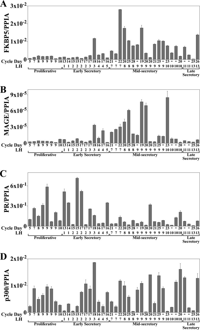
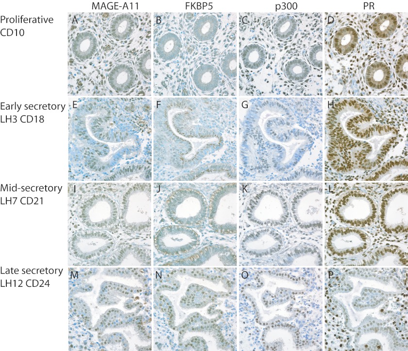
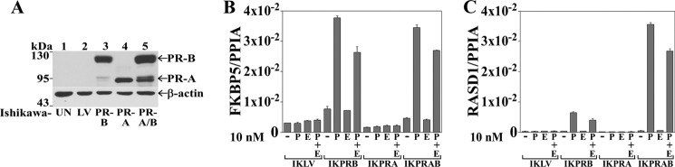
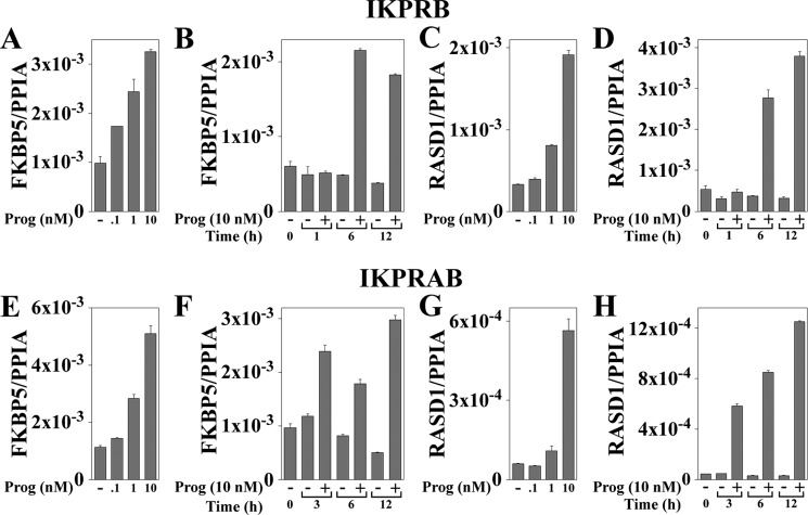
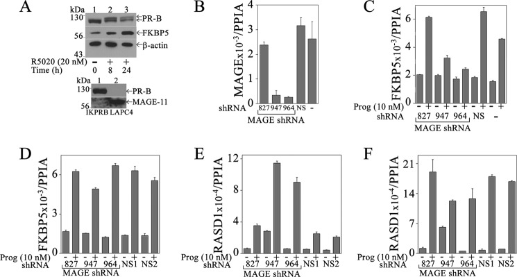
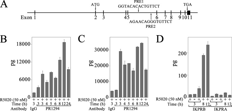
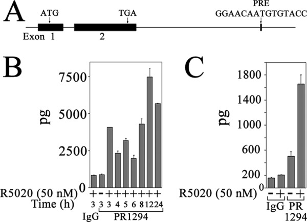
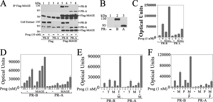
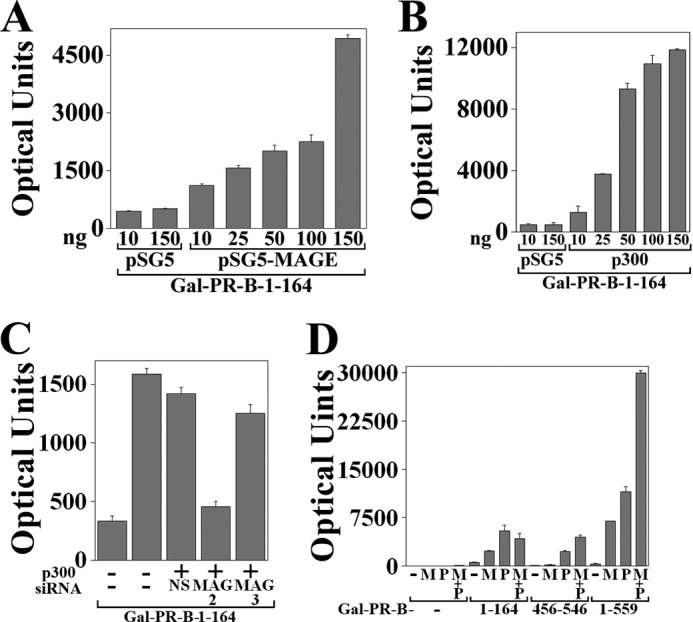
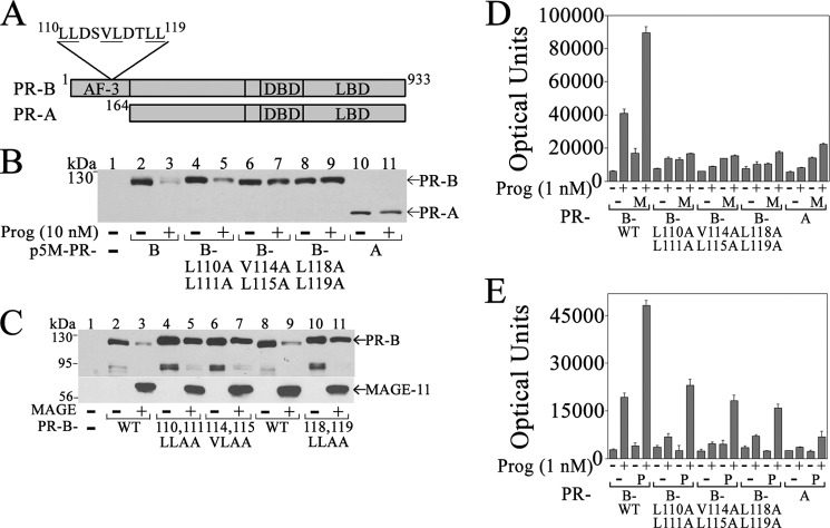
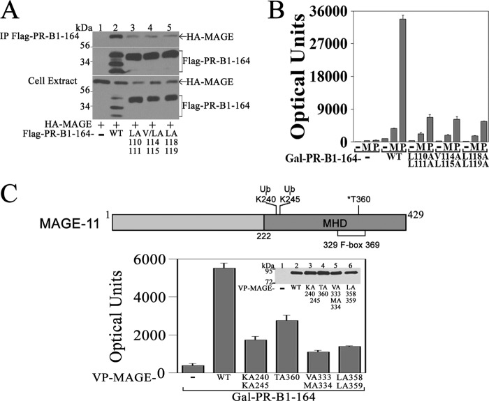
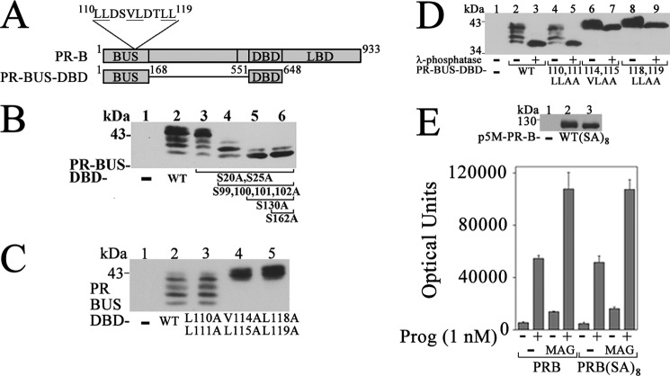
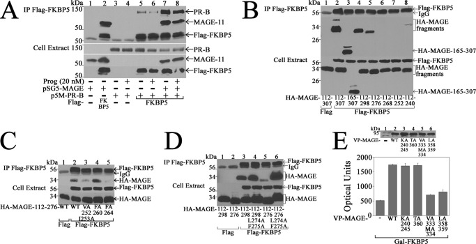
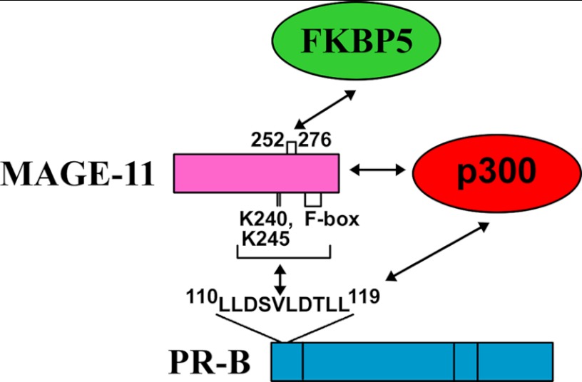
Progesterone acting through the progesterone receptor (PR) and its coregulators prepares the human endometrium for receptivity to embryo implantation and maintains pregnancy. The menstrual cycle-dependent expression of melanoma antigen-A11 (MAGE-11) in the mid-secretory human endometrium suggested a novel function in human PR signaling. Here we show that MAGE-11 is an isoform-specific coregulator responsible for the greater transcriptional activity of human PR-B relative to PR-A. PR was recruited to progesterone response regions of progesterone-regulated FK506-binding protein 5 (FKBP5) immunophilin and small Ras family G protein cell growth inhibitor RASD1 genes. Expression of MAGE-11 lentivirus shRNA in human endometrial Ishikawa cells expressing PR-B showed that MAGE-11 is required for isoform-specific PR-B up-regulation of FKBP5. In contrast, MAGE-11 was not required for progesterone up-regulation of RASD1 in endometrial cells expressing the PR-A/B heterodimer. Target gene specificity of PR-B depended on the synergistic actions of MAGE-11 and p300 mediated by the unique PR-B NH(2)-terminal (110)LLXXVLXXLL(119) motif that interacts with the MAGE-11 F-box region in a phosphorylation- and ubiquitinylation-dependent manner. A progesterone-dependent mechanism is proposed in which MAGE-11 and p300 increase PR-B up-regulation of the FKBP5 gene. MAGE-11 down-regulates PR-B, similar to the effects of progesterone, and interacts with FKBP5 to stabilize a complex with PR-B. We conclude that the coregulator function of MAGE-11 extends to isoform-specific regulation of PR-B during the cyclic development of the human endometrium.


Figures

FIGURE 1.
FIGURE 1.
FKBP5, MAGE-11, PR, and p300 mRNA levels in normal human endometrium through the menstrual cycle. Quantitative real-time RT-PCR of FKBP5 (A), MAGE-11 (B), PR (C), and p300 (D) mRNA was performed using human endometrium tissue from normal cycling women at different menstrual cycle days (CD) relative to days after the luteinizing hormone (LH) surge. RNA levels were normalized to the peptidylprolyl isomerase A (PPIA) housekeeping gene. Samples from individual subjects (N or M) include proliferative phase CD5 (N24), CD7 (N81), CD8 (N55), CD9 (N99, N38, N41, and N48), and CD10 (N57). Early secretory samples are LH1 CD13 (N63), LH1 CD14 (N7), LH2 CD15 (N21 and N64), LH2 CD17 (N54 and N19), LH3 CD17 (N73), LH3 CD18 (N93), LH4 CD16 (N50), LH4 CD17 (N17), and LH5 CD16 (N42). Mid-secretory samples are LH7 CD21 (N30), LH7 (N45), LH7 CD22 (N104), LH8 CD20 (N86), LH8 CD25 (N51), LH8 CD28 (N89), LH9 (N61), LH9 CD19 (N56), LH9 CD20 (N60, N58), LH9 CD21 (N34), LH9 CD25 (N85), LH9 (MA1–2), LH10 CD23 (N40), LH10 (N8 and N78), and LH10 CD20 (N80). Late secretory samples are LH11 (N10 and N44), LH13 CD25 (M146), and LH13 CD26 (N109). Error bars, S.E.
FIGURE 2.
FIGURE 2.
Immunostaining of MAGE-11, FKBP5, p300, and PR in normal human endometrium through the menstrual cycle. Immunostaining was performed as described under “Experimental Procedures” on serial sections of human endometrium from different menstrual cycle days (CD) relative to days after the LH surge. Tissue sections from normal subjects (N) included proliferative phase CD10 (N57) (A–D), early secretory LH3 CD18 (N93) (E–H), mid-secretory LH7 CD21 (N30) (I–L), and late secretory LH12 CD24 (N207) (M–P). Positive brown reaction product is shown against toluidine blue counterstain. Original magnification was ×60.
FIGURE 3.
FIGURE 3.
Isoform-specific PR-B up-regulation of FKBP5 in Ishikawa cells. A, immunoblot of extracts from untransfected Ishikawa cells (UN), IKLV3 cells stably transfected with control vector (LV), IKPRB1 cells stably expressing PR-B, IKPRA6 cells stably expressing PR-A, and IKPRAB36 cells stably expressing PR-A and -B (PR-A/B) (80 μg of protein/lane). The transblot was probed using PR sc-7208 and β-actin antibodies. B and C, quantitative real-time RT-PCR of FKBP5 (B) and RASD1 (C) mRNA was performed with IKLV, IKPRB, IKPRA, and IKPRAB cells incubated for 12 h with and without 10 nm progesterone (P) with and without 10 nm 17β-estradiol (E). FKBP5 and RASD1 mRNA was normalized to peptidylprolyl isomerase A (PPIA). Error bars, S.E.
FIGURE 4.
FIGURE 4.
Dose and time dependence of FKBP5 and RASD1 mRNA induction by progesterone in IKPRB and IKPRAB cells. IKPRB (A–D) and IKPRAB cells (E–H) were treated for 1–12 h in charcoal-stripped serum-containing medium with and without 0.1–10 nm progesterone as indicated. FKBP5 (A, B, E, and F) and RASD1 mRNA levels (C, D, G, and H) were analyzed by quantitative real-time RT-PCR and normalized to peptidylprolyl isomerase A (PPIA). For progesterone dose-response assays, IKPRB cells were treated for 6 h (A and C) and IKPRAB cells for 12 h (E and G). Error bars, S.E.
FIGURE 5.
FIGURE 5.
Requirement for MAGE-11 in PR-B up-regulation of FKBP5 but not RASD1. A, top, IKPRB cells were treated in charcoal-stripped serum-containing medium with 20 nm R5020 for 0, 8, and 24 h. Cell extracts (40 μg of protein for FKBP5 and β-actin, 0.1 mg protein for PR-B) were probed using FKBP5, PR, and β-actin antibodies. Bottom, IKPRB cell extracts (0.1 mg of protein/lane; lane 1) and LAPC-4 cell extracts (40 μg protein/lane; lane 2) prepared in immunoblot lysis buffer were probed on transblots using PR sc-7208 (1:250 dilution) and FLAG-MAGE antibody-1 (10 μg/ml) plus MAGE-11-(94–108) antibody (5 μg/ml). IKPRB cells (B, C, and E) and IKPRAB cells (D and F) were selected using puromycin for lentivirus expression of MAGE-11 shRNA-827, -947, and -964, nonspecific (NS) shRNA, or no virus addition (−). mRNA was assayed by quantitative real-time RT-PCR as described under “Experimental Procedures.” MAGE-11 mRNA was assayed in IKPRB cells incubated without progesterone (B). FKBP5 (C) and RASD1 mRNA (E) was assayed in IKPRB cells incubated for 6 h with and without 10 nm progesterone. FKBP5 (D) and RASD1 mRNA (F) was assayed in IKPRAB cells incubated for 12 h with and without 10 nm progesterone. Error bars, S.E.
FIGURE 6.
FIGURE 6.
PR recruitment to progesterone response regions of human FKBP5 gene. A, structure of human FKBP5 gene, which contains 11 exons, of which 10 are coding exons. Shown is the initiating ATG in exon 2, PRE-1 and PRE-2 sequences in intron 5, and TGA stop codon in exon 11. B–D, IKPRB and IKPRA cells were incubated with and without 50 nm R5020 and 0.1 μg/ml EGF for 3–24 h. DNA was cross-linked and extracted for ChIP as described under “Experimental Procedures.” Protein-DNA complexes were immunoprecipitated using 10 μg of mouse monoclonal PR antibody 1294 and assayed using FKBP5 PRE-1 (B) and PRE-2 (C) primers in IKPRB cells and FKBP5 PRE-1 primers in IKPRB and IKPRA cells (D). Immunoprecipitation with mouse IgG (10 μg/sample) served as negative control. Error bars, S.E.
FIGURE 7.
FIGURE 7.
PR recruitment to progesterone response region of human RASD1 gene. A, the structure of the human RASD1 gene contains two coding exons. Shown is the initiating ATG in exon 1, TGA stop codon in exon 2, and PRE sequence in intron 2. B, IKPRB cells were incubated with and without 50 nm R5020 and 0.1 μg/ml EGF from 3 to 24 h. DNA was cross-linked and extracted for ChIP as described under “Experimental Procedures.” Protein-DNA complexes were immunoprecipitated using 10 μg of mouse monoclonal PR antibody 1294 and assayed for RASD1 PRE as described under “Experimental Procedures.” C, IKPRAB cells were incubated for 2 h with and without 50 nm R5020. DNA was cross-linked and extracted for ChIP, and protein-DNA complexes were immunoprecipitated using 5 μg of control mouse monoclonal IgG or 5 μg of mouse monoclonal PR antibody 1294. Error bars, S.E.
FIGURE 8.
FIGURE 8.
Interaction between PR-B and MAGE-11 up-regulates FKBP5 progesterone response region. A, p5M-PR-B and -A (0.5 μg) were expressed with 1 μg of pCMV-FLAG empty vector or 1 μg of pCMV-FLAG-MAGE in COS cells. The day after transfection, cells were incubated for 24 h in serum-free, phenol red-free medium with and without 10 nm progesterone and 50 ng/ml EGF. Immunoprecipitates (IP) and cell extracts (30 μg of protein/lane) were probed on the transblot using FLAG and PR sc-7208 antibodies. B, pCMV5 empty vector (−) and p5M-PR-B and -A (6 μg) were expressed in COS cells incubated for 24 h in serum-free, phenol red-free medium in the absence of progesterone. Cell extracts (40 μg of protein/lane) were probed on the transblot using PR sc-7208 antibody. C–F, p5M-PR-B and p5M-PR-A (25 ng) were expressed in CV1 cells with 4 μg of pIE2-Luc FKBP5 luciferase reporter vector and the following DNAs: 0.5 μg of pSG5 empty vector (−) and pSG5-MAGE in cells incubated with and without 1 nm progesterone (C); 0.5 μg of pSG5 (−) and pSG5-MAGE in cells incubated with or without 0.01–10 nm progesterone (D); 1.25 μg of pSG5 (−), 1 μg of pSG5-TIF2 (T), and 0.25 μg of pSG5-MAGE (M) in cells incubated with and without 1 nm progesterone (E); and 0.6 μg of pSG5, 0.1 μg of pSG5-MAGE (M), and/or 0.5 μg of pSG5-HA-p300 (P) in cells incubated with and without 1 nm progesterone (F). Luciferase activity (mean ± S.E.) is representative of three independent experiments. Error bars, S.E.
FIGURE 9.
FIGURE 9.
Activation of unique PR-B NH2-terminal region by MAGE-11 and p300. A and B, GAL-PR-B-(1–164) (50 ng) was expressed in HeLa cells with 0.1 μg of 5XGAL4Luc3 and 10 or 150 ng of pSG5 empty vector, 10–150 ng of pSG5-MAGE (A), and 10–150 ng of pSG5-HA-p300 (B), and luciferase activity was measured. C, GAL-PR-B-(1–164) (50 ng) was expressed in HeLa cells using Lipofectamine 2000 with 0.1 μg of 5XGAL4Luc3, 0.1 μg of pSG5 empty vector (−), or 0.1 μg of pSG5-HA-p300 with and without 5 nm nonspecific siRNA-3 (NS) and MAGE-11 siRNA-2 or -3. D, GALO empty vector (−) (50 ng) or 50 ng of GAL-PR-B-(1–164) or GAL-PR-B-(456–546) or 10 ng of GAL-PR-B-(1–559) were expressed in HeLa cells with 0.1 μg of 5XGAL4Luc3 and 25 ng of pSG5 empty vector (−) or pSG5-MAGE (M) with and without pSG5-HA-p300 (P). Error bars, S.E.
FIGURE 10.
FIGURE 10.
Dependence on PR-B NH2-terminal 110LLXXVLXXLL119 motif for down-regulation and transcriptional activation by MAGE-11. A, human PR-B NH2-terminal 164 amino acid residues contain activation function 3 (AF-3) and an 110LLXXVLXXLL119 motif not present in PR-A. PR-A and -B have an identical DNA-binding domain (DBD) and ligand-binding domain (LBD). B, p5M-PR-B and L110A,L111A, V114A,L115A, and L118A,L119A mutants and p5M-PR-A (6 μg) were expressed in COS cells with and without 10 nm progesterone. The transblot of cell extracts (30 μg of protein/lane) was probed using PR sc-7208 antibody. C, p5M-PR-B wild type (WT) and L110A,L111A, V114A,L115A, and L118A,L119A mutants (0.5 μg) were expressed in COS cells with and without 1 μg of pSG5-MAGE in the absence of progesterone. The transblot of cell extracts (30 μg protein/lane) was probed using PR sc-7208 (1:250 dilution) and FLAG-MAGE-1 antibodies (0.5 μg/ml). D and E, PR-B WT; L110A,L111A, V114A,L115A and L118A,L119A mutants; and PR-A (25 ng) were expressed in CV1 cells with 3 μg of pIE2-Luc FKBP5 luciferase reporter vector and 0.5 μg of pSG5 empty vector (−) or 0.5 μg of pSG5-MAGE (M) (D) or 0.5 μg of pSG5-HA-p300 (P) (E). Cells were incubated with and without 1 nm progesterone. Error bars, S.E.
FIGURE 11.
FIGURE 11.
Interaction domains in PR-B and MAGE-11. A, pCMV-FLAG empty vector (−) (5 μg) and 5 μg of pCMV-FLAG-PR-B-(1–164) wild type (WT) and L110A,L111A, V114A,L115A, and L118A,L119A mutants were expressed in COS cells with 4 μg of pSG5-HA-MAGE. Cells were incubated with 1 μm MG132 for 24 h before harvest. Immunoprecipitates (IP) and cell extracts (30 μg of protein/lane) were probed on the transblot using HA and FLAG antibodies. B, GALO empty vector (−) or GAL-PR-B-(1–164) WT or L110A,L111A, V114A,L115A, and L118A,L119A mutants (50 ng) were expressed in HeLa cells with 0.1 μg of 5XGAL4Luc3 and 0.1 μg of pSG5 empty vector (−), pSG5-MAGE (M), or pSG5-HA-p300 (P). Luciferase activity (mean ± S.E.) is representative of three independent experiments. C, top, schematic diagram of full-length human MAGE-11 with Lys-240 (K240) and Lys-245 (K245) monoubiquitinylation (Ub) sites and cell cycle checkpoint kinase Chk1 phosphorylation site Thr-360 (T360) in F-box residues 329–369 in the MAGE homology domain (MHD). Bottom, GAL-PR-B-(1–164) (50 ng) was expressed in HeLa cells with 0.1 μg of 5XGAL4Luc3 and 0.1 μg of VP-MAGE WT or K240A,K245A, T360A, V333A,M334A, or L358A,L359A mutants. Inset, VP16 empty vector (−); VP-MAGE WT; and K240A,K245A, T360A, V333A,M334A, and L358A,L359A mutants (10 μg) were expressed in COS cells. Cells were incubated with 1 μm MG132 prior to harvest. The transblot of cell extracts (60 μg of protein/lane) was probed using VP16 antibody. Error bars, S.E.
FIGURE 12.
FIGURE 12.
MAGE-11 effects independent of PR-B terminal phosphorylation. A, schematic diagram of human PR-B, which contains the NH2-terminal 110LLXXVLXXLL119 motif, DNA-binding domain (DBD), ligand-binding domain (LBD), and PR-B upstream segment (BUS) residues 1–168 fused to DBD in PR-BUS-DBD. B, p5M-PR-BUS-DBD wild-type (WT) and serial mutants containing S20A,S25A with and without S99A,S100A,S101A,S102A with and without S130A with and without S162A (10 μg) were expressed in COS cells incubated with 1 μm MG132 for 24 h before harvest. Cell extracts (35 μg of protein) were probed on the transblot using PR DBD antibody 269–1547. C, pCMV5 empty vector (−); p5M-PR-BUS-DBD WT; and L110A,L111A, V114A,L115A, and L118A,L119A mutants (10 μg) were expressed in COS cells incubated with 1 μm MG132 for 24 h before harvest. Cell extracts (60 μg of protein/lane) were probed on the transblot using PR DBD antibody 269–1547. D, pCMV5 empty vector (−); p5M-PR-BUS-DBD WT; and L110A,L111A, V114A,L115A, and L118A,L119A mutants (10 μg) were expressed in COS cells incubated with 1 μm MG132. Cell extracts were untreated or treated with λ-phosphatase (40 μg protein/lane) and probed on the transblot using PR DBD antibody 269–1547. E, top, pCMV5 empty vector (−), p5M-PR-B WT, and p5M-PR-B-(SA)8, containing S20A,S25A,S99A,S100A,S101A,S102A,S130A,S162A mutations (6 μg) were expressed in COS cells. Cell extracts (10 μg of protein/lane) were probed on the transblot using PR antibody. Bottom, p5M-PR-B WT and p5M-PR-B-(SA)8 (25 ng) were expressed in CV1 cells with 3 μg of pIE2-Luc FKBP5 luciferase reporter vector and 0.5 μg of pSG5 empty vector (−) or 0.5 μg of pSG5-MAGE. Luciferase activity was determined after incubation with and without 1 nm progesterone. Error bars, S.E.
FIGURE 13.
FIGURE 13.
MAGE-11 stabilizes a complex with PR-B and FKBP5. In A–D, pCMV-FLAG empty vector (3 or 4 μg) (−) and 3 and 4 μg of FLAG-FKBP5 were expressed in COS cells with the following DNAs. A, 2 μg of pSG5-MAGE with and without 0.5 μg of p5M-PR-B was expressed in cells incubated with and without 20 nm progesterone and 50 ng/ml EGF. Immunoprecipitates (IP) and cell extracts (20 μg of protein/lane) were probed using PR sc-7208 and FLAG-MAGE-11 antibodies. B, 2 μg of pSG5-HA-MAGE-(112–307) and -(165–307) and 6 μg of pSG5-HA-MAGE-(112–298), -(112–276), -(112–268), -(112–252), and -(112–240) were expressed in cells incubated with 1 μm MG132 and 0.1 μg/ml EGF for 24 and 1 h before harvest. Immunoprecipitates and cell extracts (60 μg/ml) were probed on transblots using FLAG and HA antibodies. C, pSG5-HA-MAGE-(112–276); wild type (WT); and V252A,I253A, F260A, and F264A mutants (6 μg) were expressed in COS cells incubated for 24 and 1 h before harvest with 1 μm MG132 and 0.1 μg/ml EGF. Immunoprecipitates and cell extracts (60 μg/ml) were probed on transblots using FLAG and HA antibodies. D, pSG5-HA-MAGE-(112–298) with and without L274A,F275A, and pSG5-HA-MAGE-(112–276) with and without L274A,F275A (6 μg) were expressed in COS cells incubated with 1 μm MG132 and 0.1 μg/ml EGF for 24 and 1 h before harvest. Immunoprecipitates and cell extracts (60 μg of protein/lane) were probed on the transblot with HA and FLAG antibodies. E, top, VP16 empty vector (−); VP-MAGE WT; and K240A,K245A, T360A, V333A,M334A, and L358A,L359A mutants (10 μg) were expressed in COS cells incubated with 1 μm MG132 24 h before harvest. Cell extracts (50 μg of protein/lane) were analyzed using VP16 antibody. Bottom, GAL-FKBP5 (0.1 μg) was expressed in HeLa cells with 0.1 μg of 5XGAL4Luc3 and 0.1 μg of VP16 empty vector (−); VP-MAGE WT; and K240A,K245A, T360A, V333A,M334A, and L358A,L359A mutants, and luciferase activity was measured. Error bars, S.E.
FIGURE 14.
FIGURE 14.
Schematic diagram of PR-B, MAGE-11, FKBP5, and p300 transcriptional complex. MAGE-11 F-box and lysine ubiquitinylation sites interact with the PR-B NH2-terminal 110LLXXVLXXLL119 motif region absent in PR-A. MAGE-11 residues 252–276 and residues outside this region interact with FKBP5 to stabilize a complex with PR-B. MAGE-11 interaction with p300 acetyltransferase increases PR-B transcriptional activity.

Similar articles

Cited by

References

    1. Carson D. D., Bagchi I., Dey S. K., Enders A. C., Fazleabas A. T., Lessey B. A., Yoshinaga K. (2000) Embryo implantation. Dev. Biol. 223, 217–237 - PubMed
    1. Ishikawa H., Ishi K., Serna V. A., Kakazu R., Bulun S. E., Kurita T. (2010) Progesterone is essential for maintenance and growth of uterine leiomyoma. Endocrinology 151, 2433–2442 - PMC - PubMed
    1. Yin P., Lin Z., Reierstad S., Wu J., Ishikawa H., Marsh E. E., Innes J., Cheng Y., Pearson K., Coon J. S., 5th, Kim J. J., Chakravarti D., Bulun S. E. (2010) Transcription factor KLF11 integrates progesterone receptor signaling and proliferation in uterine leiomyoma cells. Cancer Res. 70, 1722–1730 - PMC - PubMed
    1. Chwalisz K., Perez M. C., Demanno D., Winkel C., Schubert G., Elger W. (2005) Selective progesterone receptor modulator development and use in the treatment of leiomyomata and endometriosis. Endocr. Rev. 26, 423–438 - PubMed
    1. Challis J. R. G., Matthews S. G., Gibb W., Lye S. J. (2000) Endocrine and paracrine regulation of birth at term and preterm. Endocr. Rev. 21, 514–550 - PubMed

Publication types

MeSH terms