. 2012 Aug 28;109(35):14001-6.
doi: 10.1073/pnas.1207730109. Epub 2012 Aug 13.

Structural investigations of a Podoviridae streptococcus phage C1, implications for the mechanism of viral entry

Affiliations

Structural investigations of a Podoviridae streptococcus phage C1, implications for the mechanism of viral entry

Anastasia A Aksyuk et al. Proc Natl Acad Sci U S A. .

Abstract

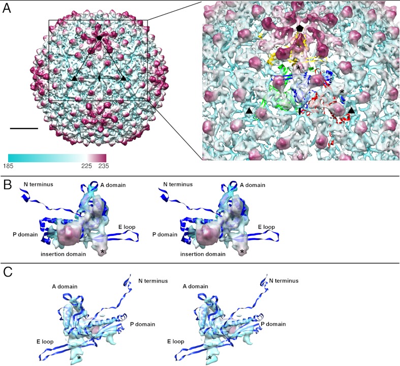
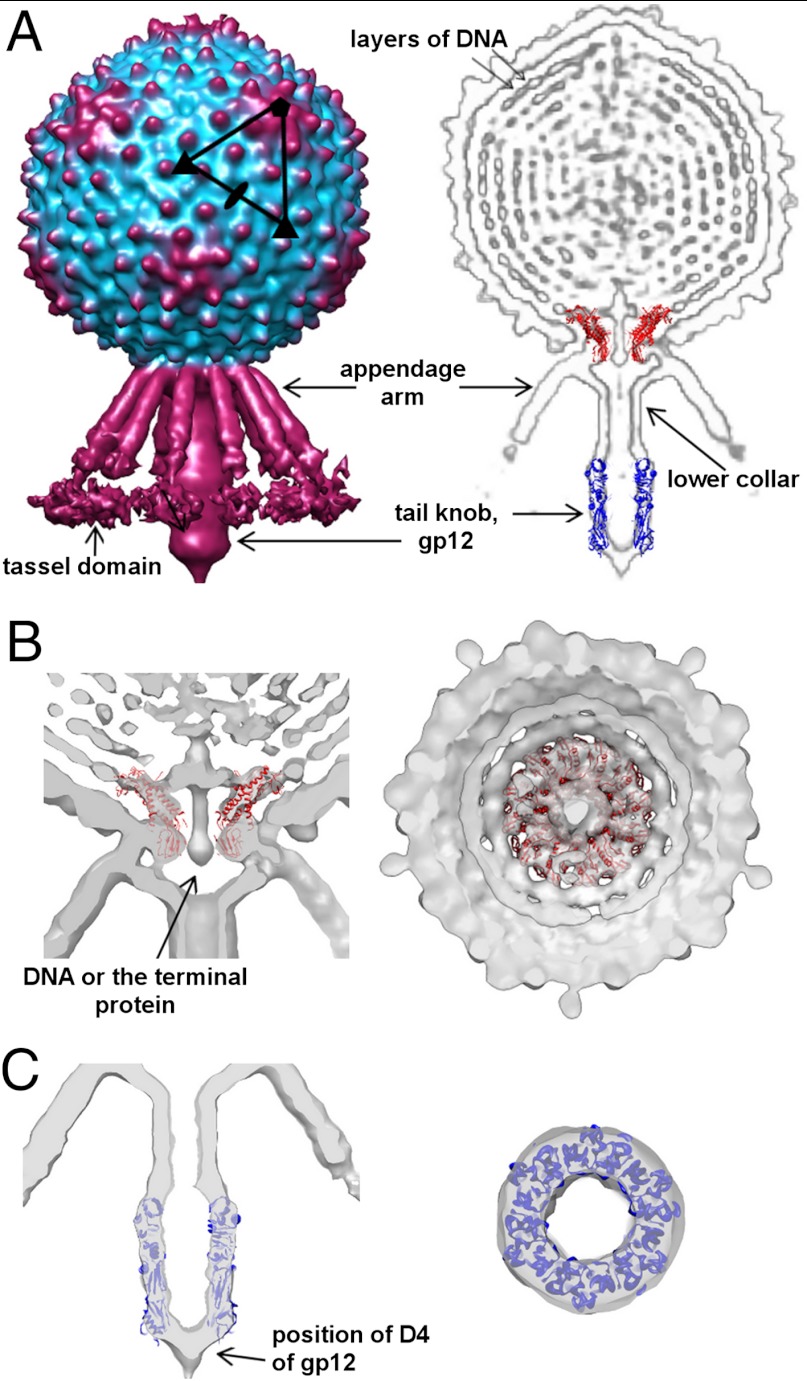
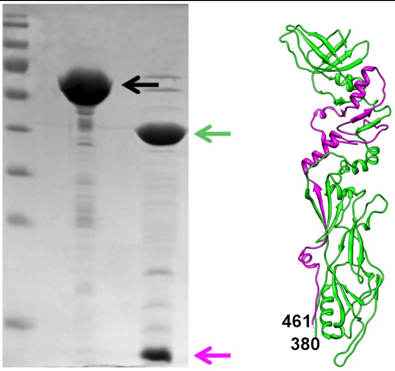
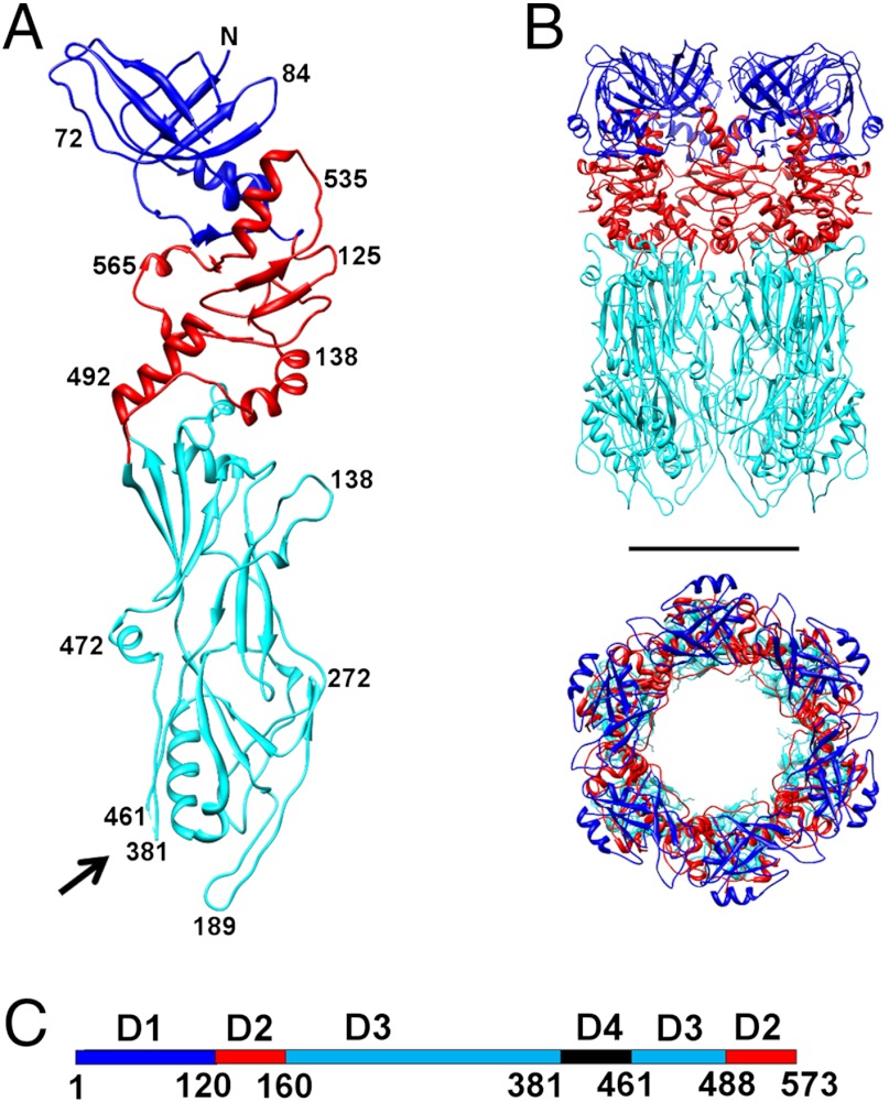
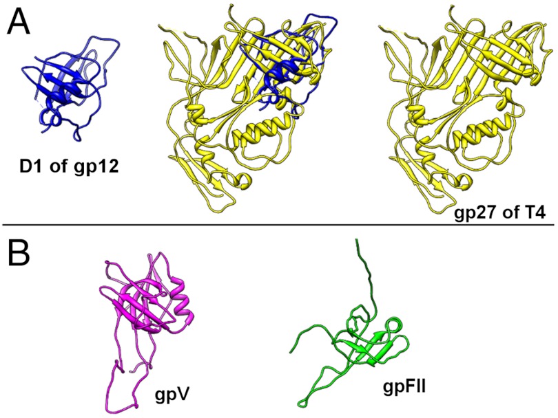
The Podoviridae phage C1 was one of the earliest isolated bacteriophages and the first virus documented to be active against streptococci. The icosahedral and asymmetric reconstructions of the virus were calculated using cryo-electron microscopy. The capsid protein has an HK97 fold arranged into a T = 4 icosahedral lattice. The C1 tail is terminated with a ϕ29-like knob, surrounded by a skirt of twelve long appendages with novel morphology. Several C1 structural proteins have been identified, including a candidate for an appendage. The crystal structure of the knob has an N-terminal domain with a fold observed previously in tube forming proteins of Siphoviridae and Myoviridae phages. The structure of C1 suggests the mechanisms by which the virus digests the cell wall and ejects its genome. Although there is little sequence similarity to other phages, conservation of the structural proteins demonstrates a common origin of the head and tail, but more recent evolution of the appendages.


Conflict of interest statement

The authors declare no conflict of interest.

Figures

Fig. 1.
Fig. 1.
Icosahedral reconstruction of the C1 capsid. (A) The surface of the capsid colored radially from cyan to magenta. The fit of HK97 subunits into four C1 capsid protein densities that form an asymmetric unit is shown on the right. The “*” symbol shows the position of two quasi-2-fold axes in the asymmetric unit. A scale bar corresponds to 100 Å. (B) A stereo view of the capsid protein density segmented from the map. (C) A stereo view of the capsid protein density segmented from the map. The orientations shown in B and C are related by 180° rotation.
Fig. 2.
Fig. 2.
Asymmetric reconstruction of the C1 virion. (A) Surface representation of the C1 virion (Left) and cross-section through the bacteriophage, showing different components of the virus (Right). The structure of the φ29 connector and the C1 major tail protein was fitted into the electron density. (B) Two orthogonal cross-sections through the cryo-EM density map, showing the fit of the φ29 connector. (C) Two orthogonal cross-sections through the cryo-EM density showing the fit of gp12 into the knob.
Fig. 3.
Fig. 3.
Trypsin resistant fragment of gp12. The lanes on the SDS-gel show the marker, full length gp12 (black arrow) and gp12 after trypsin digestion (green and magenta arrows). Corresponding fragments are colored in green and magenta in the gp12 structure shown on the right.
Fig. 4.
Fig. 4.
Structure of the major tail protein, gp12. (A) A monomer of gp12 with domains D1, D2 and D3 colored in blue, red and cyan, respectively. Black arrow points to the location of D4 where the protein was cleaved by trypsin. (B) Side and top views of the gp12 hexamer. Scale bar is 50 Å long. (C) Location of the domains on the linear sequence of gp12, using the same color scheme as in A and B.
Fig. 5.
Fig. 5.
Structural similarity of the C1 major tail protein N-terminal domain (D1) to Siphoviridae and Myoviridae proteins. (A) Structural alignment of gp12 D1 to gp27 from bacteriophage T4. (B) Structures of gpV and gpFII from bacteriophage lambda that have the same fold as D1 of the C1 major tail protein.

References

    1. Clark PF, Clark AS. A “bacteriophage” active against a hemolytic streptococcus. J Bacteriol. 1926;11:89.
    1. Evans AC. Streptococcus bacteriophage: A study of four serological types. Public Health Rep. 1934;49:1386–1401.
    1. Diaz E, Lopez R, Garcia JL. Chimeric phage-bacterial enzymes: A clue to the modular evolution of genes. Proc Natl Acad Sci USA. 1990;87:8125–8129. - PMC - PubMed
    1. Loeffler JM, Nelson D, Fischetti VA. Rapid killing of Streptococcus pneumoniae with a bacteriophage cell wall hydrolase. Science. 2001;294:2170–2172. - PubMed
    1. Schuch R, Nelson D, Fischetti VA. A bacteriolytic agent that detects and kills Bacillus anthracis. Nature. 2002;418:884–889. - PubMed

Publication types

MeSH terms

LinkOut - more resources