. 2012 Aug 28;109(35):13996-4000.
doi: 10.1073/pnas.1210275109. Epub 2012 Aug 13.

Structure and assembly of a paramyxovirus matrix protein

Affiliations

Structure and assembly of a paramyxovirus matrix protein

Anthony J Battisti et al. Proc Natl Acad Sci U S A. .

Abstract

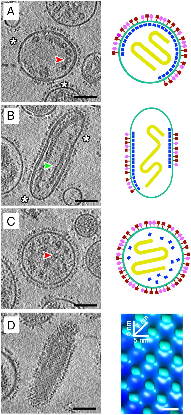
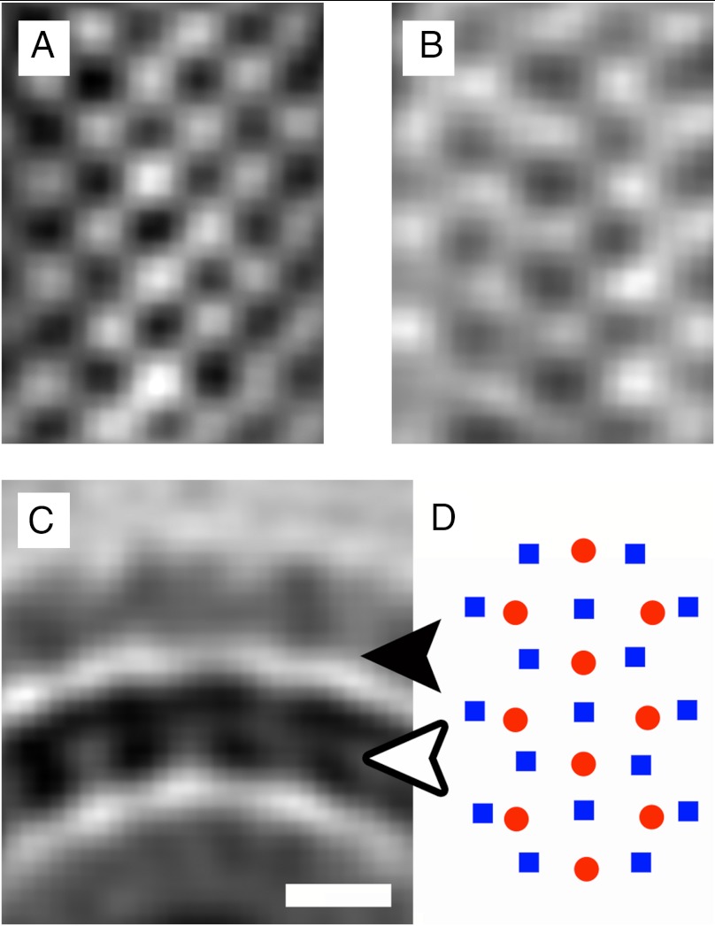
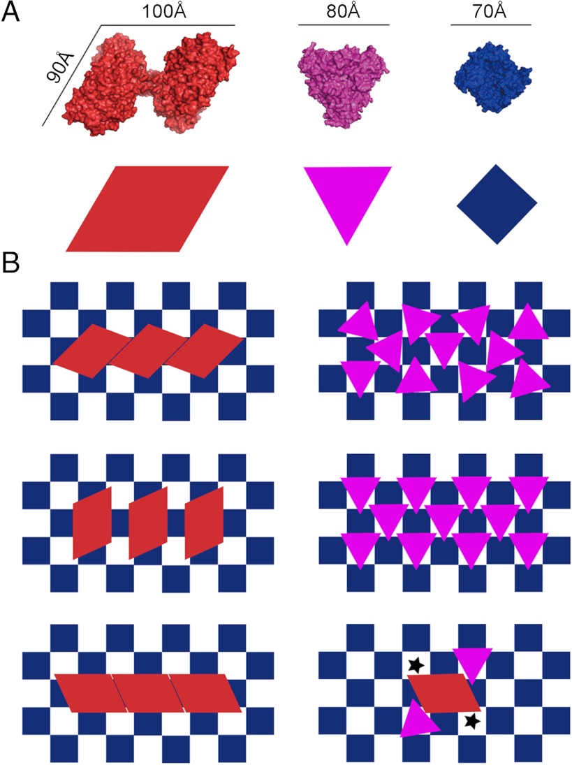
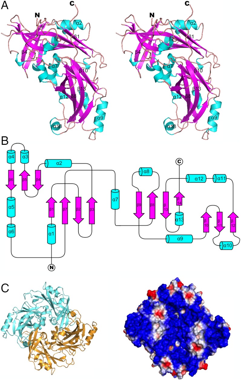
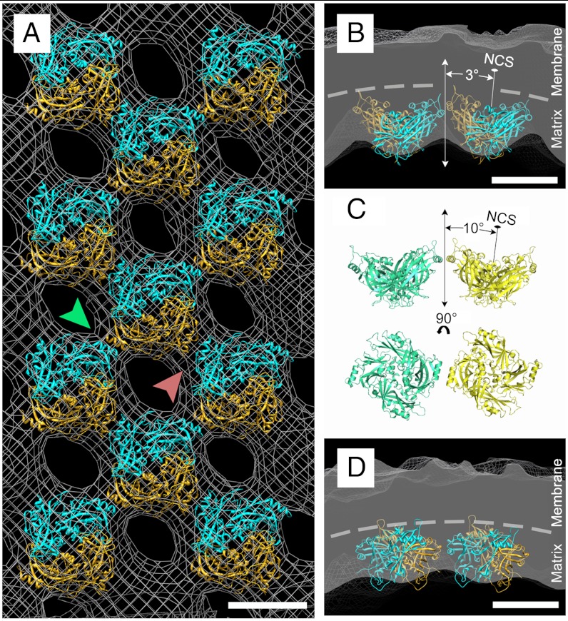
Many pleomorphic, lipid-enveloped viruses encode matrix proteins that direct their assembly and budding, but the mechanism of this process is unclear. We have combined X-ray crystallography and cryoelectron tomography to show that the matrix protein of Newcastle disease virus, a paramyxovirus and relative of measles virus, forms dimers that assemble into pseudotetrameric arrays that generate the membrane curvature necessary for virus budding. We show that the glycoproteins are anchored in the gaps between the matrix proteins and that the helical nucleocapsids are associated in register with the matrix arrays. About 90% of virions lack matrix arrays, suggesting that, in agreement with previous biological observations, the matrix protein needs to dissociate from the viral membrane during maturation, as is required for fusion and release of the nucleocapsid into the host's cytoplasm. Structure and sequence conservation imply that other paramyxovirus matrix proteins function similarly.


Conflict of interest statement

The authors declare no conflict of interest.

Figures

Fig. 1.
Fig. 1.
Tomographic and diagrammatic representations of NDV virions. (A) A tomographic section through the center of a nearly spherical virion, which has an approximately 4–5 nm thick matrix layer lining the inner surface of the viral membrane (Left). The white asterisks indicate the absence of glycoproteins and nucleocapsids where the virus lacks a matrix layer. The red arrowhead indicates a portion of the nucleocapsid as viewed end-on. The virion is also depicted schematically (Right). The HN and F glycoproteins are colored in red and magenta, respectively. The nucleocapsid is depicted in yellow and the matrix protein and membrane are colored dark blue and light blue, respectively. (B) A tomographic section through the center of an elongated virion (Left) and the corresponding schematic representation (Right). The white asterisks indicate the absence of glycoproteins and nucleocapsids where the virus lacks a matrix layer. The green arrowhead indicates a portion of the nucleocapsid that lies in plane. (C) A tomographic section through the center of a spherical virion that lacks an organized matrix protein layer (Left). The red arrowhead indicates the nucleocapsid as viewed end-on. Presumably, the matrix layer has dissociated from the viral membrane (Right). (D) The array of matrix proteins is evident in tomographic sections tangential to the viral membrane for some virions (Left). Subtomographic averaging generated an improved structure for a portion of the matrix protein layer (Right). The averaged map, as viewed from the inside of the virion, is colored according to the radius of a cylinder such that the density attributed to the membrane is light blue and the matrix protein subunits are dark blue. The on-edge and diagonal dimensions of a matrix protein subunit are indicated. For all the tomographic sections, strong density is black and the scale bar represents 75 nm. The tomographic images (AC, Left) represent the average of three layers of voxels over a thickness of approximately 4.5 nm. The tangential section (D, Left) represents the average of seven layers of voxels over a thickness of 10.5 nm and is from the same virion represented in B.
Fig. 2.
Fig. 2.
Periodic glycoprotein densities. (A) A section through the matrix protein array, which lines the inside of the viral membrane in the averaged tomographic map. (B) A section through the averaged map indicating an array on the extracellular side of the membrane. The extracellular densities were averaged assuming the same periodicities that were determined for the matrix protein array. The high densities in B oppose the low-density regions in A, indicating that the HN and F glycoproteins are anchored in the spaces between adjacent proteins in the matrix layer. (C) A cross-section of the averaged map, orthogonal to the ordered arrays. The white and black arrows indicate the planes from which A and B are derived, respectively. (D) A schematic diagram showing the relative locations of the matrix protein dimers (blue squares) and intercalating extracellular densities (red circles). For AC, black represents high density.
Fig. 3.
Fig. 3.
The relationship between matrix and glycoproteins. (A) The crystal structures of NDV HN (red) (25), prefusion parainfluenza virus F (magenta) (6), and NDV M (navy blue) (present results) are shown (Top) and represented schematically (Bottom). The structures are shown such that the viral membrane would be in the plane of the page. (B) Three possible packing arrangements (Top, Middle, and Bottom) of the HN protein (Left) and F protein (Right) relative to the spaces between the matrix proteins. The size of the HN globular head would restrict the placement of other HN proteins in some neighboring spaces. Because the globular head of F is smaller than that of HN, the placement of the F glycoprotein within the array would not be restricted. (Lower Right) The HN and F glycoproteins as they might be situated relative to the matrix layer. Black stars indicate spaces between matrix proteins that could not be occupied by a glycoprotein because of steric hindrance.
Fig. 4.
Fig. 4.
The matrix protein structure from X-ray crystallography. (A) The ribbon drawing of the monomeric structure shown as a stereo diagram. The NDV matrix protein monomer consists of two similarly folded β-sandwich domains (magenta) that are flanked by a number of α-helices (cyan). (B) A schematic diagram representing the secondary structure elements of the matrix protein monomer. (C) The surface of the matrix protein dimer that faces the viral membrane is positively charged and has a nearly square envelope. The two monomers (blue and gold) are shown as a ribbon diagram (Left). The surface charge is represented on a space-filling model (Right), with the positively charged regions shown in blue and the negatively charged regions in red.
Fig. 5.
Fig. 5.
The matrix protein environment in the virus. (A) Fit of the dimeric matrix protein crystal structure (cyan and gold ribbon diagram) into the matrix protein density (gray mesh), which was generated by subtomographic averaging. The matrix protein array lies in the plane of the page. The green arrow indicates the contacts between helix α9 of adjacent dimers. The red arrow indicates the contacts between helix α2 of adjacent dimers. The scale bar represents 5 nm. (B and D) Cross-sections of the viral membrane and matrix layer. B shows the contacts between adjacent dimers generated by helix α9. The dashed line demarcates the membrane and matrix layer interface and illustrates the membrane curvature generated by the matrix protein array. D shows the contacts between adjacent monomers generated by helix α2. The scale bars represent 5 nm. (C) Contacts between matrix dimers similar to those shown in B are made between neighboring dimers in the monoclinic crystal structure. The NCS dimer axes related by a crystallographic twofold subtend an angle of 20° between them. Similar interdimer contacts are made between neighboring dimers in the virus (B), which subtend an angle of about 6° between them.

References

    1. Lamb RA, Parks GD. Paramyxoviridae: The viruses and their replication. In: Knipe DM, Howley PM, editors. Fields Virology. 5th Ed. Philadelphia: Lippincott Williams and Wilkins; 2007. pp. 1449–1496.
    1. McGinnes LW, Pantua H, Reitter J, Morrison TG. Newcastle disease virus: Propagation, quantification, and storage. Curr Protoc Microbiol. 2006 Chapter 15:Unit 15F.2. - PubMed
    1. National Select Agent Registry. http://www.selectagents.gov. Accessed August 2012.
    1. Scheid A, Caliguiri LA, Compans RW, Choppin PW. Isolation of paramyxovirus glycoproteins. Association of both hemagglutinating and neuraminidase activities with the larger SV5 glycoprotein. Virology. 1972;50:640–652. - PubMed
    1. Yin HS, Paterson RG, Wen X, Lamb RA, Jardetzky TS. Structure of the uncleaved ectodomain of the paramyxovirus (hPIV3) fusion protein. Proc Natl Acad Sci USA. 2005;102:9288–9293. - PMC - PubMed

Publication types

MeSH terms

LinkOut - more resources