. 2012 Aug 28;109(35):13990-5.
doi: 10.1073/pnas.1203219109. Epub 2012 Aug 13.

Structural divergence of paralogous S components from ECF-type ABC transporters

Affiliations

Structural divergence of paralogous S components from ECF-type ABC transporters

Ronnie P-A Berntsson et al. Proc Natl Acad Sci U S A. .

Abstract

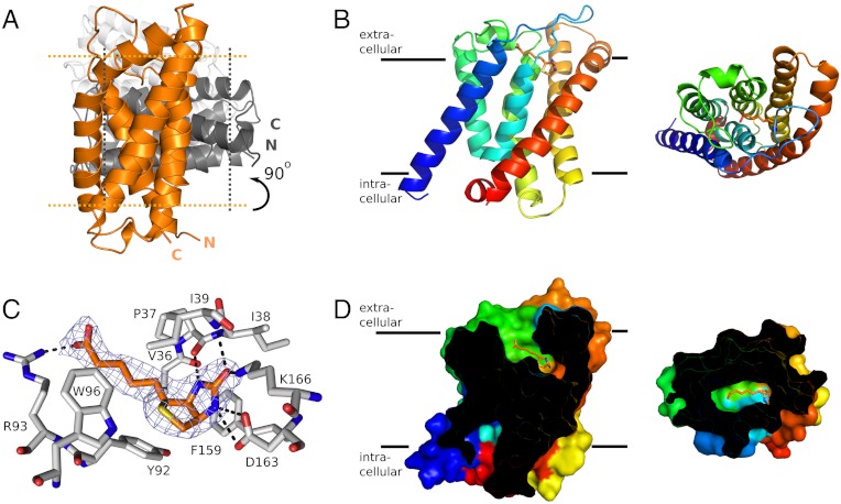
Energy coupling factor (ECF) proteins are ATP-binding cassette transporters involved in the import of micronutrients in prokaryotes. They consist of two nucleotide-binding subunits and the integral membrane subunit EcfT, which together form the ECF module and a second integral membrane subunit that captures the substrate (the S component). Different S components, unrelated in sequence and specific for different ligands, can interact with the same ECF module. Here, we present a high-resolution crystal structure at 2.1 Å of the biotin-specific S component BioY from Lactococcus lactis. BioY shares only 16% sequence identity with the thiamin-specific S component ThiT from the same organism, of which we recently solved a crystal structure. Consistent with the lack of sequence similarity, BioY and ThiT display large structural differences (rmsd = 5.1 Å), but the divergence is not equally distributed over the molecules: The S components contain a structurally conserved N-terminal domain that is involved in the interaction with the ECF module and a highly divergent C-terminal domain that binds the substrate. The domain structure explains how the S components with large overall structural differences can interact with the same ECF module while at the same time specifically bind very different substrates with subnanomolar affinity. Solitary BioY (in the absence of the ECF module) is monomeric in detergent solution and binds D-biotin with a high affinity but does not transport the substrate across the membrane.


Conflict of interest statement

The authors declare no conflict of interest.

Figures

Fig. 1.
Fig. 1.
(A) Cartoon showing the relative orientation of the three molecules of BioY in the asymmetric unit of the BioY crystals. The three molecules are colored differently (orange, dark gray, and light gray) and the approximate membrane boundaries are indicated with dotted lines for the orange and dark gray molecules. The trimeric arrangement is incompatible with a membrane-embedded oligomer. (B) A monomer of BioY in secondary structure cartoon representation colored from blue (N terminus) to red (C terminus). The bound biotin molecule is shown in stick representation with carbon atoms in orange. The Left and Right views are from the plain of the membrane and along the membrane normal (from the outside), respectively. (C) Binding site of biotin. The biotin molecule is shown in orange and the interacting residues from BioY in gray. Electron density for biotin (2FO-FC map contoured at 1.5σ) in blue mesh. (D) Sliced surface representation of BioY showing the binding cavity, with the bound biotin shown in orange. Coloring and viewpoints as in B.
Fig. 2.
Fig. 2.
Superimposed structures of BioY (orange), ThiT (gray), and RibU (yellow) viewed from the plane of the membrane (A) and from the outside of the cell (B, direction of view perpendicular to the membrane plane). The structures have been superimposed on helices 1 and 3 in order to highlight the structural similarities of helices 1–3 and the differences of helices 4–6. Loops 1 are indicated in thick lines. Helices 1–6 are marked with H1–H6.
Fig. 3.
Fig. 3.
Biotin binding to BioY. (A) Titration of 10 nM BioY with D-biotin. The intrinsic protein fluorescence was measured (excitation wavelength 280 nm, emission wavelength 360 nm). Inset: fluorescence spectrum of 300 nM BioY in the absence of biotin (solid line) and in the presence of a saturating amount of biotin (1 mM, dotted line). (B) Biotin binding to BioYRC. Titration of 50 nM BioY from Rhodobacter capsulatus with D-biotin. The intrinsic protein fluorescence was measured (excitation wavelength 280 nm, emission wavelength 349 nm). Inset: fluorescence spectrum of BioY in the absence of biotin (solid line) and in the presence of a saturating amount of biotin (100 nM, dotted line).
Fig. 4.
Fig. 4.
Biotin binding to liposomes containing purified and reconstituted BioY from L. lactis (A) and BioYRC from R. capsulatus (B), respectively. Biotin binding was measured in the presence (black circles) or absence (white circles) of membrane gradients of proton gradient, sodium ions, and a membrane potential (-120 mV). Black and white triangles represent the same experiment using liposomes without BioY. After 10.5 min an excess (1 mM) of unlabeled biotin was added. 160 μg lipids (approximately 1.6 μg of BioY) was used per time point. Squares in B: counterflow experiment. Proteoliposomes were loaded with 15 μM unlabeled biotin before 100-fold dilution into buffer containing 20 nM labeled biotin to start the experiment. All experiments were performed in duplicate or triplicate, and the error bars represent the spread in the data. The amounts of biotin bound to the liposomes in (B) is lower than in (A) because the BioYRC is less stable in detergent solution (prone to aggregation) causing lower reconstitution efficiencies of functional protein.

References

    1. Rodionov DA, Hebbeln P, Gelfand MS, Eitinger T. Comparative and functional genomic analysis of prokaryotic nickel and cobalt uptake transporters: Evidence for a novel group of ATP-binding cassette transporters. J Bacteriol. 2006;188:317–327. - PMC - PubMed
    1. Hebbeln P, Rodionov DA, Alfandega A, Eitinger T. Biotin uptake in prokaryotes by solute transporters with an optional ATP-binding cassette-containing module. Proc Natl Acad Sci USA. 2007;104:2909–2914. - PMC - PubMed
    1. Rodionov DA, et al. A novel class of modular transporters for vitamins in prokaryotes. J Bacteriol. 2009;191:42–51. - PMC - PubMed
    1. Erkens GB, Majsnerowska M, Beek ter J, Slotboom D-J. Energy coupling factor-type ABC transporters for vitamin uptake in prokaryotes. Biochemistry. 2012;51:4390–4396. - PubMed
    1. Beek ter J, Duurkens R, Erkens G, Slobtoom DJ. Quaternary structure and functional unit of Energy Coupling Factor (ECF)-type transporters. J Biol Chem. 2010;286:5471–5475. - PMC - PubMed

Publication types

MeSH terms

Associated data

LinkOut - more resources