. 2012 Sep 11;109(37):15012-7.
doi: 10.1073/pnas.1207035109. Epub 2012 Aug 13.

Retinal prosthetic strategy with the capacity to restore normal vision

Affiliations

Retinal prosthetic strategy with the capacity to restore normal vision

Sheila Nirenberg et al. Proc Natl Acad Sci U S A. .

Abstract

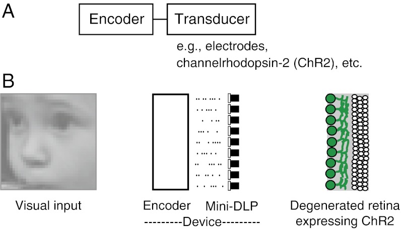
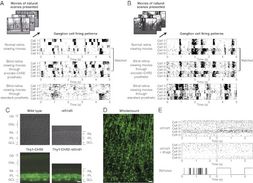
Retinal prosthetics offer hope for patients with retinal degenerative diseases. There are 20-25 million people worldwide who are blind or facing blindness due to these diseases, and they have few treatment options. Drug therapies are able to help a small fraction of the population, but for the vast majority, their best hope is through prosthetic devices [reviewed in Chader et al. (2009) Prog Brain Res 175:317-332]. Current prosthetics, however, are still very limited in the vision that they provide: for example, they allow for perception of spots of light and high-contrast edges, but not natural images. Efforts to improve prosthetic capabilities have focused largely on increasing the resolution of the device's stimulators (either electrodes or optogenetic transducers). Here, we show that a second factor is also critical: driving the stimulators with the retina's neural code. Using the mouse as a model system, we generated a prosthetic system that incorporates the code. This dramatically increased the system's capabilities--well beyond what can be achieved just by increasing resolution. Furthermore, the results show, using 9,800 optogenetically stimulated ganglion cell responses, that the combined effect of using the code and high-resolution stimulation is able to bring prosthetic capabilities into the realm of normal image representation.


Conflict of interest statement

Conflict of interest statement: S.N. and C.P. have a patent application filed through Cornell University.

Figures

Fig. 1.
Fig. 1.
Schematic of the prosthetic. (A) The two components: an encoder and a transducer. The encoder converts the image into the code used by the retinal output cells, the ganglion cells; the transducer drives the ganglion cells to fire spike patterns as the code specifies. As indicated, the transducers can be electrodes, optogenetic stimulators such as ChR2, etc. (B) The steps from visual input to retinal output for a blind retina. The input enters a device that contains the encoder and a stimulator (a mini-DLP). The encoder converts the input into patterns of electrical pulses, analogous to the patterns of spikes that would be produced by the normal retina to the same visual input. The patterns of electrical pulses are then converted into patterns of light pulses (via the DLP) to drive the ChR2 in the ganglion cells. For a schematic of the setup as used in the in vitro and in vivo experiments, see Fig. S5.
Fig. 2.
Fig. 2.
Blind retinas produce the same output as the normal retina. (A) (Top) Ganglion cell-firing patterns from a normal, wild-type retina when it was presented with movies of natural scenes (landscapes, faces, people walking, etc). Several cells are shown, including both ON transient and ON sustained cells. (For longer stretches of data, see Figs. S1 and S4.) (Middle) Ganglion cell firing patterns from a blind retina (Thy1-ChR2 rd1/rd1 retina) when it was presented with the same movies, but through the encoder-ChR2 prosthetic. As shown, the prosthetic confers on the blind retina the ability to produce firing patterns that closely match those of the normal retina. (Bottom) Ganglion cell firing patterns from a blind retina (Thy1-ChR2 rd1/rd1 retina) when it was presented with the same movies, but through the standard optogenetic prosthetic (just ChR2, no encoder). To allow comparison with the middle set of recordings, the movies were presented with the same stimulator (same mini-DLP, same wavelength, same peak intensity), so that the only difference between the Middle and Bottom recordings was the use of the encoder. As shown, although the standard approach is very effective in driving the ganglion cells, the firing patterns are not the normal patterns. The same receptive field locations were used for all panels to allow comparison of firing patterns (white circles on the movie images). (B) Same as in A, but using a movie with different image statistics and different ganglion cell types (OFF and ON-OFF types), indicating the robustness of the results. (C) Retinal cross sections from wild-type, rd1/rd1, Thy1-ChR2, and Thy1-ChR2 rd1/rd1 mice, respectively. The retina from the Thy1-ChR2 rd1/rd1 animal shows both the severe degeneration characteristic of rd1/rd1 animals (21, 22) (the absence of the photoreceptor layer) and the targeted expression of ChR2 to the ganglion cells. (Scale bar, 20 μm.) (D) Whole-mount view of the retina showing the ChR2-expressing cells. (Scale bar, 50 μm.) (E) Recordings from rd1/rd1 retinas that do not express ChR2. These retinas were stimulated using the same stimulator as in A (same mini-DLP, same wavelength, same peak intensity). (Top Left) Ganglion cell responses when the retina was stimulated with the encoded movies. (Top Right) Ganglion cell responses when the retina was stimulated with a periodic flash. As shown, and as expected, because these retinas have no photoreceptor outer segments and no ChR2, there is no response to either stimulus, just baseline firing. Bottom, Left and Right, are same as Top, Left and Right, but with the addition of the neurotransmitter blockers APB, CPP, and NBQX, as in ref. ; again, no stimulus-dependent responses were observed. For all recordings from degenerated retinas in this paper, i.e., for Figs. 2–4, the retinas were rendered blind using both methods: the use of rd1/rd1 animals and the blockers, the latter included for compulsivity.
Fig. 3.
Fig. 3.
The output of blind retinas carries the same amount of information, and the same quality of information, as the output of normal retinas, as measured using confusion matrices. (A) (Top) Confusion matrices generated from the responses of a normal wild-type retina when presented with movies of natural scenes, which include faces, people walking, landscapes, etc. (Middle) Confusion matrices generated from the responses of a blind retina (Thy1-ChR2 rd1/rd1 retina) when it was presented with the same movies, but through the encoder-ChR2 prosthetic. (Bottom) Confusion matrices generated from the responses of a blind retina (Thy1-ChR2 rd1/rd1 retina) when it was presented with the same movies, but through the standard optogenetic prosthetic. (B) Same as A, but for a different set of movies. As shown in both A and B, the confusion matrices generated from the responses of the blind retinas treated with the encoder-ChR2 prosthetic closely match those of the normal wild-type retinas: the data in the right-most matrices (the population performances) lie along the diagonal line, indicating correct identification of the stimuli. See Fig. S7 for the same analysis performed with an array of bin sizes.
Fig. 4.
Fig. 4.
Images reconstructed from the spike trains of the blind retinas. Although the brain does not necessarily reconstruct images, reconstructions serve as a convenient way to compare methods and give an approximation of the level of visual restoration possible with each approach. (A) Original image (a baby's face). (B) Image reconstructed from the responses of the encoder. (C) Image reconstructed from the responses of the blind retina when it was presented with the face through the encoder-ChR2 prosthetic. (D) Image reconstructed from the responses of the blind retina when it was presented with the face through the standard optogenetic prosthetic. The reconstructions were carried out on a processing cluster in blocks of 10 × 10 or 7 × 7 checks. The decoding was performed using maximum likelihood; that is, for each block, we found the array of gray values that maximized the probability of the observed responses (following ref. for high dimensional searches). The number and density of cells used match that of the normal mouse retina—∼9,800 ganglion cell responses per image (see Materials and Methods for numbers).
Fig. 5.
Fig. 5.
Behavioral performance: optomotor tracking occurs with the encoder-ChR2 method. (A) Baseline condition (no stimulus present). Blind animals show a drift in eye position when no stimulus is present, similar to blind humans; the direction of the drift is downward. (B) Response to drifting gratings presented using the standard optogenetic method, that is, where the stimulus was presented unencoded. As shown, no tracking occurs, just the downward drift. (C) Response to drifting gratings presented using the encoder-ChR2 method, where the stimulus was presented in its encoded form (i.e., using ON cell encoders; see main text). As shown, when the stimulus was converted into the code used by the ganglion cells, the animal became able to track it. (AC, Top) Representative example of a raw eye-position trace. (AC, Middle) Smooth component (saccades and movement artifacts removed). (AC, Bottom) Average trajectory across all trials (n = 15, 14, and 15 trials, respectively).

Similar articles

Cited by

References

    1. Chopdar A, Chakravarthy U, Verma D. Age related macular degeneration. BMJ. 2003;326:485–488. - PMC - PubMed
    1. Resnikoff S, Pascolini D, Mariotti SP, Pokharel GP. 2008. in Bull World Health Organ 86(1):63–70.
    1. Gordois A, Pezzullo L, Cutler H. The Global Economic Cost of Visual Impairment. Canberra, Australia: Access Economics Pty Limited; 2010.
    1. Chader GJ, Weiland J, Humayun MS. Artificial vision: Needs, functioning, and testing of a retinal electronic prosthesis. Prog Brain Res. 2009;175:317–332. - PubMed
    1. Zrenner E, et al. 2009. Subretinal microelectrode arrays allow blind retinitis pigmentosa patients to recognize letters and combine them to words. Proc 2nd Int Conf IEEE Biomed Eng Inf, pp 1–4.

Publication types

Substances

LinkOut - more resources