. 2012 Aug 28;109(35):14146-51.
doi: 10.1073/pnas.1202165109. Epub 2012 Aug 13.

Endothelial Semaphorin 7A promotes neutrophil migration during hypoxia

Affiliations

Endothelial Semaphorin 7A promotes neutrophil migration during hypoxia

Julio César Morote-Garcia et al. Proc Natl Acad Sci U S A. .

Abstract

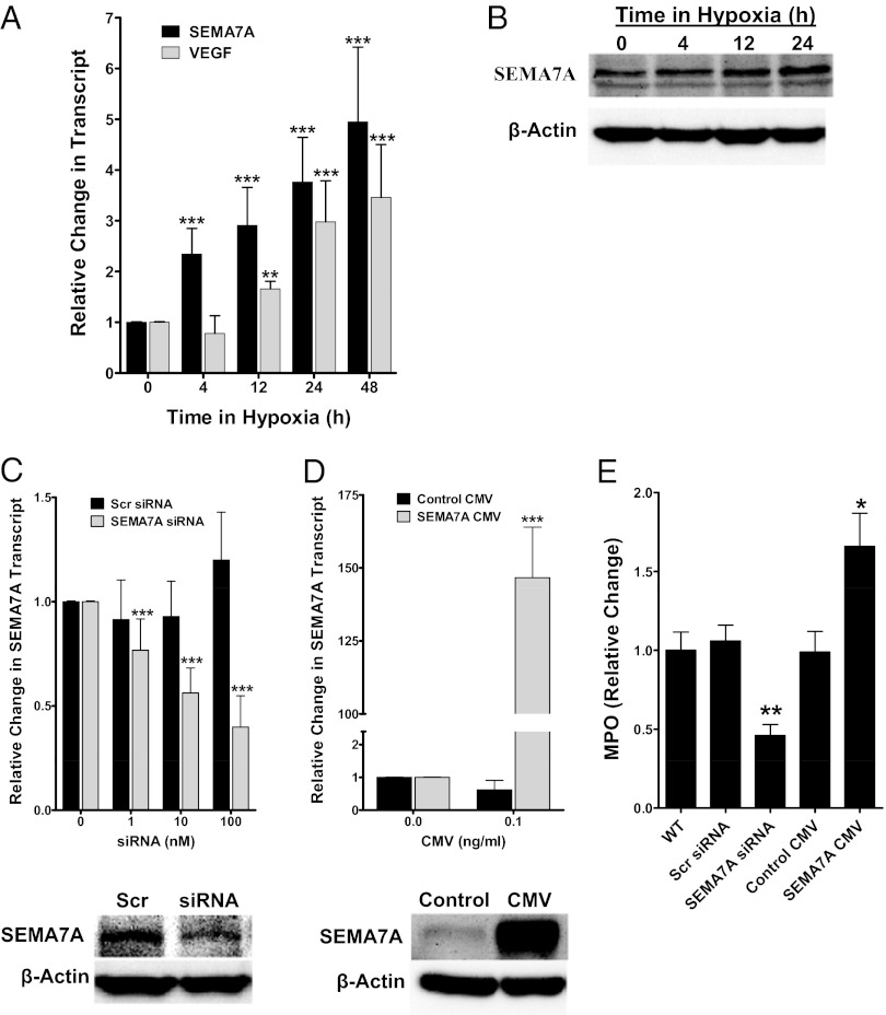
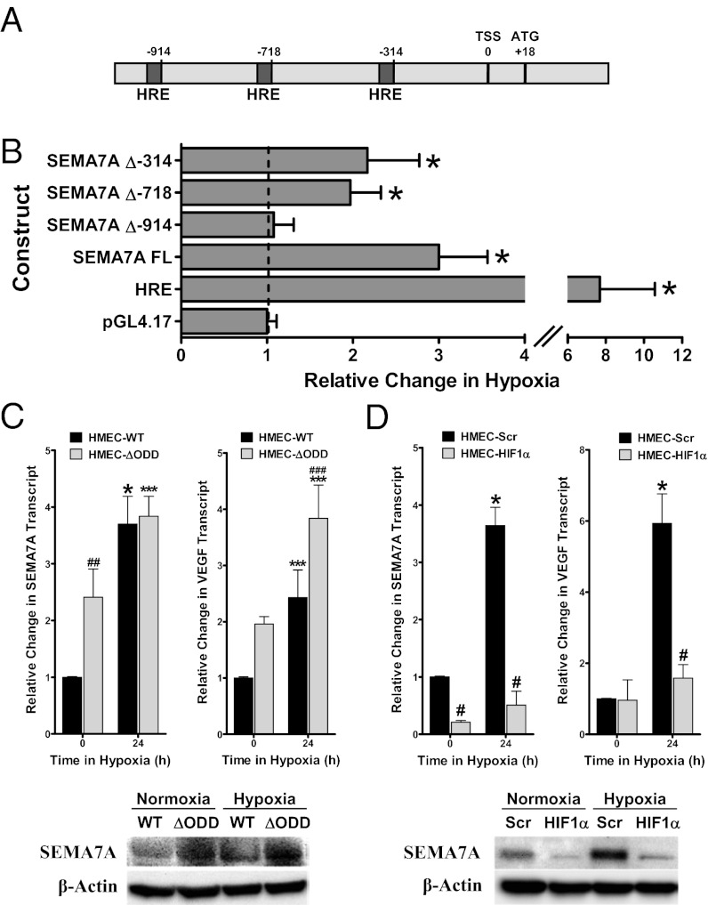
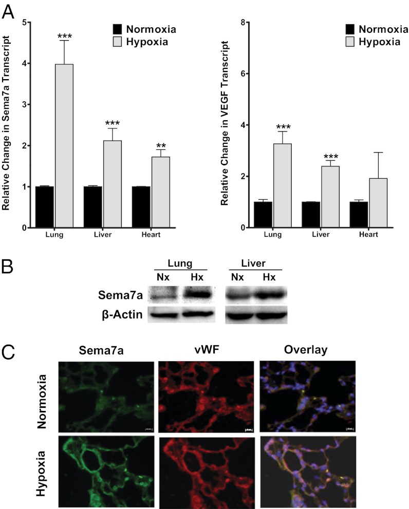
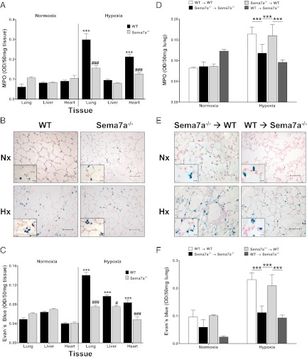
Recent studies identified basic biological principles that are shared by the immune and the nervous system. One of these analogies applies to the orchestration of cellular migration where guidance proteins that serve as a stop signal for axonal migration can also serve as a stop signal for the migration of immune-competent cells. The control of leukocyte migration is of key interest during conditions associated with inflammatory tissue changes such as tissue hypoxia or hypoxic inflammation. Semaphorins are members of these axon guidance molecules. Previously unknown, we report here the expression and induction of semaphorin 7A (SEMA7A) on endothelium through hypoxia-inducible factor 1α during hypoxia. This induction of SEMA7A translates into increased transmigration of polymorphonuclear neutrophil granulocytes across endothelial cells. Extension of these findings demonstrated an attenuated extravasation of polymorphonuclear neutrophil granulocytes in Sema7a-deficient mice from the vasculature during hypoxia. Studies using chimeric animals identified the expression of Sema7A on nonhematopoietic tissue to be the underlying cause of the observed results. Taken together, our findings demonstrate that neuronal guidance proteins do not only serve as a stop signal for leukocyte migration but also can propagate the extravasation of leukocytes from the vascular space. Future anti-inflammatory strategies might be based on this finding.


Conflict of interest statement

The authors declare no conflict of interest.

Figures

Fig. 1.
Fig. 1.
Regulation of SEMA7A during hypoxia and role in PMN transmigration in vitro. (A) Relative change in SEMA7A-mRNA transcript after exposure of HMEC-1 cells to indicated time points of hypoxia (2% oxygen). VEGF was used as positive control for hypoxia exposure. Data are expressed as fold change in transcript over normoxia ± SD at each indicated time and have been calculated relative to the internal housekeeping gene (β-actin) (***P < 0.001 and **P < 0.01 by two-tailed Student’s t test). (B) Induction of SEMA7A protein in endothelial HMEC-1 cells exposed to indicated time points of hypoxia. (C and D) Analysis of SEMA7A expression by RT-PCR and Western blot in HMEC-1 cells transfected with (C) specific siRNA against SEMA7A (SEMA7A siRNA) or negative scrambled control (Scr siRNA) (***P < 0.001 by two-tailed Student’s t test) or (D) with empty CMV (Control CMV) or SEMA7A-CMV (***P < 0.001 by two-tailed Student’s t test). (E) Transendothelial migration assay performed in HMEC-1 cells that were previously transfected with SEMA7A siRNA, SEMA7A-CMV, or respective controls. Data are presented as percentage of myeloperoxidase activity in comparison with the respective control cells (WT) (**P < 0.01 and *P < 0.05 indicate differences between SEMA7A-siRNA/CMV and their respective transfection controls by two-tailed Student’s t test). Data are representative of at least five independent experiments. In B and C, one representative Western blot of three independent experiments is depicted.
Fig. 2.
Fig. 2.
Role of HIF-1α in regulation of SEMA7A during hypoxia in vitro. (A) Graphic representation of the putative SEMA7A promoter. Three potential hypoxia-responsive elements (HRE) were identified at positions −918 to −914 (DNA consensus motif 5′-CACGT-3′), −722 to −718 (5′-CACGG-3′), and −318 to −314 (5′-CACGA-3′), relative to the transcription start site (TSS). (B) Full-length and indicated constructs containing mutations in one of the HRE were transfected into HMEC-1 cells and assayed for luciferase activity after exposure to hypoxia for 24 h. Results depict the fold change in relative luminescence in hypoxia relative to normoxic controls. An HRE plasmid containing four tandem HREs is shown as a hypoxia-positive control. Data shown are pooled from n = 6 and are normalized for background vector (empty pGL4.17) and total protein and are presented as mean ± SD, where ***P < 0.001 indicates significance between individual plasmids and empty pGL4.17 plasmid by two-tailed Student’s t test. (C) Confluent monolayers of control (HMEC-WT) or oxygen-stable HIF-1α–expressing (HMEC-ΔODD) HMEC-1 cell lines or (D) HMEC-1 with psiRNA repression of HIF-1α (HMEC-HIF1α) or control transfected cells (HMEC-Scr) were exposed to 24 h of hypoxia (2% oxygen). SEMA7A transcript levels were afterward determined by RT-PCR and Western blot. ***P < 0.001 and *P < 0.05 indicate significance between normoxia and hypoxia; ###P < 0.001, ##P < 0.01, and #P < 0.05 indicate differences between different cell types by one-factor ANOVA. Data are representative of at least five independent experiments. In C and D, one representative Western blot of three independent experiments is depicted.
Fig. 3.
Fig. 3.
Regulation of Sema7a during hypoxia in vivo. (A) Sema7a-mRNA levels were examined in lung, liver, and heart from mice subjected either to normoxia or normobaric hypoxia (8% oxygen) for 4 h. Levels of VEGF-mRNA were also assayed as positive control for hypoxia exposure. Data are pooled from two independent experiments with n ≥ 5 mice (***P < 0.001 and **P < 0.01 indicate differences between normoxia and hypoxia by two-tailed Student’s t test). (B) Analysis of Sema7a protein in lung and liver from mice after normoxia or hypoxia exposure. Protein samples were pooled (n = 6) before being electrophoresed. (C) Pulmonary immunohistochemistry for murine semaphorin 7A and von Willebrand’s factor during hypoxia. Lung from mice exposed to normobaric hypoxia or normoxia for 4 h were harvested and embedded in paraffin. Sections were stained with specific antibodies against murine Sema7a (green) and the endothelial marker von Willebrand’s factor (red). Colocalization of Sema7a with von Willebrand’s factor (vWF) is seen as yellow in the Overlay image. DAPI was used for nuclear counterstain (blue) (magnification 200×). One representative picture from four different lung sections is displayed. (Scale bars, 50 μm.)
Fig. 4.
Fig. 4.
Role of Sema7a during hypoxia in vivo. (A) Influx of PMNs into lung, liver, and heart was evaluated by MPO measurements after exposure of wild type (WT) and Sema7a−/− mice to normoxia or normobaric hypoxia (8% oxygen) for 4 h. Data are expressed as mean ± SD units of MPO/mg protein (***P < 0.001 indicates differences between normoxia and hypoxia; ###P < 0.001 between wild-type and Sema7a−/− organs by one-factor ANOVA). Data are from one experiment with n = 6 mice per group. (B) Staining of PMNs in histological sections of pulmonary tissue of WT and Sema7a−/− mice after hypoxia exposure (magnification 400×; detail 1,000×). A representative picture from three different lung sections is displayed. (C) Vascular leak was assessed by i.v. injection of Evan’s blue in wild type (WT) and Sema7a−/− mice before exposure to normoxia or normobaric hypoxia. Animals were afterward euthanized, and Evan’s blue concentrations were determined in lung, liver, and heart. Data are expressed as mean ± SD. Data from Evan’s blue OD/50-mg wet tissue are pooled from six animals per condition (***P < 0.001 indicates differences between normoxia and hypoxia; ###P < 0.001 and #P < 0.05 indicate differences between wild-type and Sema7a−/− organs by one-factor ANOVA). (D) MPO measurement in lung of chimeric animals 4 h after exposure to normoxia or normobaric hypoxia. Mean values of two independent experiments with n ≥ 5 mice per group ± SD are represented (***P < 0.001 reflects differences between indicated chimeric animals by one-factor ANOVA). (E) Staining of PMNs in histological sections of pulmonary tissue of myeloid- and tissue-specific chimeric mice (magnification 400×; detail 1,000×). A representative picture from three different lung sections is displayed. (F) Evan’s blue pulmonary extravasation in chimeric animals 4 h after exposure to normoxia or normobaric hypoxia. Data are expressed as mean ± SD. Data from Evan’s blue OD/50-mg wet tissue are pooled from two independent experiments with n ≥ 5 mice per condition (***P < 0.001 reflects differences between indicated chimeric animals by one-factor ANOVA). [Scale bars, 50 μm (400×) and 10 μm (1,000×).]

Similar articles

Cited by

References

    1. Eltzschig HK, Carmeliet P. Hypoxia and inflammation. N Engl J Med. 2011;364:656–665. - PMC - PubMed
    1. Walmsley SR, et al. Hypoxia-induced neutrophil survival is mediated by HIF-1alpha-dependent NF-kappaB activity. J Exp Med. 2005;201(1):105–115. - PMC - PubMed
    1. Wu JY, et al. The neuronal repellent Slit inhibits leukocyte chemotaxis induced by chemotactic factors. Nature. 2001;410:948–952. - PMC - PubMed
    1. Mirakaj V, et al. Repulsive guidance molecule-A (RGM-A) inhibits leukocyte migration and mitigates inflammation. Proc Natl Acad Sci USA. 2011;108:6555–6560. - PMC - PubMed
    1. Rosenberger P, et al. Hypoxia-inducible factor-dependent induction of netrin-1 dampens inflammation caused by hypoxia. Nat Immunol. 2009;10:195–202. - PubMed

MeSH terms

LinkOut - more resources