. 2012 Aug 28;109(35):14019-23.
doi: 10.1073/pnas.1212545109. Epub 2012 Aug 13.

An image-based screen identifies a small molecule regulator of megakaryopoiesis

Affiliations

An image-based screen identifies a small molecule regulator of megakaryopoiesis

Anthony E Boitano et al. Proc Natl Acad Sci U S A. .

Abstract

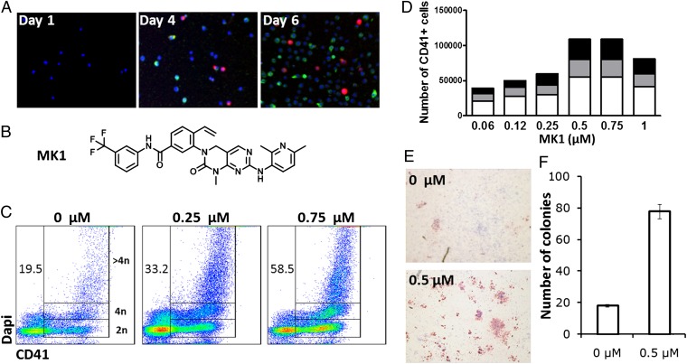
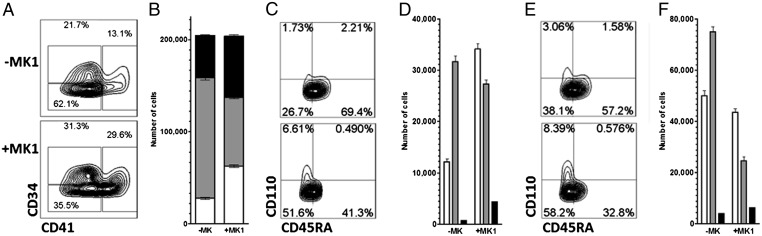
Molecules that control the lineage commitment of hematopoietic stem cells (HSCs) may allow the expansion of enriched progenitor populations for both research and therapeutic uses. In an effort to better understand and control the differentiation of HSCs to megakaryocytes, we carried out an image-based screen of a library of 50,000 heterocycles using primary human CD34(+) cells. A class of naphthyridinone derivatives was identified that induces the differentiation of common myeloid progenitors (CMP) to megakaryocytes. Kinase profiling and subsequent functional assays revealed that these compounds act through inhibition of platelet-derived growth factor receptor (PDGFR) signaling in CMPs. Such molecules may ultimately have clinical utility in the treatment of thrombocytopenia.


Conflict of interest statement

The authors declare no conflict of interest.

Figures

Fig. 1.
Fig. 1.
Screen of primary human mPB CD34+ cells identifies MK1 as an inducer of megakaryopoiesis. (A) Expression of CD41 (red) and CD71 (green) in mPB CD34+ cells cultured for 6 d in HSC expansion media. (B) Structure of MK1. (C) DNA content of mPB CD34+ cells cultured for 10 d in the presence of MK1 at the indicated concentrations. CD41+ cells containing 2N, 4N, and >4N are gated. Numbers represent the percentage of CD41+ cells. (D) Number of 2N CD41+ cells (white), 4N CD41+ cells (gray), and >4N CD41+ cells (black) from 5,000 mPB CD34+ cells cultured for 10 d in the presence of MK1 at the indicated concentrations. (E) CFU-Mk (red) generated from mPB CD34+ cells cultured in the presence or absence of MK1 (0.5 μM) for 8 d (F) Total CFU-Mk present in 5,000 mPB CD34+ cells expanded for 8 d in the presence or absence of MK1 (0.5 μM). Mean and SD are plotted from three individual plates within the same experiment (P < 0.01). Data are representative of a least three independent experiments.
Fig. 2.
Fig. 2.
MK1 promotes megakaryocyte differentiation at the expense of the granulocyte and monocyte lineages. (A) Phenotype of the progeny of 5,000 mPB CD34+ cells cultured for 10 d in the presence or absence of MK1 (0.75 μM). Megakaryocytes (CD41), stem and progenitor cells (CD41CD34+), and single lineage progenitors/mature cells (CD41CD34) are gated. (B) Absolute number of megakaryocytes (white, P < 0.01), stem and progenitor cells (black), and single lineage progenitors/mature cells (gray) in A. (C) Phenotype of the stem and progenitor cells (CD41CD34+) gated in A: CMPs (CD41CD34+CD110CD45RA), GMPs (CD41CD34+CD110CD45RA+), and MEPs (CD41CD34+CD110+CD45RA) are gated. (D) Absolute numbers of CMPs (white), GMPs (gray), and MEPs (black) in C (P < 0.01). (E) Phenotype of single lineage progenitors/mature cells (CD41CD34) gated in A: Granulocyte or monocyte progenitors (CD41CD34CD110CD45RA+), erythroid progenitors (CD41CD34CD110+CD45RA), and mature cells (CD41CD34CD110CD45RA) are gated. (F) Absolute number of mature cells (white), granulocyte or monocyte progenitors (gray), and erythroid progenitors (black) in E (P < 0.02). Data are representative of at least three independent experiments.
Fig. 3.
Fig. 3.
MK1 is a PDGFR antagonist. (A) Correlation (R2 = 0.82) between EC50 for megakaryopoiesis and IC50 for PDGFR inhibition. (B) Absolute number of CD41+ cells derived from 1,000 mPB CD34+ cells cultured for 10 d in the presence of vehicle (PBS), neutralizing antibodies against PDGF-AA, PDGF-BB, and PDGF-CC (all at 200 μg/mL) or a combination of all three antibodies (each at 200 μg/mL). All combinations have P < 0.01 when related to vehicle. (C) DNA content of mPB CD34+ cells cultured for 10 d in the presence or absence of MK1 (0.75 μM) or neutralizing antibodies against PDGF-CC (200 μg/mL). Total CD41+ cells and CD41+ cells containing 2N, 4N, and >4N are gated. (D) CFU-Mk (red) generated from mPB CD34+ cells cultured in the presence or absence of MK1 (0.75 μM) or neutralizing antibodies against PDGF-CC (200 μg/mL) for 8 d. (E) Total CFU-Mk present in 5,000 mPB CD34+ cells expanded for 8 d in the presence or absence of MK1 (0.5 μM) or neutralizing antibodies against PDGF-CC (200 μg/mL). Mean and SD are plotted from three individual plates within the same experiment. Data are representative of at least three independent experiments.

References

    1. Kondo M, et al. Biology of hematopoietic stem cells and progenitors: Implications for clinical application. Annu Rev Immunol. 2003;21:759–806. - PubMed
    1. Lyssiotis CA, et al. Chemical control of stem cell fate and developmental potential. Angew Chem Int Ed Engl. 2011;50:200–242. - PubMed
    1. Peled T, et al. Pre-clinical development of cord blood-derived progenitor cell graft expanded ex vivo with cytokines and the polyamine copper chelator tetraethylenepentamine. Cytotherapy. 2004;6:344–355. - PubMed
    1. Boitano AE, et al. Aryl hydrocarbon receptor antagonists promote the expansion of human hematopoietic stem cells. Science. 2010;329:1345–1348. - PMC - PubMed
    1. Eapen M, et al. Outcomes of transplantation of unrelated donor umbilical cord blood and bone marrow in children with acute leukaemia: A comparison study. Lancet. 2007;369:1947–1954. - PubMed

Publication types

MeSH terms

Substances