

Physicochemical properties determine nanomaterial cellular uptake, transport, and fate
- PMID: 22891796
- PMCID: PMC4414238
- DOI: 10.1021/ar300031y
Physicochemical properties determine nanomaterial cellular uptake, transport, and fate
Abstract
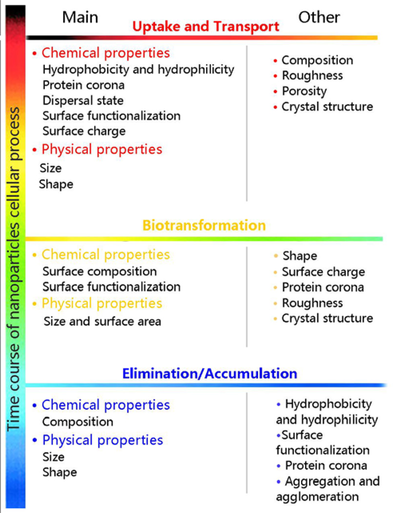
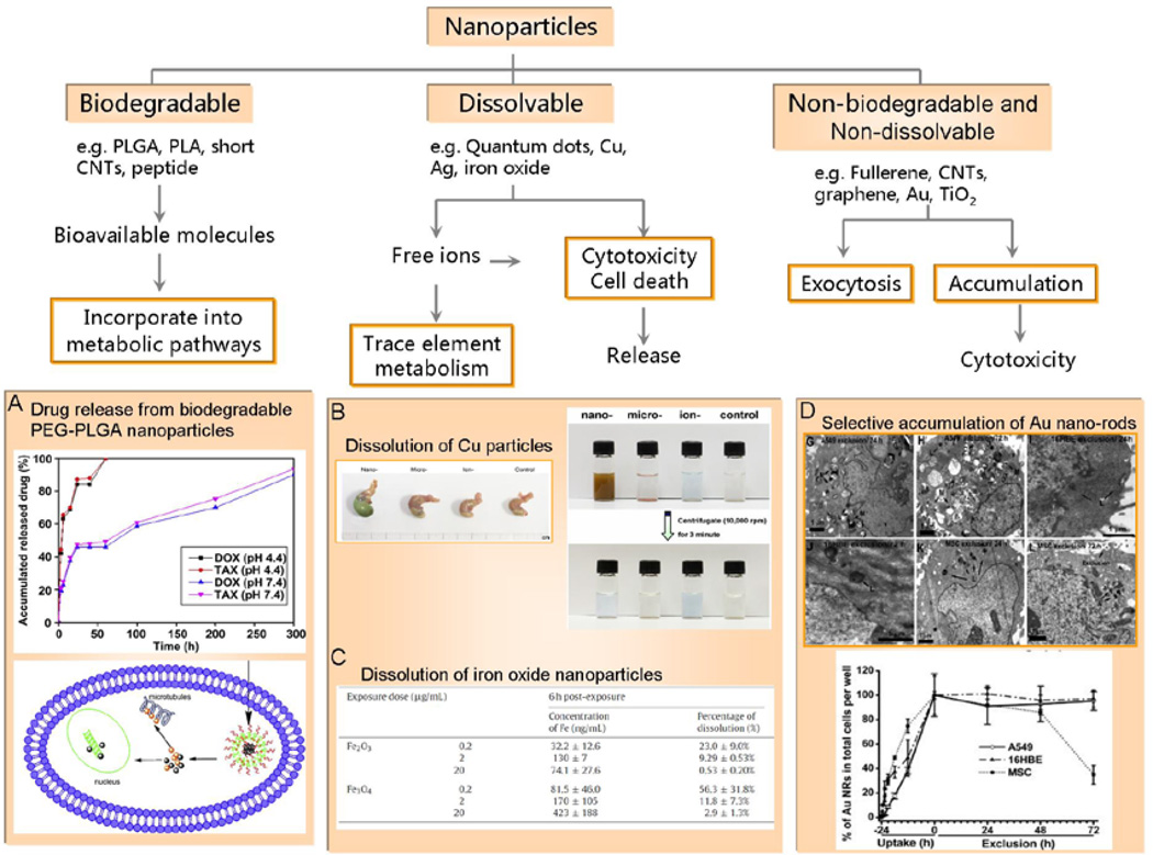
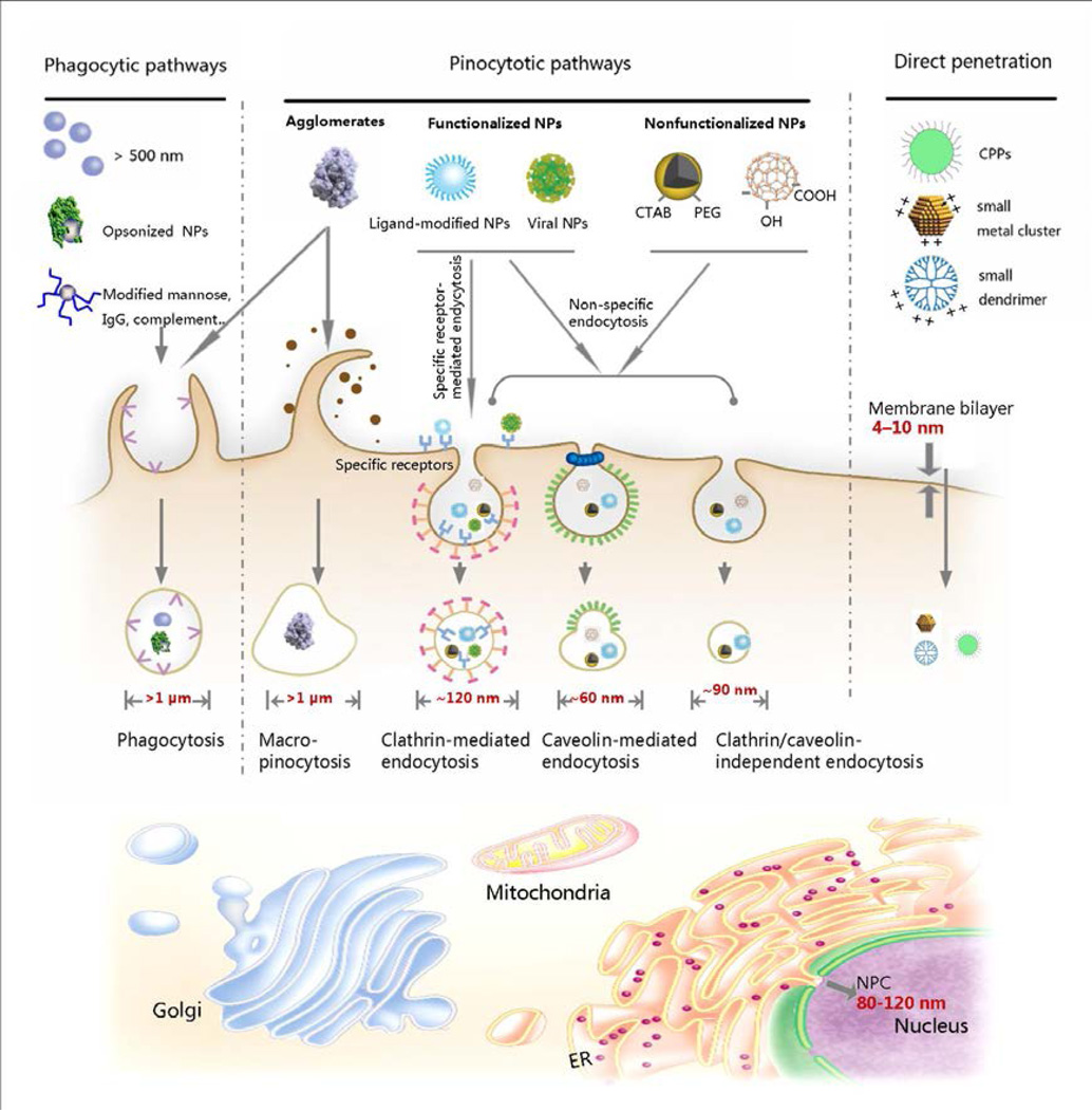
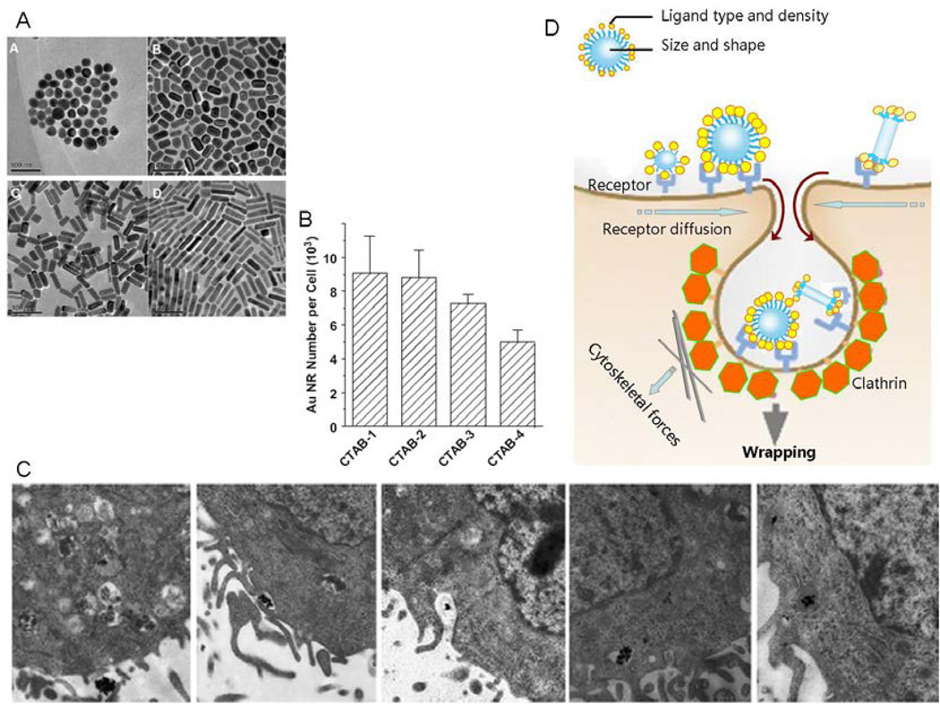
Although a growing number of innovations have emerged in the fields of nanobiotechnology and nanomedicine, new engineered nanomaterials (ENMs) with novel physicochemical properties are posing novel challenges to understand the full spectrum of interactions at the nano-bio interface. Because these could include potentially hazardous interactions, researchers need a comprehensive understanding of toxicological properties of nanomaterials and their safer design. In depth research is needed to understand how nanomaterial properties influence bioavailability, transport, fate, cellular uptake, and catalysis of injurious biological responses. Toxicity of ENMs differ with their size and surface properties, and those connections hold true across a spectrum of in vitro to in vivo nano-bio interfaces. In addition, the in vitro results provide a basis for modeling the biokinetics and in vivo behavior of ENMs. Nonetheless, we must use caution in interpreting in vitro toxicity results too literally because of dosimetry differences between in vitro and in vivo systems as well the increased complexity of an in vivo environment. In this Account, we describe the impact of ENM physicochemical properties on cellular bioprocessing based on the research performed in our groups. Organic, inorganic, and hybrid ENMs can be produced in various sizes, shapes and surface modifications and a range of tunable compositions that can be dynamically modified under different biological and environmental conditions. Accordingly, we cover how ENM chemical properties such as hydrophobicity and hydrophilicity, material composition, surface functionalization and charge, dispersal state, and adsorption of proteins on the surface determine ENM cellular uptake, intracellular biotransformation, and bioelimination versus bioaccumulation. We review how physical properties such as size, aspect ratio, and surface area of ENMs influence the interactions of these materials with biological systems, thereby affecting their hazard potential. We discuss our actual experimental findings and show how these properties can be tuned to control the uptake, biotransformation, fate, and hazard of ENMs. This Account provides specific information about ENM biological behavior and safety issues. This research also assists the development of safer nanotherapeutics and guides the design of new materials that can execute novel functions at the nano-bio interface.
Figures
References
-
- Yan L, Zheng YB, Zhao F, Li S, Gao X, Xu B, Weiss PS, Zhao Y. Chemistry and physics of a single atomic layer: strategies and challenges for functionalization of graphene and graphene-based materials. Chem. Soc. Rev. 2011;41:97–114. - PubMed
-
- Gilbert N. Nanoparticle safety in doubt. Nature. 2009;460:937. - PubMed
-
- Zhao YL, Xing GM, Chai ZF. Nanotoxicology: Are carbon nanotubes safe? Nat. Nanotechnol. 2008;3:191–192. - PubMed
-
- Zhao F, Zhao Y, Liu Y, Chang X, Chen C. Cellular uptake, intracellular trafficking, and cytotoxicity of nanomaterials. Small. 2011;7:1322–1337. - PubMed
-
- Xia T, Kovochich M, Liong M, Zink JI, Nel AE. Cationic polystyrene nanosphere toxicity depends on cell-specific endocytic and mitochondrial injury pathways. ACS Nano. 2008;2:85–96. - PubMed
Publication types
MeSH terms
Grants and funding
LinkOut - more resources
Full Text Sources
