. 2012 Dec;14(12):1836-48.
doi: 10.1111/cmi.12004. Epub 2012 Sep 4.

Identification of a cis-acting DNA-protein interaction implicated in singular var gene choice in Plasmodium falciparum

Affiliations
Free PMC article

Identification of a cis-acting DNA-protein interaction implicated in singular var gene choice in Plasmodium falciparum

Nicolas M B Brancucci et al. Cell Microbiol. 2012 Dec.
Free PMC article

Abstract

Plasmodium falciparum is responsible for the most severe form of malaria in humans. Antigenic variation of P. falciparum erythrocyte membrane protein 1 leads to immune evasion and occurs through switches in mutually exclusive var gene transcription. The recent progress in Plasmodium epigenetics notwithstanding, the mechanisms by which singularity of var activation is achieved are unknown. Here, we employed a functional approach to dissect the role of var gene upstream regions in mutually exclusive activation. Besides identifying sequence elements involved in activation and initiation of transcription, we mapped a region downstream of the transcriptional start site that is required to maintain singular var gene choice. Activation of promoters lacking this sequence occurs no longer in competition with endogenous var genes. Within this region we pinpointed a sequence-specific DNA-protein interaction involving a cis-acting sequence motif that is conserved in the majority of var loci. These results suggest an important role for this interaction in mutually exclusive locus recognition. Our findings are furthermore consistent with a novel mechanism for the control of singular gene choice in eukaryotes. In addition to their importance in P. falciparum antigenic variation, our results may also help to explain similar processes in other systems.


Figures

Fig. 1
Fig. 1
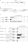
Functional var promoter mapping by serial deletion analysis. A. Schematic map of pBC. The PFL1960w upsC upstream sequence controls transcription of hdhfr-gfp. The approximate position of the TSS is indicated (Deitsch et al., 1999). The bsd resistance cassette selects for stably transfected parasites. The var intron is indicated by a bold dashed line. pBC descendants were obtained by replacing the upsC promoter with truncated sequences using BglII and NotI. hsp86 5′, hsp86 promoter; Pb DT 3′, Plasmodium berghei dhfr-thymidylate synthase terminator; rep20, 0.5 kb TARE6 repeat element; hrp2 3′; histidine-rich protein 2 terminator. B. Activities of full-length and truncated promoters in WR-selected parasites. Deletions are represented by dashed lines. Numbers represent nucleotide positions in relation to the ATG. Successful WR selection is indicated by check marks. Values represent relative hdhfr-gfp transcripts normalized against transcription of PF13_0170 (glutaminyl-tRNA synthetase, putative) and plasmid copy number. Values represent the average of three independent experiments (two replicates for 3D7/pBC1 and 3D7/pBC2) (mean ± SEM). n.a., not applicable.
Fig. 2
Fig. 2
An upsC UAS element activates the minimal promoter Kmin. A. Schematic map of pBKmin-RI. The position of the kahrp TSS is indicated (Lanzer et al., 1992). B. Schematic map of pBKmin-RI concatamers integrated into the endogenous kahrp locus (PFB0100c). C. Comparison of relative transcript levels produced by the episomal (hdhfr-gfp transcripts; grey bar) and chromosomal (kahrp transcripts; black bar) Kmin promoters, and the kahrp wild-type promoter (hdhfr-gfp transcripts; grey bar) in 3D7/pBKmin-RI parasites. Values are derived from three independent experiments and represent msp8-normalized transcripts (mean ± SEM). Values for the episomal Kmin promoter were additionally adjusted for plasmid copy number. D. Analysis of upsC-Kmin hybrid promoters. upsC insertions are depicted by bold grey lines. The rep20 element is indicated by a vertical array and the var intron by a dashed line. The graph compares relative transcript levels (msp8-normalized) produced by the episomal (hdhfr-gfp transcripts, grey bars) and chromosomal (kahrp transcripts, black bars) Kmin and upsC-Kmin hybrid promoters. Values for episomal promoters are derived from three independent experiments (mean ± SEM) and were additionally adjusted for plasmid copy number. Data for 3D7/pBKmin-RI are identical to those in Fig. 2C.
Fig. 3
Fig. 3
Transcriptional initiation from an alternative upsC upstream TSS. Identification of an alternative upsC upstream TSS (dashed arrow). Full-length and truncated promoters are schematically depicted on top. hdhfr-gfp transcript size and abundance was estimated by Northern analysis of total RNA isolated from WR-selected ring-stage parasites. Ethidium bromide-stained 18S and 28S rRNAs serve as loading control.
Fig. 4
Fig. 4
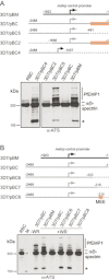
Mutually exclusive activation is mediated by a 101 bp element downstream of the TSS. A. Functional identification of a mutual exclusion element downstream of the TSS. Promoters are schematically depicted on top. pBM is a negative control construct where the mahrp1 promoter controls hdhfr-gfp transcription. upsC sequences are shown in grey. Deletions are represented by dashed lines. The orange box highlights the region required for mutually exclusive activation. PfEMP1 expression in WR-selected trophozoites was monitored by Western blot using antibodies against the conserved ATS domain of PfEMP1 (Duffy et al., 2002). The antibody cross-reacts with human spectrin. PfEMP1 is detected at various sizes above 250 kDa. The signal at 160 kDa probably represents smaller PfEMP1 species (asterisk). RBC, uninfected RBCs. B. The mutual exclusion element maps to a 101 bp region downstream of the TSS. The orange box identifies the mutual exclusion element (MEE) located at position −316 to −215. WR-selected 3D7/pBM where the mahrp promoter controls hdhfr-gfp transcription and WR-unselected 3D7/pBC carrying a silenced upsC promoter served as negative controls. RBC, uninfected RBCs. M, size standard; −WR, unselected; +WR, WR-selected.
Fig. 5
Fig. 5
Identification of a sequence-specific DNA–protein interaction implicated in mutual exclusion. A. Identification of a sequence-specific DNA–protein interaction between the 47 bp MEE2 element and an unknown nuclear factor. The 101 bp MEE sequence identified by promoter deletion analysis and the three fragments tested by EMSA (MEE1 to MEE3) are schematically depicted on top. The EMSA was carried out using radiolabelled MEE2 and parasite nuclear extract (results for MEE1 and MEE3 EMSAs were negative and are not shown). Competition was carried out in presence of a 5-, 25- and 100-fold molar excess of unlabelled DNA. B. Mutational analysis of MEE2. The EMSA was carried out using radiolabelled MEE2 and parasite nuclear extract. Competition was carried out in presence of a 25- and 100-fold molar excess of unlabelled DNA. The nucleotide sequences of wild-type and mutated MEE2 elements are indicated on the right. The ATAGATTA core motif is underlined. Mutated 8mers are highlighted in red (see Fig. S3A for competition with MEE2-mut6). The MEE2-related element upstream of PF07_0048 is shown at the bottom and differences compared with MEE2 are highlighted in red.
Fig. 6
Fig. 6
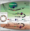
A novel model for singular var gene choice. A chromosome end cluster located in a transcriptionally permissive perinuclear region is schematically depicted on top. The unique var gene expression site (VES) recognizes a single var gene through specific interaction with unknown DNA motifs (white hexagon) and/or the MEE2 element itself (red oval). This interaction leads to dissociation of the MIF complex (blue) concomitant with the establishment of a permissive chromatin conformation (green circles) to facilitate RNA polII-dependent transcriptional initiation and/or elongation. This process involves deposition and maintenance of permissive histone modifications through modifying enzymes such as PfSET10 (Volz et al., 2012) as well as interactions between unknown transcription factors (yellow) and the UAS (green oval). Additional var genes within this subnuclear domain are excluded from the VES and protected from illegitimate transcription. Here, the function of MIF may be to block transcriptional elongation or to prevent transcriptional initiation or PIC assembly on the core promoter. var genes in heterochromatic perinculear zones that are silenced primarily through their association with H3K9me3/PfHP1 are shown below.

Similar articles

Cited by

References

    1. Baruch DI, Gormely JA, Ma C, Howard RJ, Pasloske BL. Plasmodium falciparum erythrocyte membrane protein 1 is a parasitized erythrocyte receptor for adherence to CD36, thrombospondin, and intercellular adhesion molecule 1. Proc Natl Acad Sci USA. 1996;93:3497–3502. - PMC - PubMed
    1. Beeson JG, Duffy PE. The immunology and pathogenesis of malaria during pregnancy. Curr Top Microbiol Immunol. 2005;297:187–227. - PubMed
    1. Biggs BA, Gooze L, Wycherley K, Wollish W, Southwell B, Leech JH, et al. Antigenic variation in Plasmodium falciparum. Proc Natl Acad Sci USA. 1991;88:9171–9174. - PMC - PubMed
    1. Calderwood MS, Gannoun-Zaki L, Wellems TE, Deitsch KW. Plasmodium falciparum var genes are regulated by two regions with separate promoters, one upstream of the coding region and a second within the intron. J Biol Chem. 2003;278:34125–34132. - PubMed
    1. Campbell TL, De Silva EK, Olszewski KL, Elemento O, Llinas M. Identification and genome-wide prediction of DNA binding specificities for the ApiAP2 family of regulators from the malaria parasite. PLoS Pathog. 2010;6:e1001165. - PMC - PubMed

Publication types

MeSH terms

Substances