. 2012 Sep 7;425(4):812-7.
doi: 10.1016/j.bbrc.2012.07.157. Epub 2012 Aug 7.

Regulation of the pro-inflammatory cytokine osteopontin by GIP in adipocytes--a role for the transcription factor NFAT and phosphodiesterase 3B

Affiliations

Regulation of the pro-inflammatory cytokine osteopontin by GIP in adipocytes--a role for the transcription factor NFAT and phosphodiesterase 3B

Bilal Omar et al. Biochem Biophys Res Commun. .

Abstract

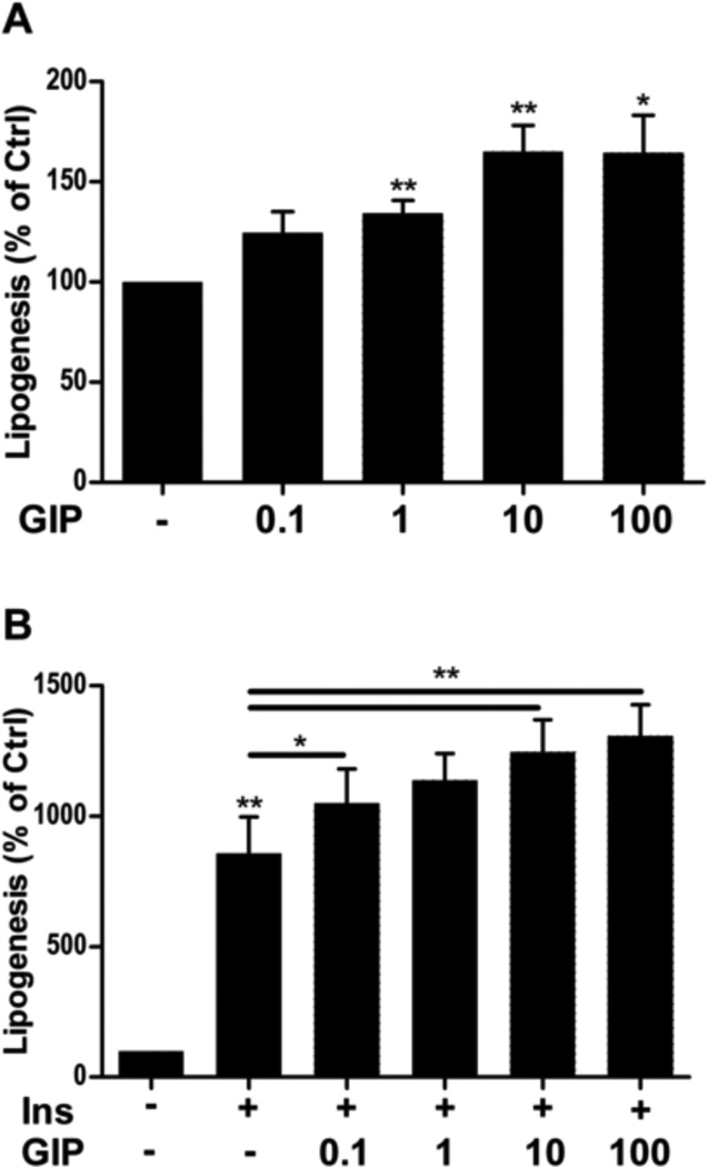
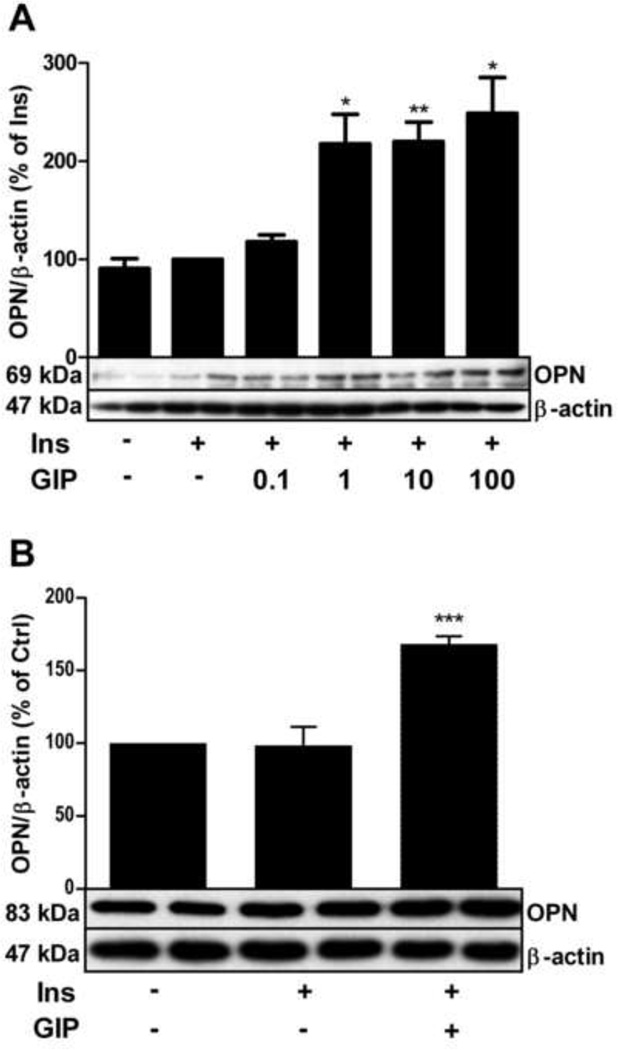
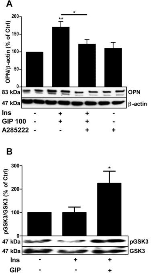
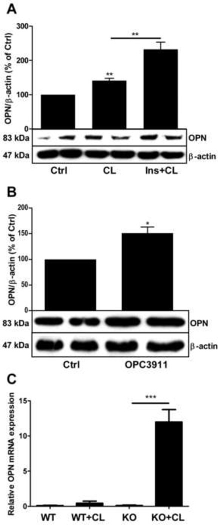
The incretin - glucose-dependent insulinotropic polypeptide (GIP) - and the pro-inflammatory cytokine osteopontin are known to have important roles in the regulation of adipose tissue functions. In this work we show that GIP stimulates lipogenesis and osteopontin expression in primary adipocytes. The GIP-induced increase in osteopontin expression was inhibited by the NFAT (the transcription factor nuclear factor of activated T-cells) inhibitor A-285222. Also, the NFAT kinase glycogen synthase kinase (GSK) 3 was upregulated by GIP. To test whether cAMP might be involved in GIP-mediated effects on osteopontin a number of strategies were used. Thus, the β3-adrenergic receptor agonist CL316,243 stimulated osteopontin expression, an effects which was mimicked by OPC3911, a specific inhibitor of phosphodiesterase 3. Furthermore, treatment of phosphodiesterase 3B knock-out mice with CL316,243 resulted in a dramatic upregulation of osteopontin in adipose tissue which was not the case in wild-type mice. In summary, we delineate mechanisms by which GIP stimulates osteopontin in adipocytes. Given the established link between osteopontin and insulin resistance, our data suggest that GIP by stimulating osteopontin expression, also could promote insulin resistance in adipocytes.


Figures

Fig. 1
Fig. 1
GIP induces lipogenesis on its own as well as potentiates insulin-induced lipogenesis in primary rat adipocytes. Primary rat adipocytes were stimulated with GIP at the indicated concentrations (nM) in the absence (A) or presence (B) of insulin (INS, 1 nM) for 30 minutes. Lipogenesis was determined as glucose incorporation into lipid relative to the untreated group. *p < 0.05, **p < 0.01. n = 5 per group. GIP: glucose-dependent insulinotropic polypeptide.
Fig. 2
Fig. 2
GIP upregulates osteopontin protein expression in primary adipocytes and 3T3-L1 adipocytes in the presence of insulin. 3T3 L1 adipocytes (A) and primary rat adipocytes (B) were stimulated with GIP at the indicated concentrations (nM) in A and 100 nM in B, in the presence or absence of insulin (INS, 1 nM) over night. Cells were lysed and osteopontin expression analyzed by western blotting. Representative blots are shown. *p < 0.05, **p < 0.01, ***p < 0.001 vs. non-treated controls. n=4 for both. GIP: glucose-dependent insulinotropic polypeptide, OPN: osteopontin
Fig. 3
Fig. 3
GIP-induced osteopontin expression is partially mediated by NFAT and involves inhibition of the NFAT export kinase GSK3. lA. Primary rat adipocytes were stimulated overnight by GIP (100 nM) in the presence of insulin (INS, 1 nM) with or without a 30 minute pre-treatment with the NFAT inhibitor A-285222 (1 µM) over night. Cells were lysed and osteopontin expression analyzed by western blotting. Representative blots are shown, n=7. B. Adipocytes were stimulated by GIP (100 nM) in presence or absence of insulin (INS, 1 nM) over night, cells were lysed and GSK3 phosphorylation was analyzed by western blotting. Representative blots are shown. .*p < 0.05, **p < 0.01 n=9. GIP: glucose-dependent insulinotropic polypeptide, GSK3: glycogen synthase kinase 3, NFAT: the transcription factor nuclear factor of activated T-cells, OPN: osteopontin
Fig. 4
Fig. 4
Osteopontin is upregulated in adipocytes treated with CL316243 (CL), a β3-adrenergic receptor agonist, by OPC 3911, a PDE3 inhibitor, and in adipose tissue from PDE3B KO mice treated with CL in vivo. A. Primary rat adipocytes were stimulated with the β3-adrenergic agonist CL316,243 (10 nM) in the presence or absence of insulin (INS, 1 nM), n=4. B. Adipocytes were incubated with or without the PDE3 inhibitor OPC 3911 (10 µM). Cells were lysed and OPN expression analyzed by western blotting. Representative blots are shown, n=4. C. Four-month old C57BL6 PDE3B knock-out mice were subcutaneously injected with 1 mg/kg of CL316243 in PBS or with PBS alone. Epididymal fat pads were collected 24 h after injections. Total mRNA was prepared and gene expression was measured using real time RT-PCR as described in methods. Gene expression levels were quantified as a ratio of target transcripts to 18S mRNA. Data are expressed as means ± SEM (n=7–8). *p < 0.05, **p < 0.01, ***p < 0.001. OPN: osteopontin, PDE3B: phosphodiesterase 3B

References

    1. Rasouli N, Kern PAJ. Adipocytokines and the metabolic complications of obesity. Clin Endocrinol Metab. 2008 Nov;93(11 Suppl 1):S64–S73. - PMC - PubMed
    1. Guilherme A, Virbasius JV, Puri V, Czech MP. Adipocyte dysfunctions linking obesity to insulin resistance and type 2 diabetes. Nat Rev Mol Cell Biol. 2008;9:367–377. - PMC - PubMed
    1. Gutierrez DA, Puglisi MJ, Hasty AH. Impact of increased adipose tissue mass on inflammation, insulin resistance and dyslipidemia. Curr Diab Rep. 2009;9:26–32. - PMC - PubMed
    1. Kahn SE, Hull RL, Utzschneider KM. Mechanisms linking obesity to insulin resistance and type 2 diabetes. Nature. 2006 Dec 14;444(7121):840–846. - PubMed
    1. Kiefer FW, Zeyda M, Todoric J, et al. Endocrinology. 2008 Mar;149(3):1350–1357. - PubMed

Publication types

MeSH terms