Comparative Study
. 2012 Nov 5;31(21):4106-23.
doi: 10.1038/emboj.2012.202. Epub 2012 Aug 14.

Upregulated function of mitochondria-associated ER membranes in Alzheimer disease

Affiliations
Comparative Study

Upregulated function of mitochondria-associated ER membranes in Alzheimer disease

Estela Area-Gomez et al. EMBO J. .

Abstract

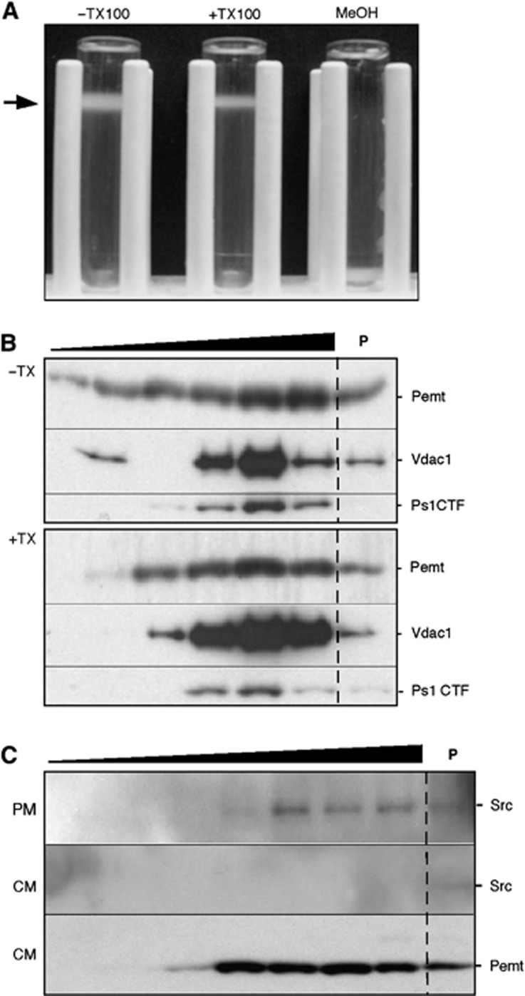
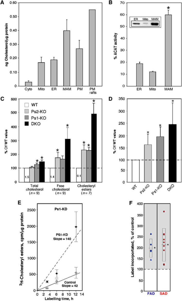
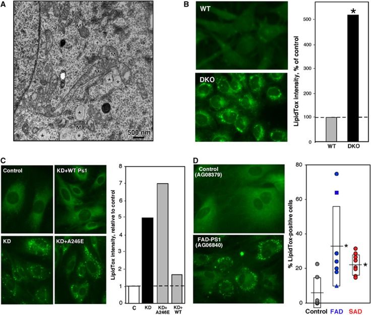
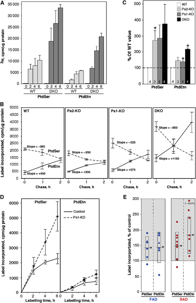
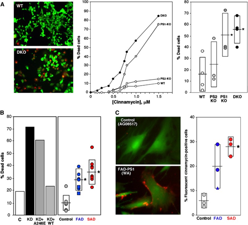
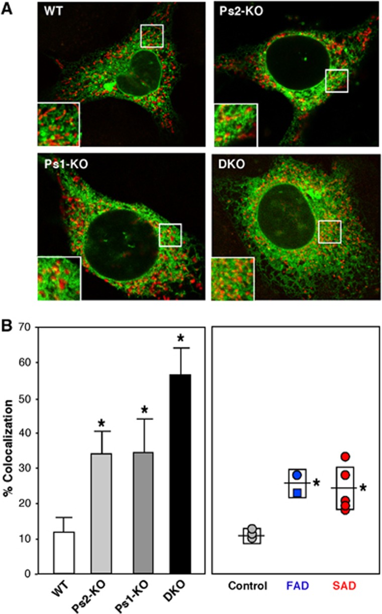
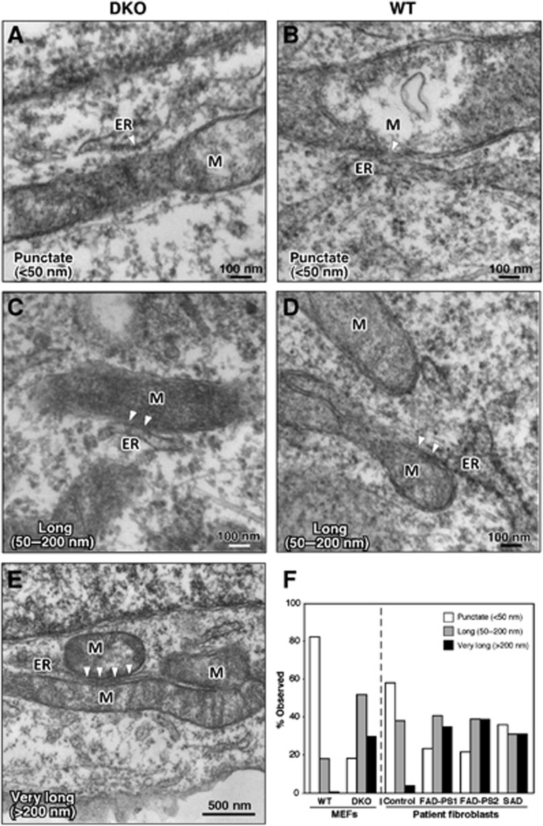
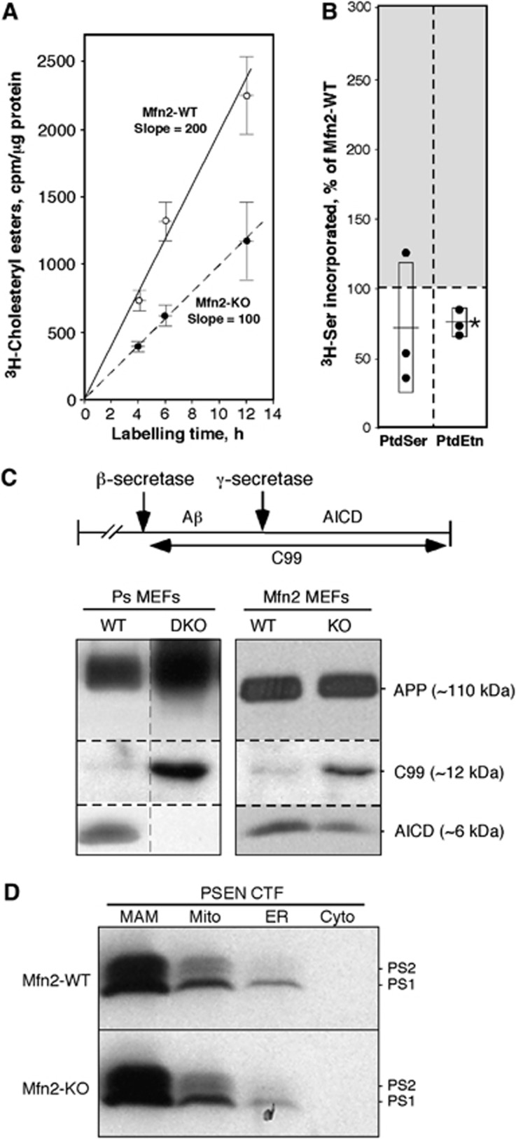
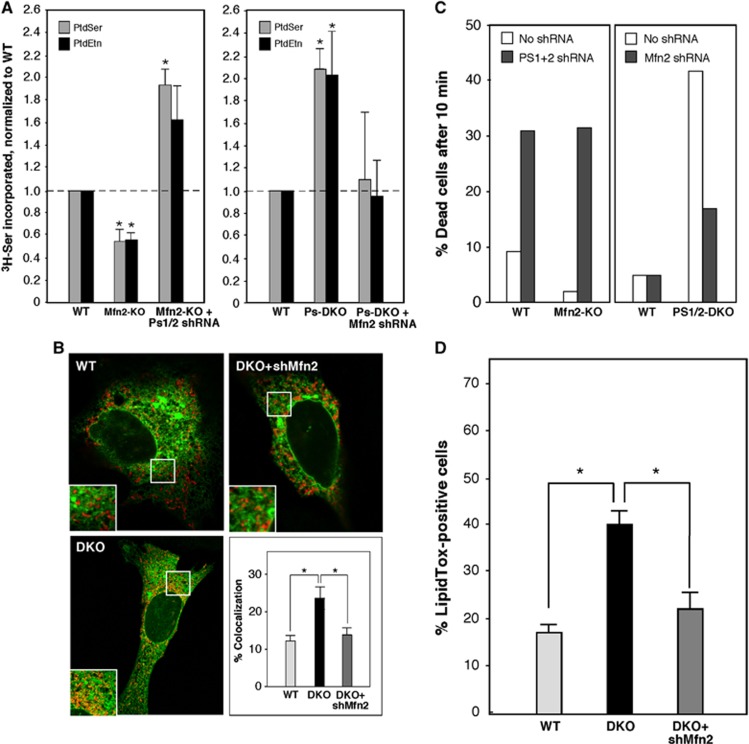
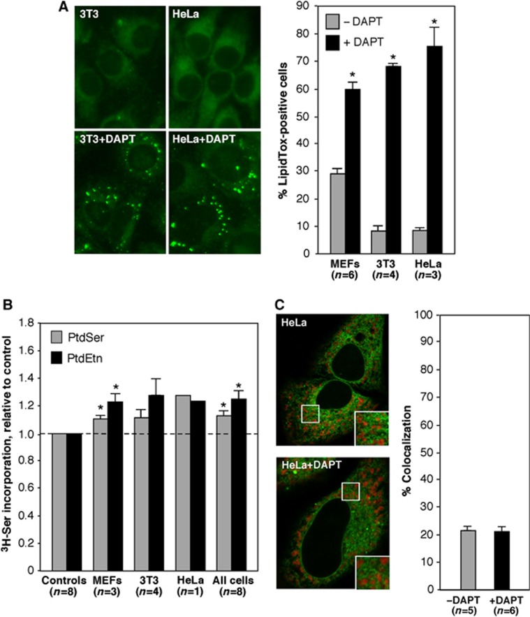
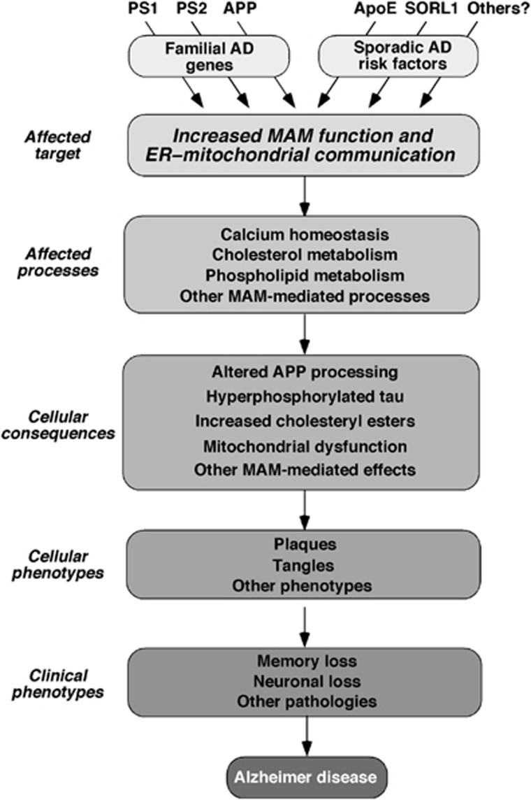
Alzheimer disease (AD) is associated with aberrant processing of the amyloid precursor protein (APP) by γ-secretase, via an unknown mechanism. We recently showed that presenilin-1 and -2, the catalytic components of γ-secretase, and γ-secretase activity itself, are highly enriched in a subcompartment of the endoplasmic reticulum (ER) that is physically and biochemically connected to mitochondria, called mitochondria-associated ER membranes (MAMs). We now show that MAM function and ER-mitochondrial communication-as measured by cholesteryl ester and phospholipid synthesis, respectively-are increased significantly in presenilin-mutant cells and in fibroblasts from patients with both the familial and sporadic forms of AD. We also show that MAM is an intracellular detergent-resistant lipid raft (LR)-like domain, consistent with the known presence of presenilins and γ-secretase activity in rafts. These findings may help explain not only the aberrant APP processing but also a number of other biochemical features of AD, including altered lipid metabolism and calcium homeostasis. We propose that upregulated MAM function at the ER-mitochondrial interface, and increased cross-talk between these two organelles, may play a hitherto unrecognized role in the pathogenesis of AD.


Conflict of interest statement

The authors declare that they have no conflict of interest.

Figures

Figure 1
Figure 1
MAM displays the features of a lipid raft. (A) Mouse liver Percoll-purified MAM treated with or without TX100 prior to centrifugation through a second Percoll gradient. The low density fraction (arrow) is detergent resistant but solubilizable by methanol (MeOH), implying that it is a DRM. (B) Western blot of fractions from a 5–30% sucrose gradient (triangle; lower density at left) of MAM isolated from a Percoll gradient (as in A). The pellet (P) denotes TX100-soluble material. (C) Western blot of gradient fractions of mouse liver PM and crude mitochondrial extract (CM) to detect Src (PM marker) and Pemt (MAM marker).
Figure 2
Figure 2
Cholesterol metabolism in PS-mutant and AD cells. (A) Total cholesterol in the indicated mouse brain fractions (n=3, except PM rafts; n=2). (B) ACAT activity in mouse brain fractions (n=4). Inset: western blot to detect ACAT protein; 20 μg protein loaded in each lane. Asterisk denotes significant difference versus ER and mito fractions (P<0.05). (C) Content of cholesterol species in PS-mutant MEFs relative to that in WT MEFs (numbers denote average amounts of the indicated cholesterol species, in ng/mg protein). (D) ACAT activity (i.e., conversion of 3H-cholesterol to 3H-cholesteryl esters) in MEFs after 6 h (n=6). (E) Kinetics of CE synthesis (performed as in D) in Ps1-KD cells (note increased slope (line of best fit, in cpm/μg/h) versus control). (F) Quantitation of 3H-CE synthesis after 6 h in fibroblasts from FAD (n=5; 4 PS1 (circles), 1 PS2 (triangle)) and SAD (n=9) patients versus paired controls. For cell lines that were evaluated multiple times (see Supplementary Table S1), the data point represents an average of the assays. Boxes with centred lines denote averages±s.d.; asterisks denote significant difference versus WT (P<0.05).
Figure 3
Figure 3
Detection of lipid droplets in PS-mutant and AD cells. (A) EM of DKO MEFs. Note the presence of features reminiscent of lipid droplets (asterisks); these were not observed in WT MEFs. M, mitochondrion. (B) Left: LipidTox staining of DKO MEFs; note punctate staining (i.e., lipid droplets) that are absent in WT MEFs. Right: Quantitation of LipidTox staining. (C) Left: LipidTox staining of Ps1-KD cells. Right: Quantitation, as in (B). Note that overexpression of human WT-PS1, but not A246E mutant PS1, reduced lipid droplet formation. C, mismatched shRNA control. (D) Example of LipidTox staining (left) and quantitation of LipidTox-positive fibroblasts from FAD (n=7) and SAD (n=9; 7 PS1 (circles), 1 PS2 (triangle), 1 APP (square)) patients versus controls (n=7) (right). See other examples in Supplementary Figure S6. Other notation as in Figure 2.
Figure 4
Figure 4
Phospholipid synthesis in PS-mutant and AD cells. (A) Synthesis of 3H-PtdSer and 3H-PtdEtn after labelling DKO MEFs with 3H-Ser for the indicated times (h) (n=3). (B) Pulse-chase. MEFs were labelled for 1 h with 3 H-Ser and chased with cold Ser for the indicated times (n=3). Note the steeper slopes (i.e., rates of 3 H-Ser incorporation) for both PtdSer (negative slopes) and PtdEtn (positive slopes), indicative of a more rapid conversion of PtdSer to PtdEtn, especially in the DKO cells. (C) Phospholipid synthesis in crude mitochondria from Ps-KO MEFs (n=3 or 4, as indicated; error bars, s.e.). (D) Kinetics of PtdSer and PtdEtn synthesis (as in A) in Ps1-KD cells. (E) Phospholipid synthesis after 6 h (as in A) in fibroblasts from FAD (n=6) and SAD (n=9) patients. Note: three of the SAD PtdEtn values were unusually high (∼600% of control) and were omitted from the statistical analyses. Other notation as in Figure 2.
Figure 5
Figure 5
Cinnamycin sensitivity in PS-mutant and AD cells. (A) Left: Example of live/dead assays (1 μM cinnamycin for 10 min at 37°C). Middle: Example of cinnamycin-sensitivity curves in PS-mutant MEFs. Right: Summary of cinnamycin sensitivity assays (1 μM Cin for 10 min) in PS-mutant MEFs. (B) Left: Example of cinnamycin sensitivity in Ps1-KD cells versus mismatch control (C). Note that overexpression of human WT PS1, but not A246E mutant PS1, could ‘rescue’ Cin sensitivity. Right: Cin/Dura sensitivity in fibroblasts from FAD (n=7) and SAD (n=8) patients versus controls (n=7). (C) Left: Example of staining of control and AD patient cells with fluorescent cinnamycin (FL-SA-Ro; orange); cells were counterstained with calcein (green) to visualize overall cell morphology. Right: Quantitation of FL-SA-Ro staining in fibroblasts from FAD (n=3) and SAD (n=3) patients compared to controls (n=3). See other examples in Supplementary Figure S7. Other notations as in Figures 2 and 3.
Figure 6
Figure 6
ER–mitochondrial colocalization in Ps-mutant and AD cells. (A) Example of confocal images of cells stained with Mito DS Red (red) and GFP-Sec61β (green). In the insets, note the large number of discrete red and green signals in the WT as compared to the Ps-mutant MEFs, which have more overlap (orange and yellow signals). (B) Quantitation of colocalization (as in A) by Image J in WT (average of 10 images ±s.d.), Ps1-KO (n=11), Ps2-KO (n=13), and DKO (n=7) MEFs (left), and in fibroblasts from FAD (n=2) and SAD (n=5) patients compared to controls (n=3) (right). Other notations as in Figures 2 and 3.
Figure 7
Figure 7
Electron microscopy of Ps-mutant and AD cells. (A, C, E) DKO MEFs. (B, D) WT MEFs. Note increased length of regions of contact between ER and mitochondria (M) (arrowheads) in DKO MEFs, and, in (E), a region of ER ‘sandwiched’ between two mitochondria. (F) Quantitation of ER–mitochondrial contact lengths in MEFs (∼40 contacts analysed) and in fibroblasts from AD patients (∼25 contacts). See also Supplementary Figure S8.
Figure 8
Figure 8
Analysis of MAM function in Mfn2-KO MEFs. (A) Kinetics of CE synthesis (performed as in Figure 2E) in Mfn2-KO MEFs (note decreased slope versus control). (B) Phospholipid synthesis after 6 h (as in Figure 4A) in Mfn2-KO cells (right) (n=3). (C) Western blot to detect APP and its C-terminal cleavage products C99 and AICD (cleavage scheme at top) in Ps1/2-DKO and Mfn2-KO MEFs (image at the left is a composite of two non-adjacent lanes from the same gel; vertical dashed line indicates where the two lanes were apposed). Note the absence of AICD in DKO MEFs, and the shift in the ratio of C99:AICD in Mfn2-KO versus WT MEFs. (D) Subcellular distribution of presenilins in Mfn2-KO versus WT MEFs. Note that the loss of Mfn2 does not alter the predominant localization of presenilins in MAM. Other notations as in Figure 2.
Figure 9
Figure 9
Rescue of phospholipid (PL) synthesis in Ps- and Mfn2-mutant MEFs. (A) Synthesis of 3H-PtdSer and 3H-PtdEtn after labelling for 6 h with 3 H-Ser in the indicated MEFs. Left: Note that PL synthesis was decreased in Mfn2-KO MEFs compared to WT but was restored upon knockdown of Ps1+Ps2 (average of four experiments ±s.e.). A control knockdown of Ps1+Ps2 in WT MEFs increased PL synthesis, as expected (not shown). Right: In the converse experiment, note that PL synthesis was increased in Ps1+Ps2 DKO MEFs compared to WT but was restored upon knockdown of Mfn2 (average of five experiments ±s.e.). A control knockdown of Mfn2 in WT MEFs reduced PL synthesis, as expected (not shown). (B) Examples (as in Figure 6A), and quantitation (average of 20 cells analysed ±s.e.), of ER–mitochondrial contacts in WT, Ps1+Ps2-DKO, and DKO cells ‘rescued’ by knockdown of Mfn2. (C) Effect of presenilins and Mfn2 on duramycin sensitivity. Note that Dura sensitivity correlates with the change in PtdEtn synthesis shown in (A). (D) Quantitation of lipid droplets in Ps1+Ps2 DKO MEFs upon knockdown of Mfn2 (average of five images ±s.e.). Other notation as in Figure 2.
Figure 10
Figure 10
MAM function in cells deficient in γ-secretase activity. (A) Detection of lipid droplets in HeLa and 3T3 cells treated with 5 μM DAPT for 24 h. Examples of LipidTox staining (left) and quantitation (right; n=number of images analysed, with a total of ∼500 cells analysed ±s.e.), as in Figure 3. Note the large increase in LipidTox staining, ranging from ∼200 to ∼700% over control. (B) Phospholipid transport in the indicated cells (as in Figure 4). Note the modest increase in PtdSer and PtdEtn synthesis, of only ∼20%. (C) Colocalization of ER and mitochondria (as in Figure 6) in HeLa cells. Note that DAPT had no effect on the degree of colocalization. Other notation as in Figure 2.
Figure 11
Figure 11
A model of pathogenesis of Alzheimer disease. In broad view, underlying genotypes that either cause AD directly (in the case of FAD) or are risk factors that predispose to developing AD (in the case of SAD) increase MAM function and/or ER–mitochondrial communication. This increase affects various MAM-mediated processes (either positively or negatively, depending on the particular gene/function involved), which in turn give rise to the various phenotypes seen in AD. See text for details.

Comment in

References

    1. Ankarcrona M, Hultenby K (2002) Presenilin-1 is located in rat mitochondria. Biochem Biophys Res Commun 295: 766–770 - PubMed
    1. Annaert WG, Levesque L, Craessaerts K, Dierinck I, Snellings G, Westaway D, St George-Hyslop P, Cordell B, Fraser P, De Strooper B (1999) Presenilin 1 controls γ-secretase processing of amyloid precursor protein in pre-golgi compartments of hippocampal neurons. J Cell Biol 147: 277–294 - PMC - PubMed
    1. Area-Gomez E, de Groof AJ, Boldogh I, Bird TD, Gibson GE, Koehler CM, Yu WH, Duff KE, Yaffe MP, Pon LA, Schon EA (2009) Presenilins are enriched in endoplasmic reticulum membranes associated with mitochondria. Am J Pathol 175: 1810–1816 - PMC - PubMed
    1. Bentahir M, Nyabi O, Verhamme J, Tolia A, Horre K, Wiltfang J, Esselmann H, De Strooper B (2006) Presenilin clinical mutations can affect γ-secretase activity by different mechanisms. J Neurochem 96: 732–742 - PubMed
    1. Bezprozvanny I, Mattson MP (2008) Neuronal calcium mishandling and the pathogenesis of Alzheimer's disease. Trends Neurosci 31: 454–463 - PMC - PubMed

Publication types

MeSH terms