. 2012 Dec;36(12):1826-34.
doi: 10.1097/PAS.0b013e31825ec07a.

The diagnostic and biological implications of laminin expression in serous tubal intraepithelial carcinoma

Affiliations

The diagnostic and biological implications of laminin expression in serous tubal intraepithelial carcinoma

Elisabetta Kuhn et al. Am J Surg Pathol. 2012 Dec.

Abstract

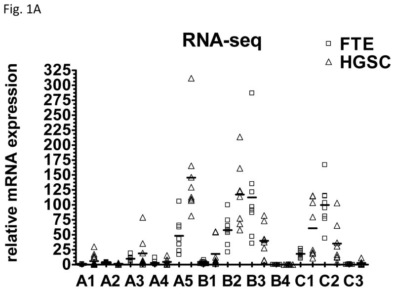
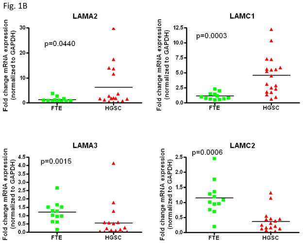
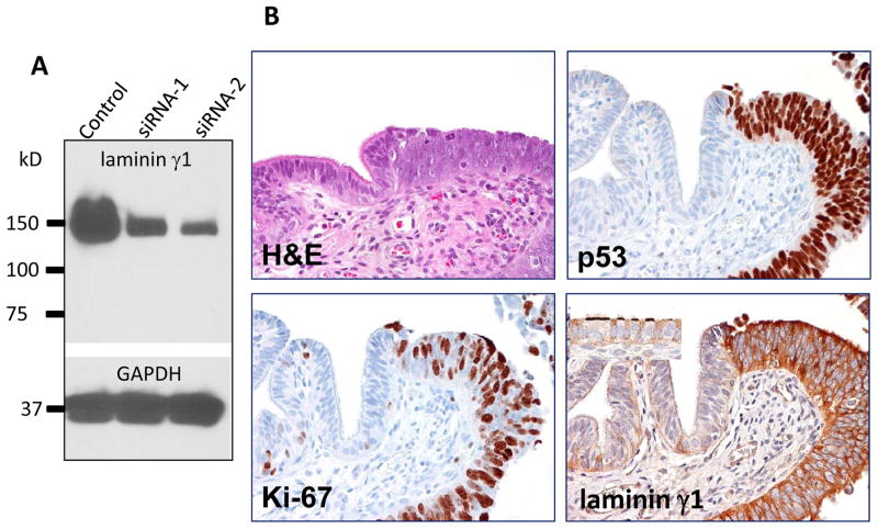
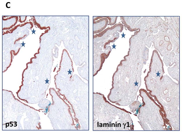
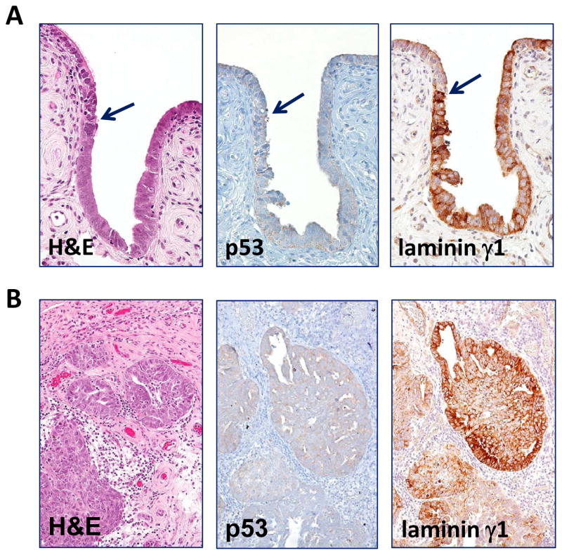
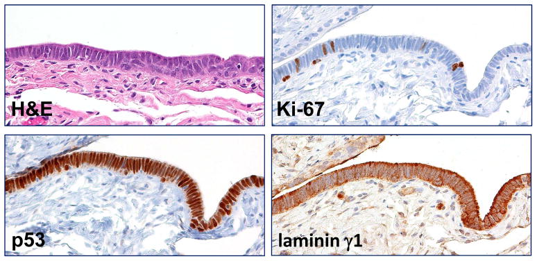
There is compelling evidence to suggest that serous tubal intraepithelial carcinoma (STIC) is the likely primary site for the development of many pelvic high-grade serous carcinomas (HGSCs). Identifying molecules that are upregulated in STIC is important not only to provide biomarkers to assist in the diagnosis of STIC but also to elucidate our understanding of the pathogenesis of HGSC. In this study, we performed RNA sequencing to compare transcriptomes between HGSC and normal fallopian tube epithelium (FTE), and we identified LAMC1 encoding laminin γ1 as one of the preferentially upregulated genes associated with HGSC. Reverse transcription polymerase chain reaction further validated LAMC1 upregulation in HGSC as compared with normal FTE. Immunohistochemical analysis was performed on 32 cases of concurrent HGSC and STIC. The latter was diagnosed on the basis of morphology, TP53 mutations, and p53 and Ki-67 immunohistochemical patterns. Laminin γ1 immunostaining intensity was found to be significantly higher in STIC and HGSC compared with adjacent FTE in all cases (P<0.001). In normal FTE, laminin γ1 immunoreactivity was predominantly localized in the basement membrane or on the apical surface of ciliated cells, whereas in STIC and HGSC cells, laminin γ1 staining was diffuse and intense throughout the cytoplasm. More importantly, strong laminin γ1 staining was detected in all 13 STICs, which lacked p53 immunoreactivity because of null mutations. These findings suggest that the overexpression of laminin γ1 immunoreactivity and alteration of its staining pattern in STICs can serve as a useful tissue biomarker, especially for those STICs that are negative for p53 and have a low Ki-67 labeling index.


Figures

Fig. 1
Fig. 1
Expression of different laminin chains in high-grade serous carcinoma tissues and samples prepared from normal fallopian tube epithelium. A. Relative mRNA levels of each chain were plotted according to tissue prepared. HGSC: high-grade serous carcinoma; FTE: normal fallopian tube epithelium. B. Relative mRNA levels of representative laminin chains were plotted.
Fig. 1
Fig. 1
Expression of different laminin chains in high-grade serous carcinoma tissues and samples prepared from normal fallopian tube epithelium. A. Relative mRNA levels of each chain were plotted according to tissue prepared. HGSC: high-grade serous carcinoma; FTE: normal fallopian tube epithelium. B. Relative mRNA levels of representative laminin chains were plotted.
Fig. 2
Fig. 2
Comparison of immunoreactivity of p53 and laminin γ1 in a fallopian tube diffusely involved by a serous tubal intraepithelial carcinoma. A. Western blot analysis shows a marked decrease in lamininγ1 protein expression after OVCAR3 cells were treated with siRNA-1 and siRNA-2 that target LAMC1 gene. GAPDH is used as a loading control. B. Immunostaining of laminin γ1, p53 and Ki-67 in a STIC. A high magnification view of a STIC and adjacent normal-appearing tubal epithelium. The STIC demonstrates a diffuse and intense p53 staining, a high Ki-67 labeling index and diffuse of laminin γ1 expression. In normal tubal epithelium, of lamininγ1 immunoreactivity is detected in basement membrane and apical surface of ciliated cells (inset) whereas of laminin γ1 immunoreactivity in STIC cells loses the “polarity” with strong staining in both cell surface and cytoplasm. C. A low magnification view of a fallopian tube section contains diffuse STIC. The distribution of laminin γ1 immunoreactivity faithfully overlapped with p53 staining. Asterisks highlight the discrete areas of tubal epithelium that is negative for both stains.
Fig. 2
Fig. 2
Comparison of immunoreactivity of p53 and laminin γ1 in a fallopian tube diffusely involved by a serous tubal intraepithelial carcinoma. A. Western blot analysis shows a marked decrease in lamininγ1 protein expression after OVCAR3 cells were treated with siRNA-1 and siRNA-2 that target LAMC1 gene. GAPDH is used as a loading control. B. Immunostaining of laminin γ1, p53 and Ki-67 in a STIC. A high magnification view of a STIC and adjacent normal-appearing tubal epithelium. The STIC demonstrates a diffuse and intense p53 staining, a high Ki-67 labeling index and diffuse of laminin γ1 expression. In normal tubal epithelium, of lamininγ1 immunoreactivity is detected in basement membrane and apical surface of ciliated cells (inset) whereas of laminin γ1 immunoreactivity in STIC cells loses the “polarity” with strong staining in both cell surface and cytoplasm. C. A low magnification view of a fallopian tube section contains diffuse STIC. The distribution of laminin γ1 immunoreactivity faithfully overlapped with p53 staining. Asterisks highlight the discrete areas of tubal epithelium that is negative for both stains.
Fig. 3
Fig. 3
Laminin γ1 immunoreactivity in p53 negative STIC and HGSC. A. A p53 negative STIC shows upregulation of laminin γ1 expression. Arrows: junction between the STIC and normal-appearing tubal epithelium. B. Laminin γ1 expression is detected in the concurrent p53 negative HGSC.
Fig. 4
Fig. 4
Expression of laminin γ1 in a serous tubal intraepithelial lesion (STIL). The lesion is composed of atypical epithelial cells with enlarged nuclei and lacks of ciliated cells. Although the p53 staining, like a STIC, is diffuse and intense, the Ki-67 labeling index is lower than established STIC with an estimation of approximately 9%. Significantly higher laminin γ1 immunoreactivity is observed in STIL than in adjacent normal-appearing tubal epithelium (asterisk).

References

    1. Barth TF, Rinaldi N, Bruderlein S, et al. Mesothelial cells in suspension expose an enriched integrin repertoire capable of capturing soluble fibronectin and laminin. Cell Commun Adhes. 2002;9:1–14. - PubMed
    1. Callahan MJ, Crum CP, Medeiros F, et al. Primary fallopian tube malignancies in BRCA-positive women undergoing surgery for ovarian cancer risk reduction. J Clin Oncol. 2007;25:3985–3990. - PubMed
    1. Carlson JW, Jarboe EA, Kindelberger D, et al. Serous tubal intraepithelial carcinoma: diagnostic reproducibility and its implications. Int J Gynecol Pathol. 2010;29:310–314. - PubMed
    1. Engbring JA, Kleinman HK. The basement membrane matrix in malignancy. The Journal of pathology. 2003;200:465–470. - PubMed
    1. Folkins AK, Jarboe EA, Roh MH, et al. Precursors to pelvic serous carcinoma and their clinical implications. Gynecol Oncol. 2009;113:391–396. - PubMed

Publication types

MeSH terms