Molecular confirmation of t(6;11)(p21;q12) renal cell carcinoma in archival paraffin-embedded material using a break-apart TFEB FISH assay expands its clinicopathologic spectrum
- PMID: 22892601
- PMCID: PMC4441265
- DOI: 10.1097/PAS.0b013e3182613d8f
Molecular confirmation of t(6;11)(p21;q12) renal cell carcinoma in archival paraffin-embedded material using a break-apart TFEB FISH assay expands its clinicopathologic spectrum
Abstract
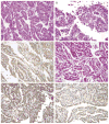
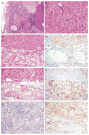
A subset of renal cell carcinomas (RCCs) is characterized by t(6;11)(p21;q12), which results in fusion of the untranslated Alpha (MALAT1) gene to the TFEB gene. Only 21 genetically confirmed cases of t(6;11) RCCs have been reported. This neoplasm typically demonstrates a distinctive biphasic morphology, comprising larger epithelioid cells and smaller cells clustered around basement membrane material; however, the full spectrum of its morphologic appearances is not known. The t(6;11) RCCs differ from most conventional RCCs in that they consistently express melanocytic immunohistochemical (IHC) markers such as HMB45 and Melan A and the cysteine protease cathepsin K but are often negative for epithelial markers such as cytokeratins. TFEB IHC has been proven to be useful to confirm the diagnosis of t(6;11) RCCs in archival material, because native TFEB is upregulated through promoter substitution by the gene fusion. However, IHC is highly fixation dependent and has been proven to be particularly difficult for TFEB. A validated fluorescence in situ hybridization (FISH) assay for molecular confirmation of the t(6;11) RCC in archival formalin-fixed, paraffin-embedded material has not been previously reported. We report herein the development of a break-apart TFEB FISH assay for the diagnosis of t(6;11)(p21;q12) RCCs. We validated the assay on 4 genetically confirmed cases and 76 relevant expected negative control cases and used the assay to report 8 new cases that expand the clinicopathologic spectrum of t(6;11) RCCs. An additional previously reported TFEB IHC-positive case was confirmed by TFEB FISH in 46-year-old archival material. In conclusion, TFEB FISH is a robust, clinically validated assay that can confirm the diagnosis of t(6;11) RCC in archival material and should allow a more comprehensive clinicopathologic delineation of this recently recognized neoplastic entity.
Conflict of interest statement
Conflicts of Interest and Source of Funding: The authors have disclosed that they have no significant relationships with, or financial interest in, any commercial companies pertaining to this article.
Figures


References
-
- Argani P, Ladanyi M. Renal carcinomas associated with Xp11.2 translocations/TFE3 gene fusions. In: Eble JN, Sauter G, Epstein J, Sesterhenn I, editors. Pathology and Genetics of Tumors of the Urinary System & Male Genital Organs. Lyon, France: IARC; 2004. pp. 37–38.
-
- Argani P, Antonescu CR, Couturier J, et al. PRCC-TFE3 renal carcinomas: Morphologic, immunohistochemical, ultrastructural, and molecular analysis of an entity associated with the t(X;1) (p11.2;q21) Am J Surg Pathol. 2002;26:1553–1566. - PubMed
-
- Argani P, Laé M, Ballard ET, et al. Translocation carcinomas of the kidney after chemotherapy in childhood. J Clin Oncol. 2006;24:1529–1534. - PubMed
Publication types
MeSH terms
Substances
Grants and funding
LinkOut - more resources
Full Text Sources
Medical

