

Reduction of N-acetylaspartate in the medial prefrontal cortex correlated with symptom severity in obsessive-compulsive disorder: meta-analyses of (1)H-MRS studies
- PMID: 22892718
- PMCID: PMC3432192
- DOI: 10.1038/tp.2012.78
Reduction of N-acetylaspartate in the medial prefrontal cortex correlated with symptom severity in obsessive-compulsive disorder: meta-analyses of (1)H-MRS studies
Abstract
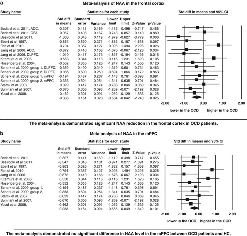
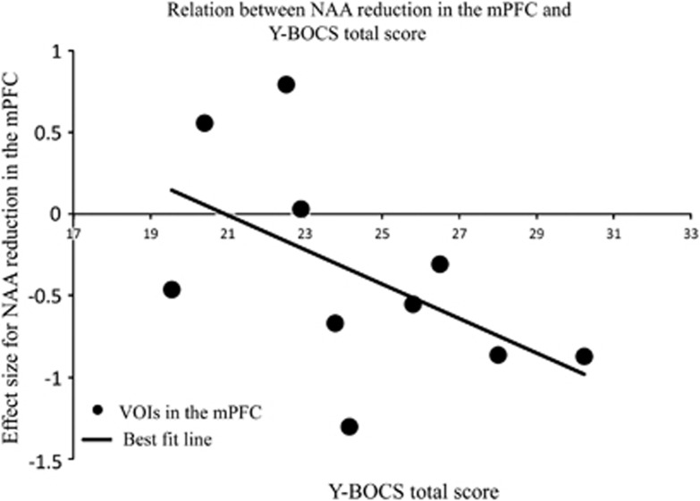
Structural and functional neuroimaging findings suggest that disturbance of the cortico-striato-thalamo-cortical (CSTC) circuits may underlie obsessive-compulsive disorder (OCD). However, some studies with (1)H-magnetic resonance spectroscopy ((1)H-MRS) reported altered level of N-acetylaspartate (NAA), they yielded inconsistency in direction and location of abnormality within CSTC circuits. We conducted a comprehensive literature search and a meta-analysis of (1)H-MRS studies in OCD. Seventeen met the inclusion criteria for a meta-analysis. Data were separated by frontal cortex region: medial prefrontal cortex (mPFC), dorsolateral prefrontal cortex, orbitofrontal cortex, basal ganglia and thalamus. The mean and s.d. of the NAA measure were calculated for each region. A random effects model integrating 16 separate datasets with 225 OCD patients and 233 healthy comparison subjects demonstrated that OCD patients exhibit decreased NAA levels in the frontal cortex (P=0.025), but no significant changes in the basal ganglia (P=0.770) or thalamus (P=0.466). Sensitivity analysis in an anatomically specified subgroup consisting of datasets examining the mPFC demonstrated marginally significant reduction of NAA (P=0.061). Meta-regression revealed that NAA reduction in the mPFC was positively correlated with symptom severity measured by Yale-Brown Obsessive Compulsive Scale (P=0.011). The specific reduction of NAA in the mPFC and significant relationship between neurochemical alteration in the mPFC and symptom severity indicate that the mPFC is one of the brain regions that directly related to abnormal behavior in the pathophysiology of OCD. The current meta-analysis indicates that cortices and sub-cortices contribute in different ways to the etiology of OCD.
Figures
References
-
- Martinot JL, Allilaire JF, Mazoyer BM, Hantouche E, Huret JD, Legaut-Demare F, et al. Obsessive-compulsive disorder: a clinical, neuropsychological and positron emission tomography study. Acta Psychiatrica Scandinavica. 1990;82:233–242. - PubMed
-
- Mcguire PK, Bench CJ, Frith CD, Marks IM, Frackowiak RS, Dolan RJ. Functional anatomy of obsessive-compulsive phenomena. Br J Psychiatry. 1994;164:459–468. - PubMed
-
- Perani D, Colombo C, Bressi S, Bonfanti A, Grassi F, Scarone S, et al. [18F]FDG PET study in obsessive-compulsive disorder. A clinical/metabolic correlation study after treatment. Br J Psychiatry. 1995;166:244–250. - PubMed
Publication types
MeSH terms
Substances
LinkOut - more resources
Full Text Sources
Medical
Miscellaneous
