. 2012:3:1000.
doi: 10.1038/ncomms2008.

FOXO3 signalling links ATM to the p53 apoptotic pathway following DNA damage

Affiliations

FOXO3 signalling links ATM to the p53 apoptotic pathway following DNA damage

Young Min Chung et al. Nat Commun. 2012.

Erratum in

  • Nat Commun. 2013;4:1520

Abstract

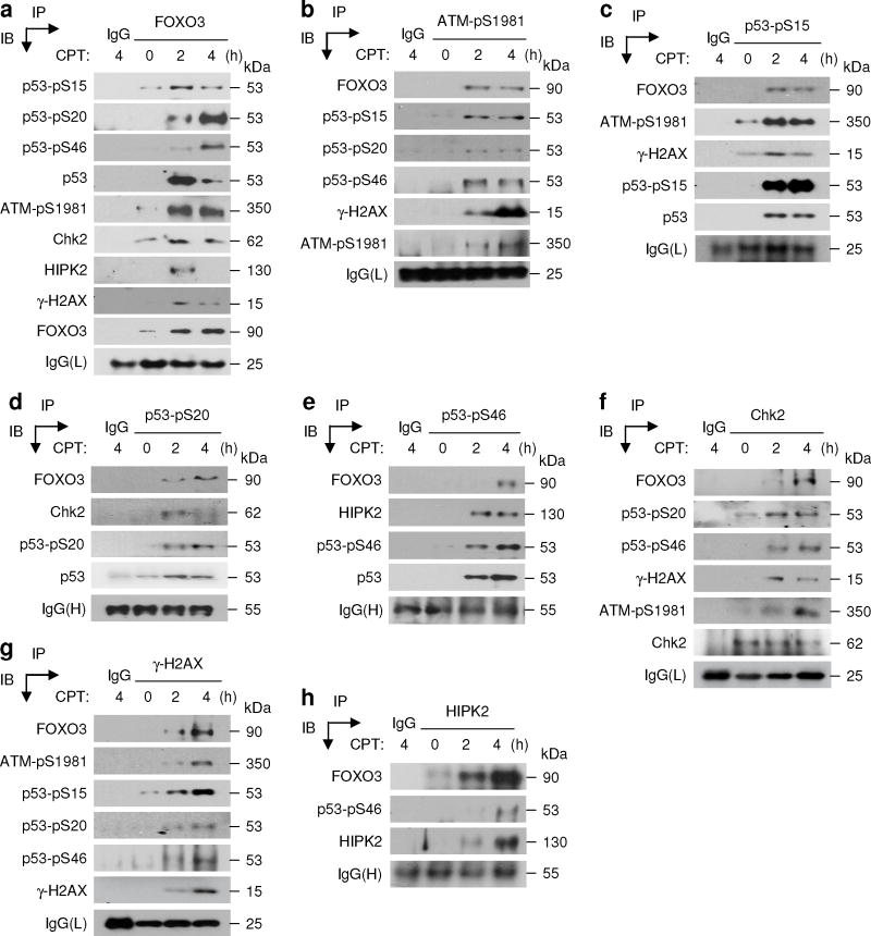
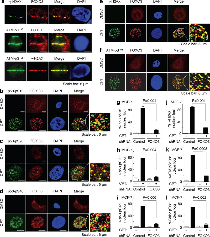
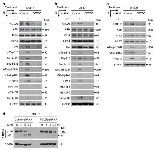
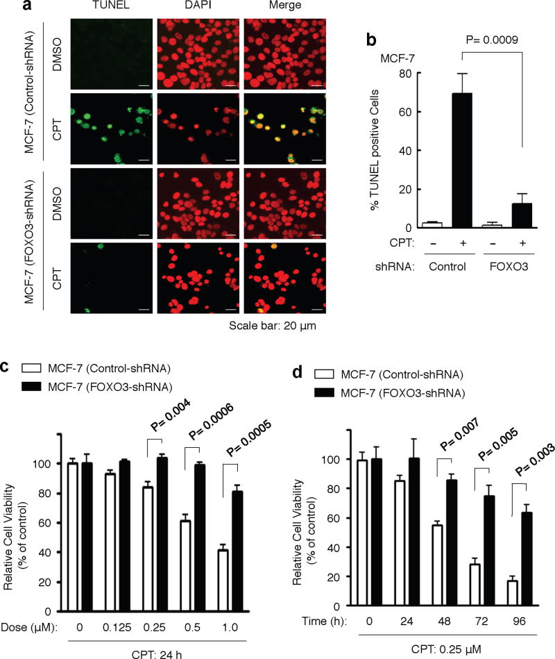
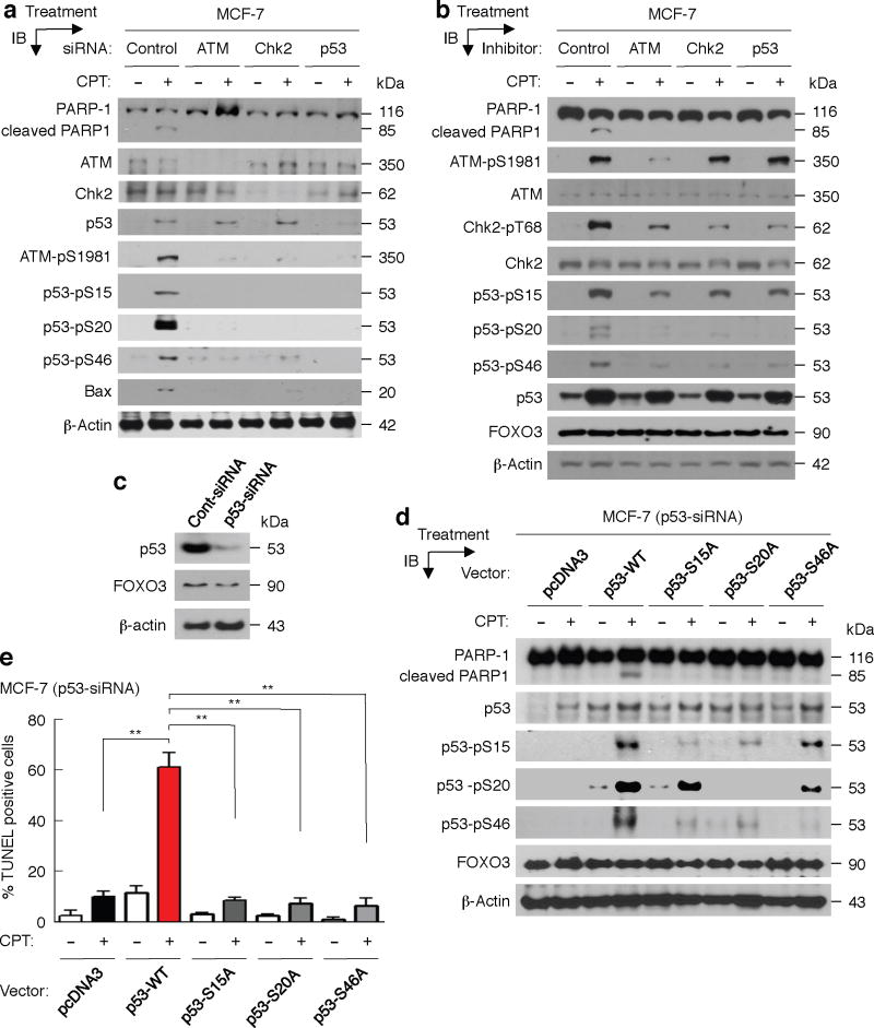
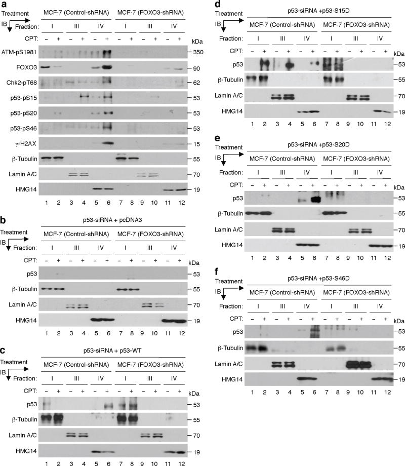
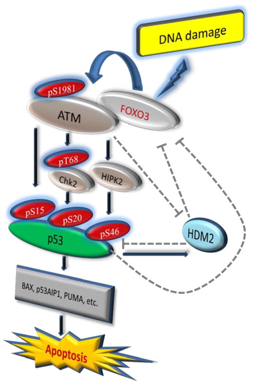
DNA damage as a result of environmental stress is recognized by sensor proteins that trigger repair mechanisms, or, if repair is unsuccessful, initiate apoptosis. Defects in DNA damage-induced apoptosis promote genomic instability and tumourigenesis. The protein ataxia-telangiectasia mutated (ATM) is activated by DNA double-strand breaks and regulates apoptosis via p53. Here we show that FOXO3 interacts with the ATM-Chk2-p53 complex, augments phosphorylation of the complex and induces the formation of nuclear foci in cells on DNA damage. FOXO3 is essential for DNA damage-induced apoptosis and conversely FOXO3 requires ATM, Chk2 and phosphorylated p53 isoforms to trigger apoptosis as a result of DNA damage. Under these conditions FOXO3 may also have a role in regulating chromatin retention of phosphorylated p53. These results suggest an essential link between FOXO3 and the ATM-Chk2-p53-mediated apoptotic programme following DNA damage.


Conflict of interest statement

Competing financial interests: The authors declare no competing financial interests.

Figures

Fig. 1
Fig. 1. FOXO3 interacts with the ATM-Chk2-p53-H2AX complex
(a) Whole cell lysates of MCF-7 treated with CPT (1 μM) for 2 or 4 hours (h) or DMSO (0, control) were subjected to immunoprecipitation (IP) with an anti-FOXO3 antibody (Ab) or an isotype IgG (control) followed by immunoblotting (IB) with Abs as indicated, or an anti-FOXO3 (IP control) or a control Ab against IgG light chain (L). (b–h) Similarly, cell lysates were subjected to IP with an Ab against ATM-pS1981 (b) or p53-pS15 (c) or p53-pS20 (d) or p53-pS46 (e) or Chk2 (f) or γ-H2AX (g) or HIPK2 (h) or an isotype IgG followed by IB with the indicated Abs or a control Ab against IgG(L) or heavy chain (H).
Fig. 2
Fig. 2. FOXO3 is essential for the formation of ATM-Chk2-p53-H2AX nuclear foci
(a) Accumulation of a fraction of FOXO3 at laser-induced damage was detected in MCF-7 cells 30 min after focused laser micro-irradiation. The cells were stained with Abs against FOXO3 and γ-H2AX or ATM-pS1981, followed by fluorescence microscopy as described in the section of Immunofluorescence under “Methods”. DAPI was used to show the nuclei, and co-localizations of FOXO3 with ATM-pS1981 were shown as the merged images. Scale bar: 6 μm. (b–f) MCF-7 cells were treated with CPT (1 μM) or DMSO control for 2 h, and co-localizations between FOXO3 and p53-pS15 (b) or p53-pS20 (c) or p53-pS46 (d) or γ-H2AX (e) or ATM-pS1981 (f) were detected using Abs as indicated and followed by fluorescence microscopy. Scale bar: 6 μm. (g–l) FOXO3 is necessary for the formation of p53-pS15, p53-pS20, and p53-pS46, γ-H2AX, ATM-pS1981, and Chk2-pT68 nuclear foci upon CPT-induced DNA damage. MCF-7 stable cell lines transfected with FOXO3-shRNA or control-shRNA were treated with CPT (1 μM) or DMSO for 2 h, and then the subcellular localizations and co-localization of FOXO3 and p53-pS15 (g) or p53-pS20 (h) or p53-pS46 (i) or γ-H2AX (j) or ATM-pS1981 (k) or Chk2-pT68 (l) were detected using specific Abs as indicated. An average of 200 cells with specific nuclear foci in each comparison was determined and shown. The images of nuclear foci of these proteins were shown in Supplementary Fig. S5. The error bars represent standard deviation, and the statistical test is the paired t-test.
Fig. 3
Fig. 3. FOXO3 is required for ATM-Chk2-p53 phosphorylation and PARP-1 cleavage
(a) MCF-7 stable cell lines transfected with FOXO3-shRNA or control-shRNA were treated with CPT (1 μM) or DMSO (negative control) for 4 h, and whole cell lysates were prepared and the levels of proteins were analyzed by IB with specific Abs as highlighted or an anti-β-actin (loading control). (b, c) A549 (b) and H1299 (c) stable cell lines transfected with FOXO3-shRNA or control-shRNA were treated with CPT (1 μM) or DMSO (negative control) for 4 h, and whole cell lysates were analyzed by IB with specific Abs as indicated or an anti-β-actin. (d) Cell lysates from MCF-7 stable cell lines (FOXO3-shRNA and control-shRNA) treated with CPT (1 μM) for an indicated time course were subjected to IB analysis using Abs against PARP-1 and β-actin (loading control).
Fig. 4
Fig. 4. FOXO3 is necessary for inducing DNA damage-induced apoptosis
(a) MCF-7 stable cell lines (FOXO3-shRNA and control-shRNA) were treated with CPT (1 μM) or DMSO (control) for 48 h, and the cells were fixed on the slides for determining cellular apoptosis by TUNEL assays (Promega). Nuclei were stained with DAPI (color-inverted to red), and merged images (yellow) were considered as apoptotic cells. Scale bar: 20 μm. (b) An average (%) of apoptotic (TUNEL-positive) cells was determined and shown in the diagram; The samples include three biological replicates, the error bars represent standard deviation, and the statistical test is the paired t-test. The significant P values between the FOXO3-shRNA group versus the control group treated with CPT are highlighted. (c, d) MCF-7 (control-shRNA) and MCF-7 (FOXO3-shRNA) cells were treated with various doses of CPT or DMSO (control) for 24 hours (c) or with a low dose CPT (0.25 μM) for a time course as indicated (d), and performed cell survival assays by the MTT assays (c) or cell counting (d). The significant P values between the FOXO3-shRNA group versus the control group treated with CPT are indicated. The number of biological replicates is three, the error bars represent standard deviation, and the statistical test is the paired t-test.
Fig. 5
Fig. 5. Phosphorylated ATM-Chk2-p53 may be needed for FOXO3-mediated apoptosis
(a) MCF-7 cells were transfected with control-siRNA (control) or siRNA (0.3 μM) targeting ATM or Chk2 or p53 and incubated for 48 hours (h), and then these cells were treated with CPT (1 μM) (+) or DMSO (−) for 48 h. Whole cell lysates were prepared from these treated cells and the levels of PARP-1 and its degraded proteins, ATM, Chk2, p53, and their phosphorylated proteins or a p53 target (Bax) were analyzed by IB with specific Abs as indicated or an anti-β-actin (loading control). (b) MCF-7 cells were first treated with ATM inhibitor (Mirin) or Chk2 inhibitor (NSC10955) or p53 inhibitor (Pifithrin), 20 μM/each, or DMSO for 6 h, and then treated with CPT (1 μM) or DMSO for 16 h. Whole cell lysates were prepared from these treated cells and the levels of PARP-1 and its degraded proteins, and the indicated proteins and β-actin were analyzed by IB analysis with specific Abs. (c) MCF-7 cells were transfected with p53-siRNA or Cont-siRNA (Control-siRNA) for 48 h. Whole cell lysates were prepared from these transfected cells and the expressions of p53 and FOXO3 (control) were determined by IB analysis with specific Abs. (d) MCF-7 (p53-siRNA) cells were transfected with pcDNA3 (control) or p53 wild-type (WT) or mutant (p53-S15A or p53-S20A or p53-46A) expression vectors (2 μg DNA/each) for 48 h, and then treated with CPT (1 μM) (+) or DMSO (−) for 48 h. Whole cell lysates were prepared from these treated cells and the levels of PARP-1 and its degraded proteins, and the indicated proteins were analyzed by IB analysis with specific Abs. (e) MCF-7 (p53-siRNA) cells were transfected with control and expression vectors for 24 h, the transfected cells were treated with CPT (1 μM) or DMSO control for 36 h, and cellular apoptosis was determined by TUNEL assays (Promega). Images of apoptotic cells were shown in Supplementary Fig. S9. An average (%) of TUNEL-positive (apoptotic) cells was determined and shown in the diagram. **, P= 0.0001.
Fig. 6
Fig. 6. FOXO3 plays a role in regulating chromatin retention of phosphorylated p53
(a) MCF-7 stable cell lines transfected with FOXO3-shRNA or control-shRNA were treated with CPT (1 μM) (+) or DMSO (−) (control) for 4 h, and cells were harvested and fractionated with Nonidet P-40 as described in the section of Chromatin Retention Assay under “Methods”. Equal amount (20 μg) of each fraction was analyzed by IB analysis with the highlighted Abs as described above. Proteins β-tubulin, lamin A/C, and HMG14 (high-mobility-group 14, a chromosome binding protein) represent the fractionation and loading controls of the cytosol (fraction I), the nucleoplasm (fraction III) and the chromatin (fraction IV), respectively. (b–f) MCF-7 (control-shRNA) and MCF-7 (FOXO3-shRNA) cells were transfected with p53 siRNA for 48 h, then transfected with pcDNA3 (negative control) (b) or the p53-WT vector (c) or the specific vectors expressing p53-S15D (d), p53-S20D (e), and p53-S46D (f) for 36 h. The transfected cells were treated with CPT (1 μM) or DMSO for 4 h, then cells were harvested and subjected to chromatin fractionation and IB analysis as described above.
Fig. 7
Fig. 7. A link between FOXO3 and the ATM-Chk2-p53-mediated apoptotic program
A schematic shows the FOXO3-dependent activation of ATM-pS1981, Chk2-pT68, HIPK2, p53-pS15, p53-pS20, p53-pS46, and the possible roles of p53 and Hdm2 in downregulation of FOXO3 as negative feedback loops, and the downstream p53 apoptotic-signaling pathway after DNA damage induced by CPT. The dashed lines denote the inhibitory pathways according to the published literature.

Similar articles

Cited by

References

    1. Shiloh Y. ATM and related protein kinases: safeguarding genome integrity. Nat Rev Cancer. 2003;3:155–168. - PubMed
    1. Kastan MB, Bartek J. Cell-cycle checkpoints and cancer. Nature. 2004;432:316–323. - PubMed
    1. Bartek J, Lukas J. Chk1 and Chk2 kinases in checkpoint control and cancer. Cancer Cell. 2003;3:421–429. - PubMed
    1. Giaccia AJ, Kastan MB. The complexity of p53 modulation: emerging patterns from divergent signals. Genes Dev. 1998;12:2973–2983. - PubMed
    1. Prives C, Hall PA. The p53 pathway. J Pathol. 1999;187:112–126. - PubMed

Publication types

MeSH terms