. 2012:3:1006.
doi: 10.1038/ncomms2005.

TREX exposes the RNA-binding domain of Nxf1 to enable mRNA export

Affiliations

TREX exposes the RNA-binding domain of Nxf1 to enable mRNA export

Nicolas Viphakone et al. Nat Commun. 2012.

Abstract

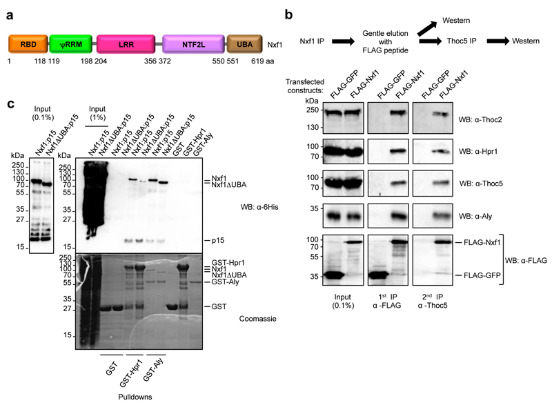
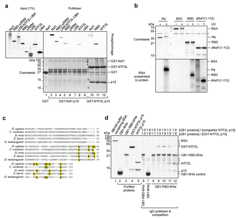
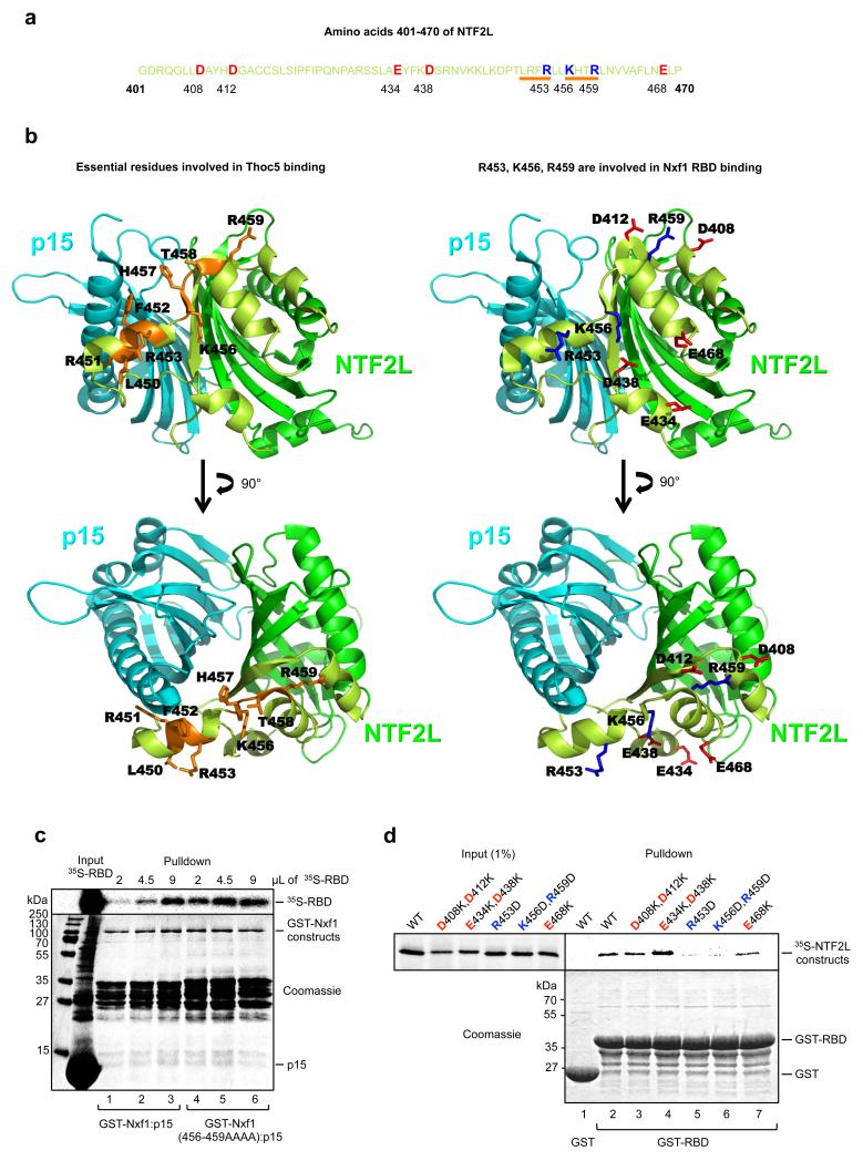
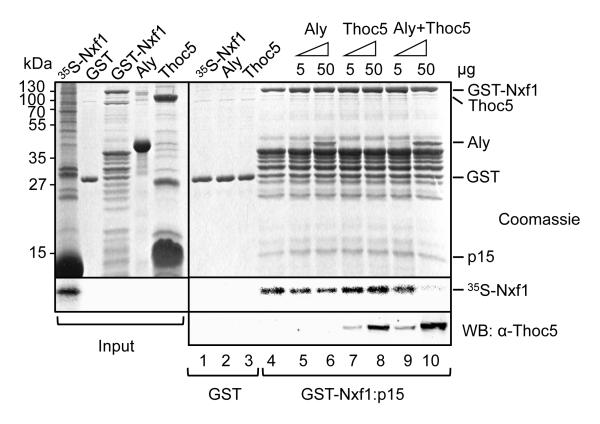
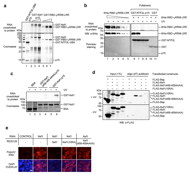
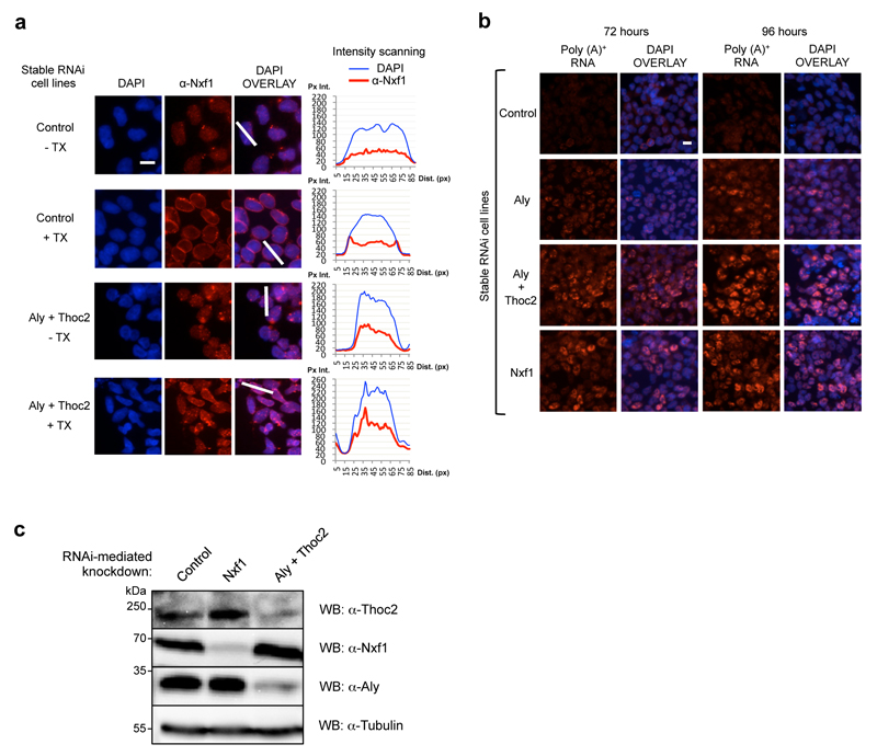
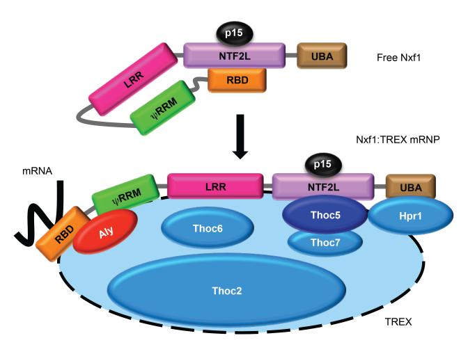
The metazoan TREX complex is recruited to mRNA during nuclear RNA processing and functions in exporting mRNA to the cytoplasm. Nxf1 is an mRNA export receptor, which binds processed mRNA and transports it through the nuclear pore complex. At present, the relationship between TREX and Nxf1 is not understood. Here we show that Nxf1 uses an intramolecular interaction to inhibit its own RNA-binding activity. When the TREX subunits Aly and Thoc5 make contact with Nxf1, Nxf1 is driven into an open conformation, exposing its RNA-binding domain, allowing RNA binding. Moreover, the combined knockdown of Aly and Thoc5 markedly reduces the amount of Nxf1 bound to mRNA in vivo and also causes a severe mRNA export block. Together, our data indicate that TREX provides a license for mRNA export by driving Nxf1 into a conformation capable of binding mRNA.


Figures

Figure 1
Figure 1. Nxf1 and TREX assemble in a complex in vivo
(a) Schematic of Nxf1 showing its domains. RBD, RNA binding domain; ψRRM, pseudo RNA recognition motif; LRR, leucine rich repeat; NTF2L, NTF2-like; UBA, ubiquitin associated. (b) Immunoprecipitation (IP) analysis of Nxf1. FLAG-Nxf1 was immunoprecipitated using FLAG antibody, eluted using FLAG peptide and then immunoprecipitated a second time using Thoc5 antibody. At each stage, samples were analysed by western blotting using the indicated antibodies. (c) The UBA domain of Nxf1 is required for interaction with Hpr1. GST-Hpr1 or GST-Aly pulldowns with the indicated Nxf1 constructs. Nxf1-GB1-6His or Nxf1ΔUBA-GB1-6His were co-expressed with p15-6His in BL21RP E. coli strain. Total extracts were prepared, cleared by centrifugation, and used in pulldown experiments with GST-Hpr1 or GST-Aly in RB100 buffer in the presence of RNase A (10 μg/mL). Pulldowns were analysed by SDS-PAGE followed by Coomassie staining or western blot using 6His antibody.
Figure 2
Figure 2. The NTF2L domain of Nxf1 sequesters the RNA Binding Domain
(a) GST pulldowns with the indicated GST fusions and domains of Nxf1 synthesised and 35S-radiolabeled in vitro in reticulocyte lysate. (b) RNA-binding activity of the indicated constructs was analysed by UV-crosslinking. dNxf1: Drosophila Melanogaster Nxf1 protein. The asterisk designates the RNase inhibitor present in the reactions. (c) The Homo sapiens N-terminal RNA binding domain sequence is aligned with the equivalent sequences from other metazoa. Within the H.sapiens sequence, 10 arginines have been shown to be essential for the RNA binding activity in vitro, poly(A)+ RNA association in vivo, and mRNA export. These are highlighted in yellow in the alignment as are equivalent residues in other Nxf1 sequences. In all metazoa, the sequences which align with the H. sapiens 35 amino acid arginine rich sequence involved in RNA binding, show a strong bias towards arginine residues as follows: H. sapiens 10/35 amino acids = 29% arginine, C.corturnix, 6/26 amino acids = 23% arginine, D.rerio 10/37 amino acids = 27% arginine, Xenopus laevis, 6/33 amino acids = 18% arginine, D.melanogaster, 6/30 = 20% arginine. (d) Binding of the NTF2L domain to the RBD is direct. Binding of the NTF2L domain to the RBD was analysed by an IgG pulldown experiment performed in RB100 buffer in the presence of BSA (lane 1) and RNase A using purified GB1-6His or GB1-RBD-6His and GST-NTF2L:p15 (lanes 7-12). Unbound proteins were washed away and subsequently either buffer (lanes 8-10) or NTF2L:p15 competitor (lanes 7, 11, 12) was added at the indicated molar ratios. The GB1 tag binds IgG beads. Purified proteins used in this assay are shown in lanes 2-5. Bound proteins were eluted and subsequently analysed by SDS-PAGE and Coomassie staining. Lane 6 shows IgG binding and elution of GB1-RBD-6His alone, performed in the same conditions as the pulldown assays. Additional bands, not present in the GB1-RBD-6His input sample, arising from the IgG beads are marked on the right hand side of the gel by brackets.
Figure 3
Figure 3. Identification of the NTF2L:RBD binding interface
(a) Sequence of the region of the NTF2L domain targeted by the mutagenesis. Targeted residues are highlighted as red for acidic and blue for basic. Orange bars indicate the most crucial residues involved in Thoc5 binding (Katahira J. et al, EMBO J., 2009 Mar 4;28(5):556-67) that also enhance Nxf1 interdomain interaction when mutated to alanines (in case of 456-459). (b) Positions of the above mentioned residues on NTF2L:p15 structure (1JKG) using the same colors as in (a). p15 is in cyan, the NTF2L domain is in dark green, targeted region within the NTF2L domain is in light green. (c) Nxf1(456-459AAAA) mutant binds Nxf1 RBD more efficiently than wild type Nxf1. GST pulldowns with the indicated fusions and increasing amounts of 35S-RBD. (d) GST pulldown experiment using GST-RBD and the indicated in vitro translated mutants of the NTF2L domain of Nxf1.
Figure 4
Figure 4. Both Aly and Thoc5 are required to disrupt Nxf1-Nxf1 interactions
Pulldown assays with GST-Nxf1:p15 and 35S-radiolabeled Nxf1 in the presence of the indicated TREX components. The lower panel is a Western blot to detect the binding of Thoc5. All of these experiments were carried out in the presence of RNase A.
Figure 5
Figure 5. The Nxf1 intramolecular interaction regulates its RNA binding activity
(a) RNA cross-linking activity for GST-TAP:p15 and GST-RBD-ψRRM-LRR. The three different Nxf1 construct protein concentrations used (from left to right) were 88, 265 and 530 nM and the RNA concentration was 6.5 μM. (b) The Nxf1 RBD fails to bind RNA when complexed with the NTF2L domain. The 6His-RBD-ψRRM-LRR protein efficiently crosslinks with RNA (top panel lanes 1-5, using 200, 100, 50, 20, 10 ng of protein). RNA binding was detected by phosphoimaging. 6His-RBD-ψRRM-LRR was detected by Western blot (middle panel) and the GST constructs were detected by Ponceau staining (bottom panel). (c) RNA cross-linking activity for GST-Nxf1(456-459AAAA):p15 and GST-Nxf1:p15. RNA binding was detected by phosphoimaging. (d) mRNP capture assay with indicated Nxf1 constructs. Bap is bacterial alkaline phosphatase control. Nxf1(10RA) is a mutant form of Nxf1 which is unable to bind RNA efficiently. (e) RNA export assay in 293T cells. Cells were transfected with the Nxf1 RNAi vector together with the indicated rescue Nxf1 expression vectors, resistant to RNAi. Cells were analysed by fluorescent in situ hybridisation (FISH) with Cy3-oligo(dT) (top panel) and images overlayed with DAPI staining of the same cells (bottom panel). All pictures were taken at the same exposure level. The white horizontal bar represents 10 μm.
Figure 6
Figure 6. Thoc5 and Aly trigger Nxf1 opening allowing RNA binding and efficient mRNA export
(a) Nxf1 remodelling assay. The indicated protein complexes were formed and UV cross-linked with a 5′-radiolabeled RNA. The top panels are phosphoimages and the bottom panels are their corresponding gels coomassie stained. (b) mRNP capture assay shows that Aly and Thoc5 knockdown prevents efficient recruitment of Nxf1 to mRNA. miRNA expression was induced for 48 hours prior to analysis. (c) Oligo(dT) FISH on stable 293 cell lines expressing miRNAs targeting the indicated genes. The time points refer to the time following induction of the miRNA expression. Cells were treated with actinomycin D for 2 hours prior to FISH to reduce nascent RNA signals. All pictures are taken at the same exposure level. (d) Nuclear envelope association of Nxf1 is impaired in vivo when both TREX components Aly and Thoc5 are depleted by RNAi. After 72 hours of induction of miRNAs expression, the indicated 293 stable RNAi cell lines were subjected to Triton X-100 extraction prior to fixation when indicated (+ TX) and the localisation of Nxf1 was analysed by immunostaining α-Nxf1 and fluorescence intensity scanning (white bars and corresponding graphs). The Y-axis of the graphs represents the pixel intensity (Px Int.) and the X-axis the distance in pixels (Dist. (px)). The white horizontal bars in (c) and (d) represent 10 μm.
Figure 7
Figure 7. Thoc2 plays a role in Nxf1 localisation and mRNA export
(a) Nuclear envelope association of Nxf1 is impaired in vivo when both TREX components Aly and Thoc2 are depleted by RNAi. Localisation of Nxf1 was analysed by immunostaining of Control or (Aly+Thoc2) knockdown cell lines after 96h of induction of miRNA expression using Nxf1 antibody. Where indicated (+ TX), cells were treated with 0.5% Triton X-100 before fixing. The Y-axis of the graphs represents the pixel intensity (Px Int.) and the X-axis the distance in pixels (Dist. (px)). (b) A strong block of bulk poly(A)+ RNA export is observed in Nxf1 RNAi and (Aly+Thoc2) RNAi cell lines. Oligo (dT) FISH on stable 293 cell lines expressing miRNAs targeting the indicated genes. The time points refer to the time following induction of miRNAs expression. Cells were treated with actinomycin D for 2 hours prior to FISH to reduce nascent RNA signals. All pictures are taken at the same exposure level. The white horizontal bars in (a) and (b) represent 10 μm. (c) Western Blot analysis of the indicated cell lines. The expression of the miRNAs is under the control of a tetracycline inducible promoter. Extracts were prepared 96 hours post-induction of miRNA expression and analysed using the indicated antibodies.
Figure 8
Figure 8. A model for the conformational change in Nxf1 induced by TREX during mRNA export
Free Nxf1:p15 sequesters its N- terminal RNA binding domain. During mRNA export, assembled TREX binds Nxf1:p15. This allows the two subunits Aly and Thoc5 to remodel Nxf1 and expose its RNA binding domain, allowing direct interaction between Nxf1 and mRNA during export. For clarity not all TREX subunits are illustrated in the figure.

References

    1. Herold A, Klymenko T, Izaurralde E. NXF1/p15 heterodimers are essential for mRNA nuclear export in Drosophila. RNA. 2001;7:1768–1780. - PMC - PubMed
    1. Herold A, Teixeira L, Izaurralde E. Genome-wide analysis of nuclear mRNA export pathways in Drosophila. Embo J. 2003;22:2472–2483. - PMC - PubMed
    1. Rodriguez-Navarro S, Hurt E. Linking gene regulation to mRNA production and export. Curr Opin Cell Biol. 2011;23:302–309. - PubMed
    1. Segref A, et al. Mex67p, a novel factor for nuclear mRNA export, binds to both poly(A)+ RNA and nuclear pores. Embo J. 1997;16:3256–3271. - PMC - PubMed
    1. Zolotukhin AS, Tan W, Bear J, Smulevitch S, Felber BK. U2AF participates in the binding of TAP (NXF1) to mRNA. J Biol Chem. 2002;277:3935–3942. - PubMed

Publication types

MeSH terms