

How to optimize the use of MRI in anatomic ACL reconstruction
- PMID: 22893266
- PMCID: PMC3685708
- DOI: 10.1007/s00167-012-2153-9
How to optimize the use of MRI in anatomic ACL reconstruction
Abstract
Purpose: Magnetic resonance imaging (MRI) is the most current diagnostic imaging procedure for suspected ACL injuries. It is an accurate, highly sensitive and specific tool for the diagnosis of ACL tears, graft tears and associated injuries. However, it can also be used for various other aspects of anatomic ACL reconstruction.
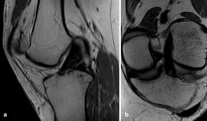
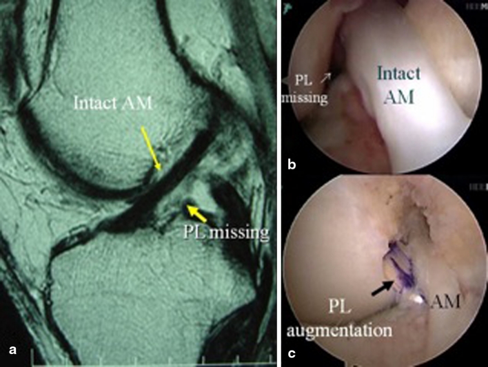
Methods: Special sequences as the oblique sagittal plane should be obtained from a parallel line to the lateral epicondyle, ensuring a proper visualization of both bundles of the ACL. Another special set of images, the oblique-coronal sequence, allows for the ACL long-axis evaluation. The coronal-oblique sequence increases the sensitivity and specificity of diagnosing isolated AM or PL bundle injuries and also helps to visualize the proximal insertion of the bundles for haemorrhage and rupture.
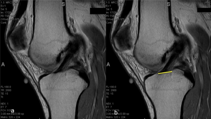
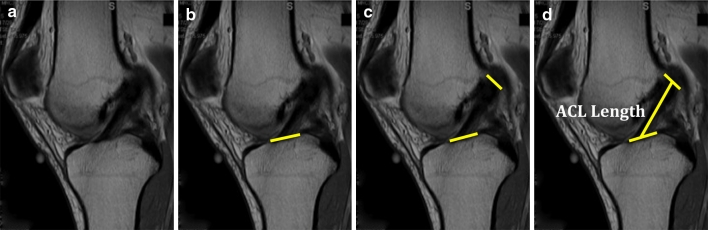
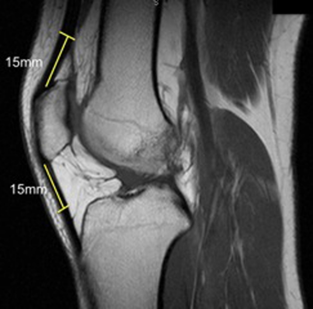
Results: Quantitative measurements can be taken from a proper MRI protocol, so as to determine the rupture pattern; measure insertion site size, inclination angle and autograft size; and evaluate for post-operative complications. These parameters help surgeons to objectively decide for a better graft and technique for an individualized approach and to evaluate the anatomic placement of the graft.
Conclusions: MRI can be used in different ways, serving as a very valuable tool in anatomic ACL reconstruction. Special protocols can provide accurate visualization of the double-bundle anatomy. Objective parameters to aid in pre-operative decisions and graft's anatomic placement evaluation can be also extracted from the MR images.
Figures


References
-
- Ahn JH, Lee SH, Yoo JC, Ha HC. Measurement of the graft angles for the anterior cruciate ligament reconstruction with transtibial technique using postoperative magnetic resonance imaging in comparative study. Knee Surg Sports Traumatol Arthrosc. 2007;15:1293–1300. doi: 10.1007/s00167-007-0389-6. - DOI - PubMed
-
- Benjaminse A, Gokeler A, van der Schans CP. Clinical diagnosis of an anterior cruciate ligament rupture: a meta-analysis. J Orthop Sports Phys Ther. 2006;36:267–288. - PubMed
Publication types
MeSH terms
LinkOut - more resources
Full Text Sources
Medical
