

Case Reports
Dermatitis herpetiformis: relevance of the physical examination to diagnosis suspicion
Affiliations
- PMID: 22893336
- PMCID: PMC3418983
Item in Clipboard
Case Reports
Dermatitis herpetiformis: relevance of the physical examination to diagnosis suspicion
Can Fam Physician.
2012 Aug.
No abstract available
Figures
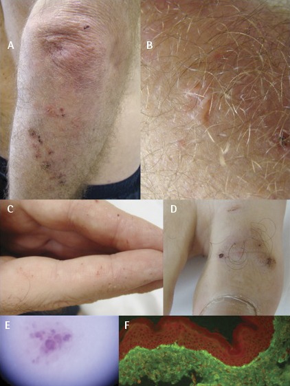
Clinical photographs of patient 1, a 44-year-old white man:
A) Excoriated papules and crusts on the forearm and extensor surface of the elbow. B) Meticulous clinical examination showed small vesicles on the skin of the elbow. C) Purpuric lesions on the volar aspect of the fingers. D) Purpuric lesions on the volar aspect of the toes. E) Dermoscopy of a petechial lesion on the finger demonstrating violaceous globules. F) Direct immunofluorescence of a lesion on the skin of the forearm. Notice the granular deposits of immunoglobulin A–fluorescein-marked complex at the dermal-epidermal junction of the skin (papillae of dermis).
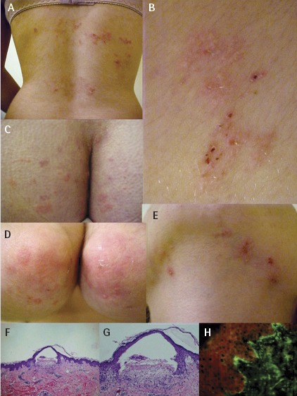
Clinical features of patient 2, a 19-year-old white woman:
A) Excoriated papules and plaques on the back. B) Detail of the excoriated papules and crusts on the back. C) Papules and urticarial plaques on the buttocks and sacral area. D) Erythematous papules and urticarial lesions on the elbows. E) Excoriated papules and plaques on the submandibular area. F) Histopathologic examination of a skin lesion on the elbow showing small vesicles (hematoxylin and eosin stain, original magnification × 100). G) Detail of the subepidermal vesicle (hematoxylin and eosin stain, original magnification × 200). H) Direct immunofluorescence of a lesion on the back. Note the granular deposits of immunoglobulin A–fluorescein-marked complex at the dermal-epidermal junction of the skin (papillary dermis).
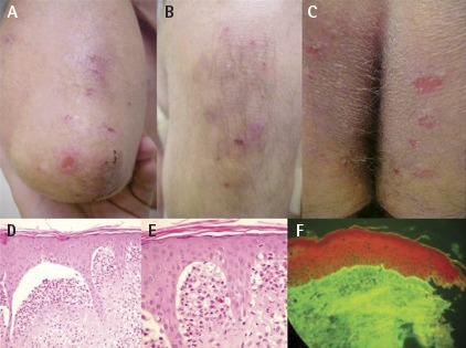
Clinical features of patient 3, a 58-year-old white man.
A) Excoriated papules and crusts on the extensor surface of the elbow; B) knee; and C) buttocks. D) Histopathologic examination of a skin lesion on the dorsum of the foot showing a small subepidermal vesicle (hematoxylin and eosin stain, original magnification × 400). E) Detail of the subepidermal vesicle leading to a microabscess filled with neutrophils (hematoxylin and eosin stain, original magnification × 1000). F) Direct immunofluorescence of the back skin. Note the granular deposits of immunoglobulin A–fluorescein-marked complex at the dermal-epidermal junction of the skin (papillary dermis).
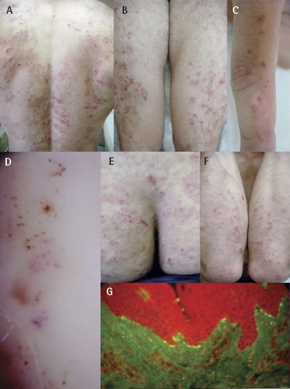
Clinical features of patient 4, a 22-year-old white man.
A) Extensive excoriated papules and crusts on the back and B) legs. C) Purpuric lesions on the volar aspect of the fingers. D) Dermoscopy of petechial lesions on the finger demonstrating violaceous globules (newest lesions) and brownish dots and globules (oldest lesions). E) Excoriated papules and residual lesions on the buttocks. F) Excoriated papules and crusts on the extensor surface of the elbow. G) Direct immunofluorescence of the forearm skin. Note the granular deposits of immunoglobulin A–fluorescein-marked complex at the dermal-epidermal junction of the skin (papillary dermis).
Similar articles
-
[Delayed diagnosis of dermatitis herpetiformis as a silent form of coeliac disease--a case report].Pol Merkur Lekarski. 2002 Jun;12(72):506-8. Pol Merkur Lekarski. 2002. PMID: 12362671 Polish.
-
[Dermatitis herpetiformis and celiac disease].Rev Med Chil. 2021 Sep;149(9):1330-1338. doi: 10.4067/S0034-98872021000901330. Rev Med Chil. 2021. PMID: 35319687 Review. Spanish.
-
Bullous lesions in a woman suffering from ulcerative colitis.Gastroenterol Nurs. 2010 Jan-Feb;33(1):58-9. doi: 10.1097/SGA.0b013e3181ca01aa. Gastroenterol Nurs. 2010. PMID: 20145453 No abstract available.
-
Physical signs for the general dental practitioner. Dermatitis herpetiformis.Dent Update. 2006 Jun;33(5):316. Dent Update. 2006. PMID: 16841614 No abstract available.
-
Atopic Dermatitis Is Associated with Dermatitis Herpetiformis and Celiac Disease in Children.J Invest Dermatol. 2021 Jan;141(1):191-193.e2. doi: 10.1016/j.jid.2020.05.091. Epub 2020 Jun 12. J Invest Dermatol. 2021. PMID: 32540248 Review. No abstract available.
Cited by
-
Immunomodulatory Plant Natural Products as Therapeutics against Inflammatory Skin Diseases.Curr Top Med Chem. 2024;24(12):1013-1034. doi: 10.2174/0115680266277952240223120435. Curr Top Med Chem. 2024. PMID: 38485678 Review.
-
Cerebellar degeneration in gluten ataxia is linked to microglial activation.Brain Commun. 2024 Mar 7;6(2):fcae078. doi: 10.1093/braincomms/fcae078. eCollection 2024. Brain Commun. 2024. PMID: 38510211 Free PMC article.
-
Dermatitis herpetiformis in an African woman.Pan Afr Med J. 2018 Jun 12;30:119. doi: 10.11604/pamj.2018.30.119.14012. eCollection 2018. Pan Afr Med J. 2018. PMID: 30364361 Free PMC article.
-
Promising Natural Products in New Drug Design, Development, and Therapy for Skin Disorders: An Overview of Scientific Evidence and Understanding Their Mechanism of Action.Drug Des Devel Ther. 2022 Jan 6;16:23-66. doi: 10.2147/DDDT.S326332. eCollection 2022. Drug Des Devel Ther. 2022. PMID: 35027818 Free PMC article. Review.
-
Dermatitis Herpetiformis: Novel Perspectives.Front Immunol. 2019 Jun 11;10:1290. doi: 10.3389/fimmu.2019.01290. eCollection 2019. Front Immunol. 2019. PMID: 31244841 Free PMC article. Review.
References
-
- Collin P, Reunala T. Recognition and management of the cutaeous manifestations of celiac disease: a guide for dermatologists. Am J Clin Dermatol. 2003;4(1):13–20. - PubMed
-
- Nicolas ME, Krause PK, Gibson LE, Murray JA. Dermatitis herpetiformis. Int J Dermatol. 2003;42(8):588–600. - PubMed
-
- McGovern TW, Bennion SD. Palmar purpura: an atypical presentation of childhood dermatitis herpetiformis. Pediatr Dermatol. 1994;11(4):319–22. - PubMed
-
- Marks R, Jones EW. Purpura in dermatitis herpetiformis. Br J Dermatol. 1971;84(4):386–8. - PubMed
-
- Moulin G, Barrut D, Franc MP, Viornery P, Knezynski S. Pseudopurpuric palmar localizations of herpetiform dermatitis [article in French] Ann Dermatol Venereol. 1983;110(2):121–6. - PubMed
Publication types
MeSH terms
LinkOut - more resources
Full Text Sources
Medical