. 2012 Sep;26(9):1005-15.
doi: 10.1007/s10822-012-9594-6. Epub 2012 Aug 15.

Structure-based virtual screening of small-molecule antagonists of platelet integrin αIIbβ3 that do not prime the receptor to bind ligand

Affiliations

Structure-based virtual screening of small-molecule antagonists of platelet integrin αIIbβ3 that do not prime the receptor to bind ligand

Ana Negri et al. J Comput Aided Mol Des. 2012 Sep.

Abstract

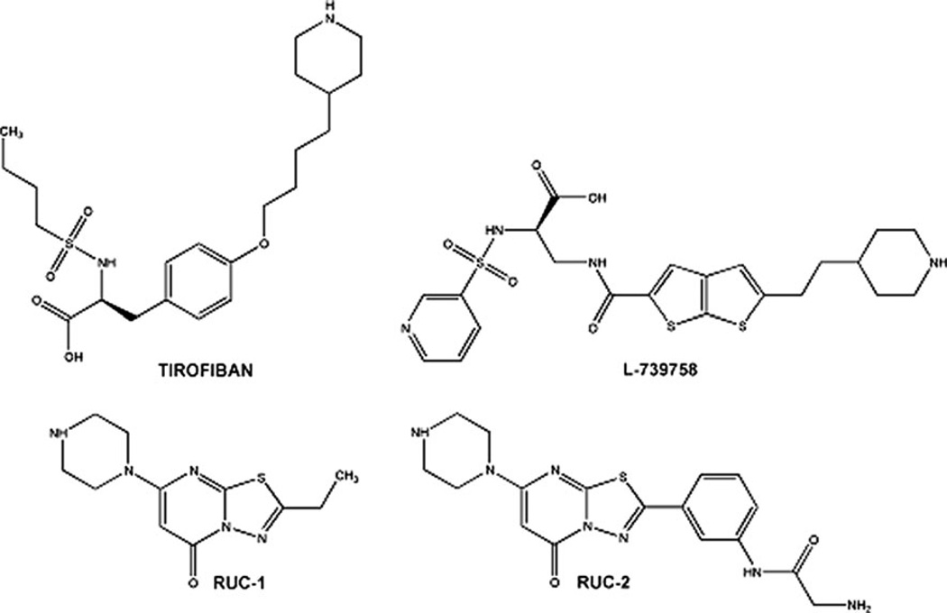
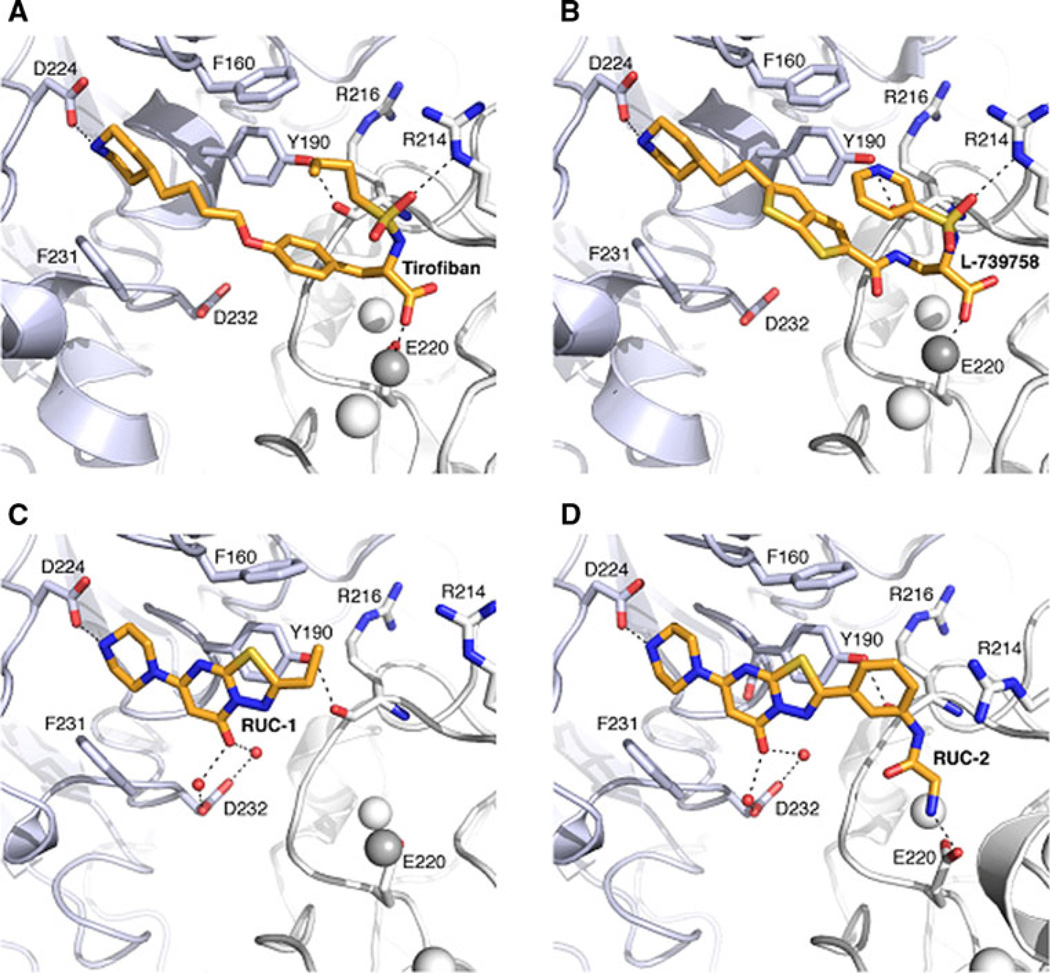
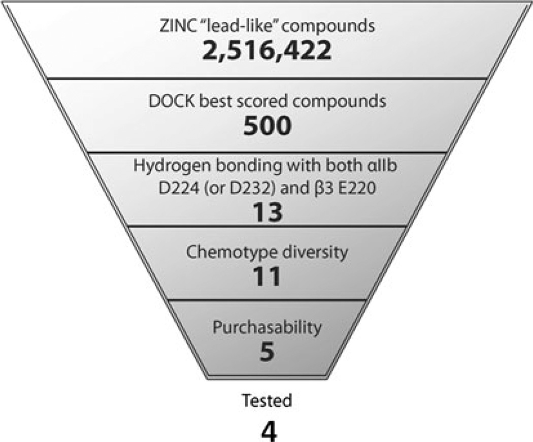
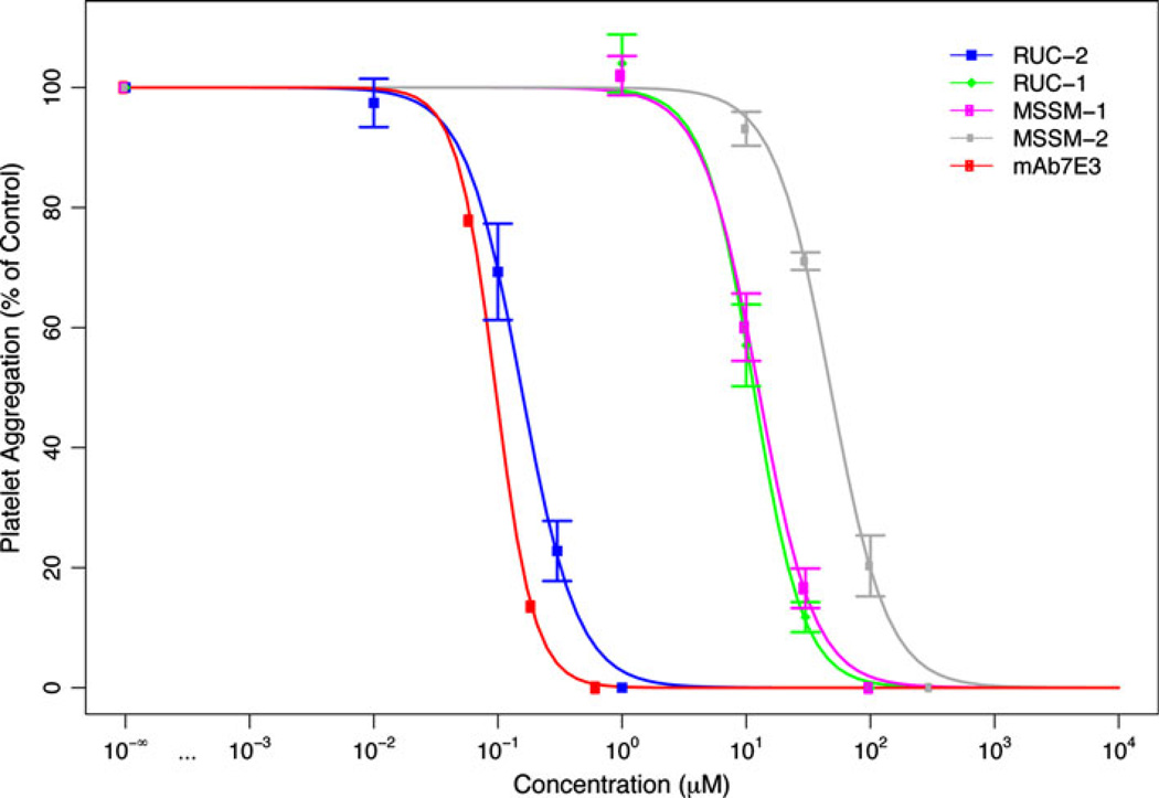
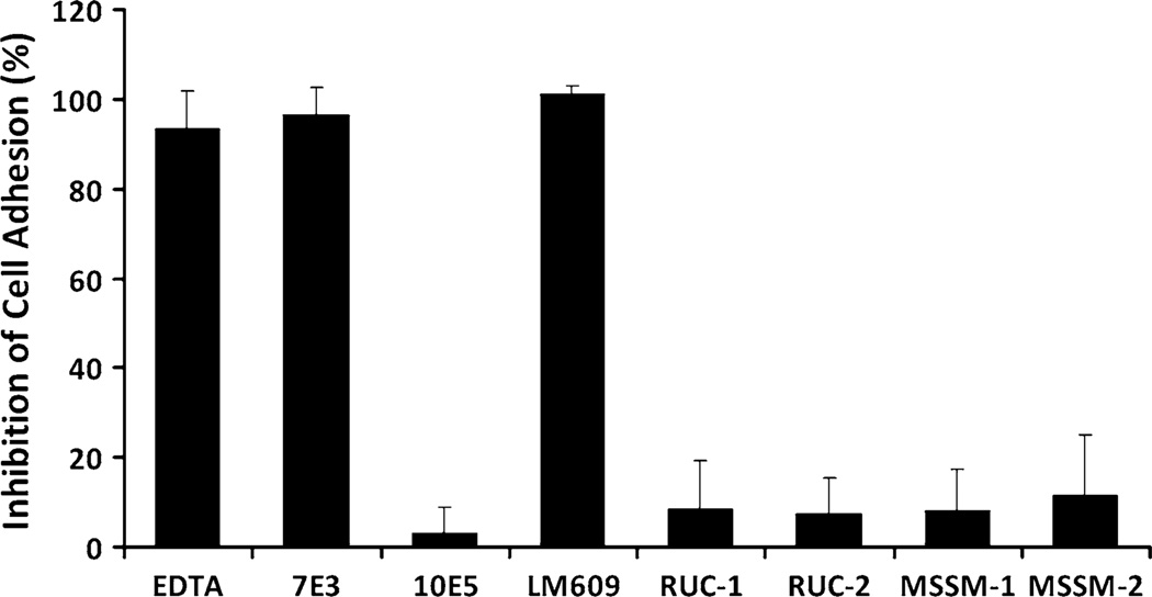
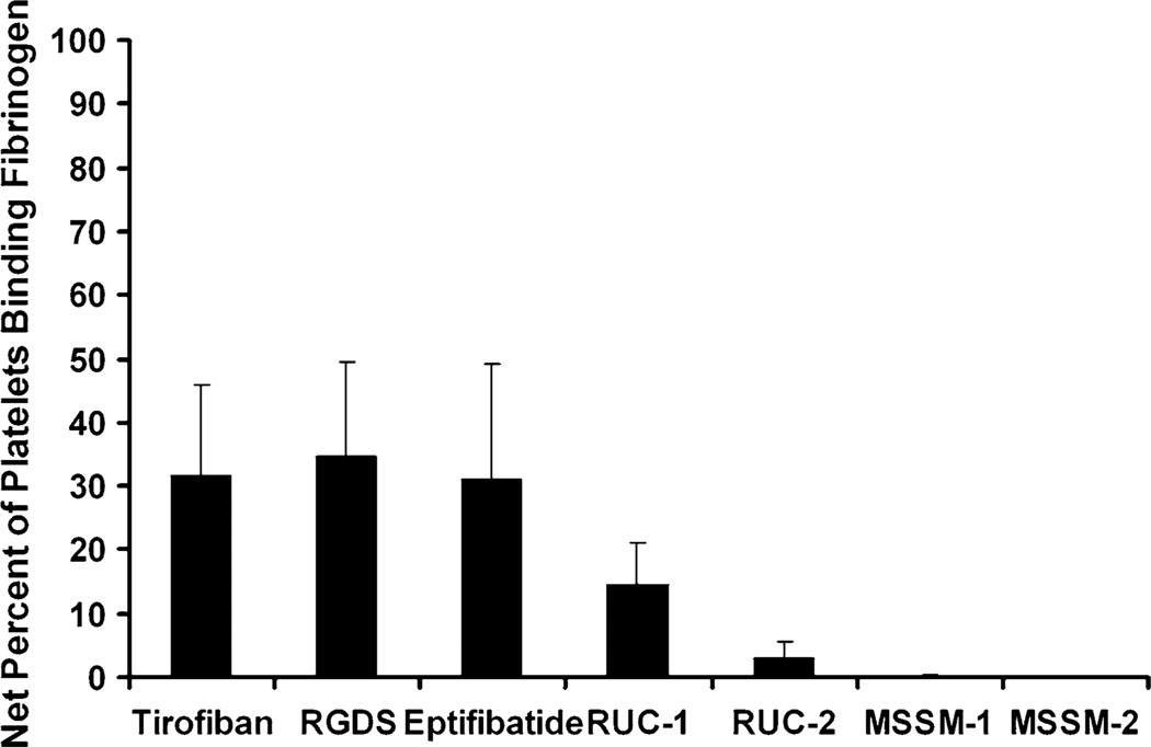
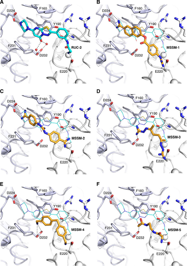
Integrin αIIbβ3 has emerged as an important therapeutic target for thrombotic vascular diseases owing to its pivotal role in mediating platelet aggregation through interaction with adhesive ligands. In the search for effective anti-thrombotic agents that can be administered orally without inducing the high-affinity ligand binding state, we recently discovered via high-throughput screening of 33,264 compounds a novel, αIIbβ3-selective inhibitor (RUC-1) of adenosine-5'-diphosphate (ADP) -induced platelet aggregation that exhibits a different chemical scaffold and mode of binding with respect to classical Arg-Gly-Asp (RGD)-mimicking αIIbβ3 antagonists. Most importantly, RUC-1 and its higher-affinity derivative, RUC-2, do not induce major conformational changes in the protein β3 subunit or prime the receptor to bind ligand. To identify additional αIIbβ3-selective chemotypes that inhibit platelet aggregation through similar mechanisms, we screened in silico over 2.5 million commercially available, 'lead-like' small molecules based on complementarity to the predicted binding mode of RUC-2 into the RUC-1-αIIbβ3 crystal structure. This first reported structure-based virtual screening application to the αIIbβ3 integrin led to the identification of 2 αIIbβ3-selective antagonists out of 4 tested, which compares favorably with the 0.003 % "hit rate" of our previous high-throughput chemical screening study. The newly identified compounds, like RUC-1 and RUC-2, showed specificity for αIIbβ3 compared to αVβ3 and did not prime the receptor to bind ligand. They thus may hold promise as αIIbβ3 antagonist therapeutic scaffolds.


Figures

Fig. 1
Fig. 1
Chemical structures of known antagonists co-crystallized with αIIbβ3 integrin
Fig. 2
Fig. 2
Crystallographic binding modes of antagonists tirofiban, L-739758, RUC-1, and RUC-2 (A, B, C and D, respectively). The αIIb and β3 subunits are shown as light blue and white cartoons, respectively. Side chains of protein residues interacting with the ligands are shown as sticks. ADMIDAS and SyMBS ions are shown as white spheres while the MIDAS ion is shown as a grey sphere. Ligands are shown as orange sticks, while black dot lines indicate hydrogen bonds
Fig. 3
Fig. 3
A workflow of the structure-based virtual screening approach applied to a model of the RUC-2/αIIbβ3 complex
Fig. 4
Fig. 4
Effect of RUC-1, RUC-2, MSSM-1 and MSSM-2 on adhesion of platelets to immobilized fibrinogen at different concentrations. Washed platelets in buffer containing 1 mM Ca2+ and 1 mM Mg2+ were allowed to adhere to microtiter wells precoated with purified fibrinogen (50 µg/ml) in the absence or presence of the indicated compounds. After 1 h at RT the wells were washed and the adherent platelets detected by lysing them with 0.1 % Triton X-100 and assaying the released acid phosphatase activity
Fig. 5
Fig. 5
Inhibition of ADP-induced platelet aggregation. Compounds were incubated at the indicated concentrations with citrated platelet-rich plasma for 15 min at 37 °C and then ADP (5 µM) was added and aggregation monitored by the change in light transmission. The initial slopes of platelet aggregation were measured and the IC50s determined by comparison with the untreated sample. Data shown are mean ± SD (n = 3), except for mAb 7E3, which is shown as the means of 2 separate experiments
Fig. 6
Fig. 6
Effect of EDTA, mAbs 7E3, 10E5, LM609, RUC-1, RUC-2, MSSM-1, and MSSM-2 on adhesion of HEK293 cells expressing αVβ3 to immobilized vitronectin. HEK293 cells expressing αVβ3 were added in buffer containing 1 mM Mg2+ to microtiter wells precoated with vitronectin (5 µg/ml). After 1 h at RT, the wells were washed and the adherent cells detected by lysing the cells with Triton X-100 and measuring released acid phosphatase activity
Fig. 7
Fig. 7
Priming effects of compounds. Tirofiban (0.5 µM), an RGDS peptide (100 µM), eptifibatide (1 µM), RUC-1 (100 µM), RUC-2 (1 µM), MSSM-1 (300 µM), or MSSM-2 (300 µM) were added to washed platelets and then the platelets were fixed with 1 % paraforrmaldeyde. After quenching the paraformaldehyde with glycine and washing, fluorescent fibrinogen (200 µg/ml) was added for 30 min at 37 °C and then, after washing again, bound fluorescent fibrinogen was detected by flow cytometry. The percentage of platelets with fluorescence values above 25 arbitrary units (AU) was recorded. The data shown is the percentage of platelets with values above 25 AU in the presence of each compound, minus the percentage in the untreated platelet sample. The latter averaged 4 ± 3 %
Fig. 8
Fig. 8
Predicted binding modes of the 5 ligands identified by the docking screens (B, C, D, E and F) compared to the RUC-2 binding pose (A). The αIIb subunit is shown as a light blue cartoon with the side chains of residues as sticks. The β3 subunit is shown in white cartoon with the side chain of E220 as sticks. ADMIDAS and SyMBS ions are shown as white spheres. The five ligands are shown as orange sticks and the co-crystallized antagonist RUC-2 is shown using cyan sticks (A) or cyan lines (B, C, D, E and F). Black dot lines indicate hydrogen bonds

References

    1. Lopez AD, Mathers CD, Ezzati M, Jamison DT, Murray CJL. Global burden of disease and risk factors, 2011/01/21 edn. Washington: World Bank; 2006. Measuring the global burden of disease and risk factors, 1990–2001. - PubMed
    1. Roger VL, Go AS, Lloyd-Jones DM, Adams RJ, Berry JD, Brown TM, Carnethon MR, Dai S, de Simone G, Ford ES, Fox CS, Fullerton HJ, Gillespie C, Greenlund KJ, Hailpern SM, Heit JA, Ho PM, Howard VJ, Kissela BM, Kittner SJ, Lackland DT, Lichtman JH, Lisabeth LD, Makuc DM, Marcus GM, Marelli A, Matchar DB, McDermott MM, Meigs JB, Moy CS, Mozaffarian D, Mussolino ME, Nichol G, Paynter NP, Rosamond WD, Sorlie PD, Stafford RS, Turan TN, Turner MB, Wong ND, Wylie-Rosett J. Heart disease and stroke statistics—2011 update: a report from the American Heart Association. Circulation. 2011;123(4):e18–e209. - PMC - PubMed
    1. Yang G, Kong L, Zhao W, Wan X, Zhai Y, Chen LC, Koplan JP. Emergence of chronic non-communicable diseases in China. Lancet. 2008;372(9650):1697–1705. - PubMed
    1. Hynes RO. Integrins: bidirectional, allosteric signaling machines. Cell. 2002;110(6):673–687. - PubMed
    1. Springer TA, Zhu J, Xiao T. Structural basis for distinctive recognition of fibrinogen gammaC peptide by the platelet integrin alphaIIbbeta3. J Cell Biol. 2008;182(4):791–800. - PMC - PubMed

Publication types

MeSH terms

Substances

LinkOut - more resources