. 2012 Dec;36(4):227-33.
doi: 10.1007/s11259-012-9535-9. Epub 2012 Aug 15.

Derivation of feline vaccine-associated fibrosarcoma cell line and its growth on chick embryo chorioallantoic membrane - a new in vivo model for veterinary oncological studies

Affiliations

Derivation of feline vaccine-associated fibrosarcoma cell line and its growth on chick embryo chorioallantoic membrane - a new in vivo model for veterinary oncological studies

K Zabielska et al. Vet Res Commun. 2012 Dec.

Abstract

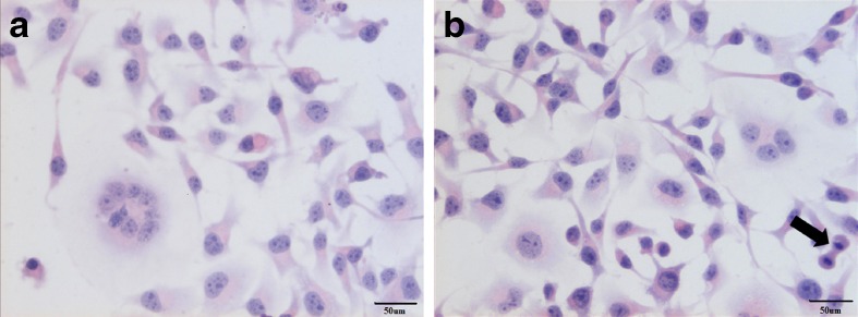
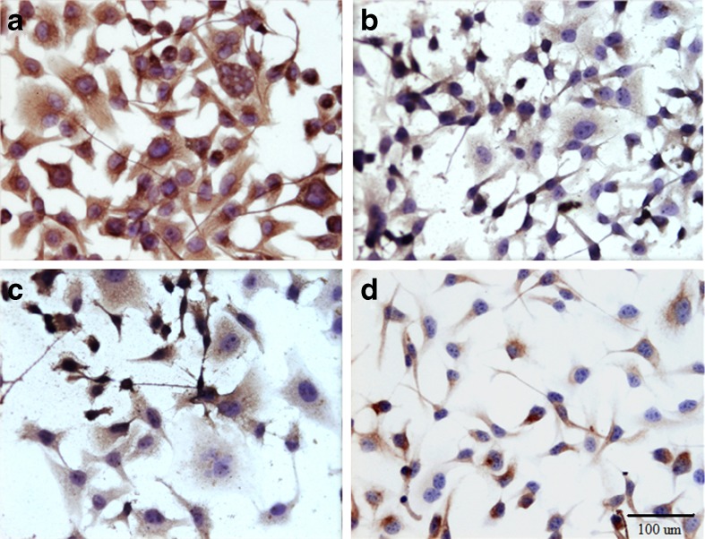
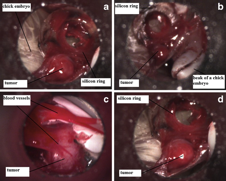
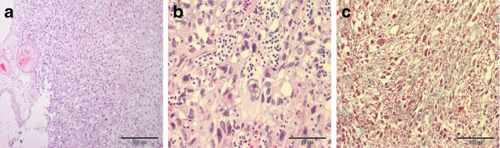
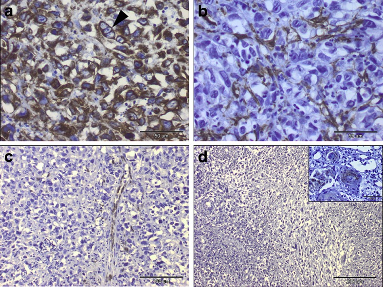
Feline vaccine associated fibrosarcomas are the second most common skin tumor in cats. Methods of treatment are: surgery, chemotherapy and radiotherapy. Nevertheless, the usage of cytostatics in feline vaccine associated sarcoma therapy is limited due to their adverse side effects, high toxicity and low biodistribution after i.v. injection. Therefore, much research on new therapeutic drugs is being conducted. In human medicine, the chick embryo chorioallantoic membrane (CAM) model is used as a cheap and easy to perform assay to assess new drug effectiveness in cancer treatment. Various human cell lines have different tumors growth on CAM. In veterinary medicine such model has not been described yet. In the present article derivation of feline vaccine associated fibrosarcoma cell line and its growth on CAM is described. The cell line and the tumor grown were confirmed by histopathological and immunohistochemical examination. As far as we believe, this is the first attempt to create such model, which may be used for further in vivo studies in veterinary oncology.


Figures

Fig. 1
Fig. 1
Feline vaccine-associated sarcoma cells stained with hematoxylin-eosin method. Multinucleated giant cells is visible on Fig. 1. original magnification 400×, and mitotic figure (black arrow)
Fig. 2
Fig. 2
Feline vaccine-associated sarcoma cells stained for a) vimentin, note a strong cytoplasmic staining reaction (brown color), original magnification 400×, b) smooth muscle actin, original magnification 400×, c) desmin, original magnification 400×, d) cytokeratin, original magnification 200×. Peroxidase-based EnVision™kit (DakoCytomation, Denmark) and haematoxylin counterstain
Fig. 3
Fig. 3
Feline fibrosarcoma on chick embryos on the 16th day of incubation (photographs taken by contractor, Welch Allyn MacroView™ Veterinary Otoscope 71032, USA)
Fig. 4
Fig. 4
a) Microscopic examination of undifferentiated sarcoma. The neoplastic cells were arranged in an irregular pattern. Haematoxylin and eosin stain, original magnification 100×. b) Sarcoma characterized by neoplastic cells with atypia, eosinophilic cytoplasm and unclear cell border, some with prominent eosinophilic nucleoli. Haematoxylin and eosin stain, original magnification 400×. c) Interstitial collagen fibres (green) separated neoplastic cells were very scant. Masson’s trichome stain, original magnification 200×
Fig. 5
Fig. 5
Immunohistochemical labelling of sarcoma: a. Intense vimentin-cytoplasmic immunostaining in neoplastic cells. Arrowhead showed a trinucleated neoplastic cell. Peroxidase-based EnVision™kit (DakoCytomation, Denmark) and hematoxylin counterstain, original magnification 400×. b. Cytoplasmic positivity of alpha-smooth muscle actin noted in some neoplastic cells (graph on the left) Peroxidase-based EnVision™kit (DakoCytomation, Denmark) and hematoxylin counterstain, original magnification 400×. c. Immunopositivity for desmin distributed among blood vessels. Peroxidase-based EnVision™kit (DakoCytomation, Denmark) and hematoxylin counterstain, original magnification 200×. d. No immunoreactivity with cytokeratin. Inset: Cytokeratin immune-staining were detected only in small foci of presumptive squamous epithelial cells within chorioallantois membrane of chicken embryo. Peroxidase-based EnVision™kit (DakoCytomation, Denmark) and hematoxylin counterstain, original magnification 100×, inset: 400×

Similar articles

Cited by

References

    1. Armstrong BP, Quigley JP, Sidebottom E. Transepithelial invasion and intramesenchymal infiltration of the chick embryo chorioallantoic membrane by tumor cell lines. J Biol Chem. 1982;42:1826–1837. - PubMed
    1. Balke M, Neumann A, Kersting C, Agelopoulos K, Geber C, Gosheger G, Buerger H, Hagedorn M. Morphologic characterization of osteosarcoma growth on the chick chorioallantoic membrane. BMC Res Notes. 2010;3:58. doi: 10.1186/1756-0500-3-58. - DOI - PMC - PubMed
    1. Brigger I, Dubernet C, Couvreur P. Nanoparticles in cancer therapy and diagnosis. Adv Drug Deliv Rev. 2002;54(5):631–651. doi: 10.1016/S0169-409X(02)00044-3. - DOI - PubMed
    1. Couto SS, Griffey SM, Duarte PC, Madawewll BR. Feline vaccine-associated fibrosarcoma: morphologic distinctions. Vet Pathol. 2002;39:33–41. doi: 10.1354/vp.39-1-33. - DOI - PubMed
    1. Dagg CP, Karnofsky DA, Roddy J. growth of transplantable human tumors in the chick embryo and hatched chick. Cancer Res. 1956;16(7):589–594. - PubMed