. 2012 Oct;10(10):1294-305.
doi: 10.1158/1541-7786.MCR-12-0275. Epub 2012 Aug 14.

Macrophages promote fibroblast growth factor receptor-driven tumor cell migration and invasion in a CXCR2-dependent manner

Affiliations

Macrophages promote fibroblast growth factor receptor-driven tumor cell migration and invasion in a CXCR2-dependent manner

Laura R Bohrer et al. Mol Cancer Res. 2012 Oct.

Abstract

Infiltration of immune cells, specifically macrophages, into the tumor microenvironment has been linked to increased mammary tumor formation and progression. Activation of growth factor receptor signaling pathways within mammary epithelial cells, such as the fibroblast growth factor receptor 1 (FGFR1) pathway, induces recruitment of macrophages to the mammary epithelium. These macrophages promote increased epithelial cell proliferation and angiogenesis. However, the specific mechanisms by which these macrophages are regulated by the preneoplastic epithelial cells and the mechanisms of action of the macrophages within the developing FGFR1-driven tumor microenvironment remain unknown. In this study, we show that activation of inducible FGFR1 in mammary glands leads to decreased activity of the TGFβ/Smad3 pathway in macrophages associated with early stage lesions. Further studies show that macrophages have increased expression of inflammatory chemokines that bind Cxcr2 following exposure to conditioned media from mammary epithelial and tumor cells in which the FGF pathway had been activated. The increase in these ligands is inhibited following activation of the TGFβ pathway, suggesting that decreased TGFβ signaling contributes to the upregulation of these chemokines. Using coculture studies, we further show that macrophages are capable of promoting epithelial and tumor cell migration and invasion through activation of Cxcr2. These results indicate that macrophage-derived Cxcr2 ligands may be important for promoting mammary tumor formation regulated by FGFR signaling. Furthermore, these results suggest that targeting Cxcr2 may represent a novel therapeutic strategy for breast cancers that are associated with high levels of infiltrating macrophages.


Figures

Figure 1
Figure 1. Exposure to conditioned media from mammary epithelial cells with activated iFGFR1 leads to regulation of inflammation-associated genes in vitro
RAW 264.7 cells were exposed to conditioned media from B/B-treated HC-11/R1 cells for 2 hours and either cells were collected for gene expression or serum free media was added for 24 hours to examine secreted protein expression or activity. A) qRT-PCR analysis of ArgI gene expression levels (left panel) and arginase activity (right panel). B) qRT-PCR analysis of IL-10 gene expression levels (left panel) and secreted protein level by ELISA (right panel). C) qRT-PCR analysis of IL-12 gene expression levels (left panel) and secreted protein level by ELISA (right panel). D) qRTPCR analysis of TGFβ1 gene expression levels (left panel) and Smad3 transcriptional activity by luciferase assay (right panel). Expression levels were normalized to cyclophilin for qRT-PCR. Error bars represent standard error of the mean (SEM). *P<0.05, **P<0.005, ***P<0.001.
Figure 2
Figure 2. Alterations in the TGFβ gene expression and Smad3 activity in macrophages isolated from mammary glands following FGFR1 activation
A) Cd11b-positive sorted cells were analyzed for expression of the indicated genes using RT-PCR. The lanes represent macrophages sorted from three MMTV-iFGFR1 transgenic (Tg) or three non-transgenic wild-type (WT) littermates and pooled. B) Expression of TGFβ1 in macrophages isolated from MMTV-iFGFR1 transgenic mice or non-transgenic wild-type littermates treated with B/B for 48 hours. Expression levels were normalized to cyclophilin. C) Non-transgenic (i,ii) and transgenic (iii,iv) mice were treated with B/B for 48 hours and mammary glands were collected. Sections were co-stained with the F4/80 and phospho-Smad3 antibodies. F4/80 positive cells (red) are found in the stroma surrounding the epithelial structures (blue=DAPI staining of nuclei). Phospho-Smad3 (green) is detected in nuclei of F4/80 positive cells in sections from the non-transgenic (arrow, ii) but not transgenic (arrow, iv), mice. Scale bars=50 μm D) Quantification of the percentage of F4/80+/phospho-Smad3+ cells. Error bars represent SEM. *P<0.05, ***P<0.0001.
Figure 3
Figure 3. Activation of iFGFR1 in mammary epithelial cells leads to increased expression of Cxcr2-binding chemokines in macrophages
A) HC-11 cells were treated with R1/RAW-CM with or without B/B. Whole cell lysates were collected and the expression of Cxcr2 and β-tubulin (loading control) was analyzed by immunoblotting. B) RAW 264.7 cells were treated with R1-CM in the presence or absence of 10 ng/ml TGFβ for 2 hours. Expression of the given genes was determined by qRT-PCR and normalized to cyclophilin (left panel). For protein expression, RAW 264.7 cells were incubated with R1-CM for 2 hours followed by serum free media for 24 hours. The conditioned media samples were analyzed using ELISAs (middle and right panel). C) RAW 264.7 cells were treated with R1-CM for the given time. Whole cell lysates were collected and the expression of pERK1/2 and ERK1/2 (loading control) was analyzed by immunoblotting. D) RAW 264.7 cells were pre-treated with DMSO (solvent control) or U0126 for 30 min. and then treated for an additional 2 hours with the same conditions in the presence of B/B R1-CM. The expression of the given genes was analyzed by qRT-PCR and normalized to cyclophilin. Error bars represent SEM. *P<0.05, **P<0.01, ***P<0.001
Figure 4
Figure 4. Cxcr2 inhibition leads to decreased macrophage-induced migration of HC-11 cells
A) Model of the migration co-culture assay. B) R1-CM, -/+ B/B, was exposed to RAW 264.7 cells for 24 hours and the conditioned media, R1/RAW-CM was used as the chemoattractant for a transwell migration assay. HC-11 cells were plated on top of the insert with ethanol (solvent control) or CXCR2 inhibitor (+ 20 nM and ++ 200nM). After 18 hours, the migrated cells on the bottom of the insert were stained with hematoxylin and counted. Error bars represent SEM. ***P<0.001.
Figure 5
Figure 5. Macrophages induce invasion of primary mammary epithelial cells grown in 3D culture
A) Primary MECs from iFGFR1 mice were grown in Matrigel in the absence or presence of BMDM. Every 2-3 days fresh media was added with the treatments: -/+ 30 nM B/B and -/+ 200 nM CXCR2 inhibitor. After 10 days of treatment, cells were fixed and immunostained with cytokeratin 8 (green) and F4/80 (red). DAPI (blue) labeled nuclei. Arrows indicate invasive structures. Left images light microscopy Scale bars=50 μm. B) For each treatment, approximately 80 structures were examined for invasion from 3 independent experiments. Error bars represent SEM. *P<0.05, **P<0.01
Figure 6
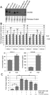
Figure 6. The migratory ability of MCF-7 cells increases with macrophage secreted CXCR2 ligands
A) Whole cell lysates were collected from the different cell lines and equal amounts were used for immunoblot analysis. The expression of CXCR2 was determined and the membrane was stained with Ponceau S for a loading control. B) THP-1 cells were exposed to conditioned media from MCF-7 cells treated with 50 ng/ml bFGF or no treatment (NT). Cells were collected after 2 or 4 hours, and RNA was isolated for qRT-PCR for the given genes that were normalized to cyclophilin (upper panel). For protein expression, THP-1 cells were incubated with MCF-CM for 2 hours and then serum free media for 24 hours. The conditioned media was analyzed using ELISAs (lower panel). C) MCF-CM, -/+ bFGF, was exposed to THP-1 cells for 24 hours, and the conditioned media, MCF/THP-CM was used as the chemoattractant for a transwell migration assay. MCF-7 cells were plated on top of the insert with ethanol (solvent control) or CXCR2 inhibitor (+ 100 nM, ++ 200 nM, and +++ 400 nM). After 18 hours, the migrated cells on the bottom of the insert were stained with hematoxylin and counted. Error bars represent SEM. *P<0.05, **P<0.01, ***P<0.001.

Similar articles

Cited by

References

    1. de Visser KE, Eichten A, Coussens LM. Paradoxical roles of the immune system during cancer development. Nat Rev Cancer. 2006;6(1):24–37. - PubMed
    1. Bingle L, Brown NJ, Lewis CE. The role of tumour-associated macrophages in tumour progression: implications for new anticancer therapies. J Pathol. 2002;196(3):254–65. - PubMed
    1. Pollard JW. Tumour-educated macrophages promote tumour progression and metastasis. Nat Rev Cancer. 2004;4(1):71–8. - PubMed
    1. Qian BZ, Pollard JW. Macrophage diversity enhances tumor progression and metastasis. Cell. 2010;141(1):39–51. - PMC - PubMed
    1. DeNardo DG, Barreto JB, Andreu P, Vasquez L, Tawfik D, Kolhatkar N, et al. CD4(+) T cells regulate pulmonary metastasis of mammary carcinomas by enhancing protumor properties of macrophages. Cancer Cell. 2009;16(2):91–102. - PMC - PubMed

Publication types

MeSH terms