

The structure and function relationship in glaucoma: implications for detection of progression and measurement of rates of change
- PMID: 22893677
- PMCID: PMC3466074
- DOI: 10.1167/iovs.12-10345
The structure and function relationship in glaucoma: implications for detection of progression and measurement of rates of change
Abstract
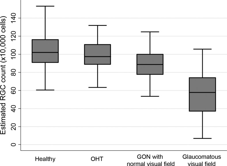
Purpose: To evaluate the relationship between change in estimated retinal ganglion cell (RGC) counts and change in measures of functional and structural damage in glaucoma, from cross-sectional data.
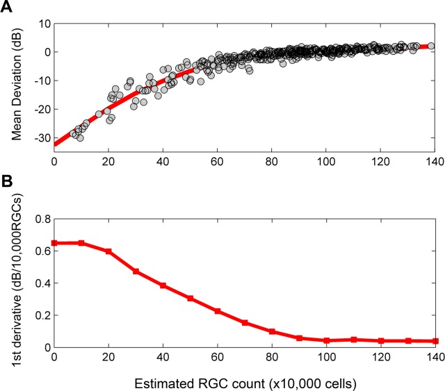
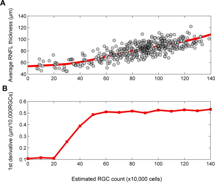
Methods: The study included 397 eyes of 397 patients with glaucoma, suspects, and healthy individuals. All eyes underwent testing with standard automated perimetry (SAP) and spectral-domain optical coherence tomography (SD-OCT). Estimates of retinal ganglion cell (RGC) counts were obtained from SAP and SD-OCT using a previously derived algorithm. Smoothing spline curves were fitted to investigate the relationship between functional/structural parameters and RGC counts. The first derivatives (i.e., slopes) of these curves were obtained to investigate the relationship between changes in these measures.
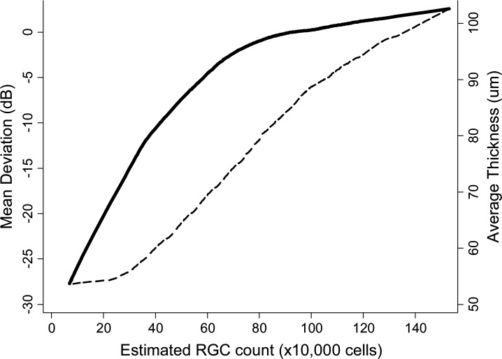
Results: A nonlinear relationship was observed between SAP mean deviation (MD) and RGC counts. The same amount of RGC loss corresponded to largely different amounts of MD change depending on the stage of the disease. For SDOCT average retinal nerve fiber layer (RNFL) thickness, a linear relationship was seen with RGC counts throughout most of the spectrum of disease, but reaching a plateau in advanced glaucoma. Changes in RGC counts for eyes with early damage corresponded to small changes in MD, but to relatively larger changes in RNFL thickness. For eyes with advanced disease, changes in RGC counts produced relatively larger changes in MD but only small or no changes in average RNFL thickness.
Conclusions: The analysis and interpretation of rates of SAP and SD-OCT change, as indicators of the velocity of neural damage in glaucoma, should take into account the severity of the disease.
Conflict of interest statement
Disclosure:
Figures
References
-
- Medeiros FA, Susanna R Jr, Singh K. Who should be treated? In: Weinreb RN, Liebmann J.eds Medical Treatment of Glaucoma. The Hague, The Netherlands: Kugler Publications; 2010:1–15
-
- Caprioli J. The importance of rates in glaucoma. Am J Ophthalmol. 2008;145:191–192 - PubMed
-
- Bengtsson B, Heijl A. A visual field index for calculation of glaucoma rate of progression. Am J Ophthalmol. 2008;145:343–353 - PubMed
-
- Artes PH, Nicolela MT, LeBlanc RP, Chauhan BC. Visual field progression in glaucoma: total versus pattern deviation analyses. Invest Ophthalmol Vis Sci. 2005;46:4600–4606 - PubMed
-
- Lee YH, Kim CS, Hong SP. Rate of visual field progression in primary open-angle glaucoma and primary angle-closure glaucoma. Korean J Ophthalmol. 2004;18:106–115 - PubMed
Publication types
MeSH terms
Grants and funding
LinkOut - more resources
Full Text Sources
Other Literature Sources
Medical
Miscellaneous
