. 2012 Aug 16:13:73.
doi: 10.1186/1471-2350-13-73.

Genetic characterization in symptomatic female DMD carriers: lack of relationship between X-inactivation, transcriptional DMD allele balancing and phenotype

Affiliations

Genetic characterization in symptomatic female DMD carriers: lack of relationship between X-inactivation, transcriptional DMD allele balancing and phenotype

Simona Brioschi et al. BMC Med Genet. .

Abstract

Background: Although Duchenne and Becker muscular dystrophies, X-linked recessive myopathies, predominantly affect males, a clinically significant proportion of females manifesting symptoms have also been reported. They represent an heterogeneous group characterized by variable degrees of muscle weakness and/or cardiac involvement. Though preferential inactivation of the normal X chromosome has long been considered the principal mechanism behind disease manifestation in these females, supporting evidence is controversial.

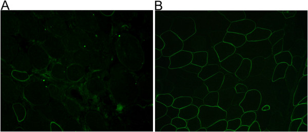
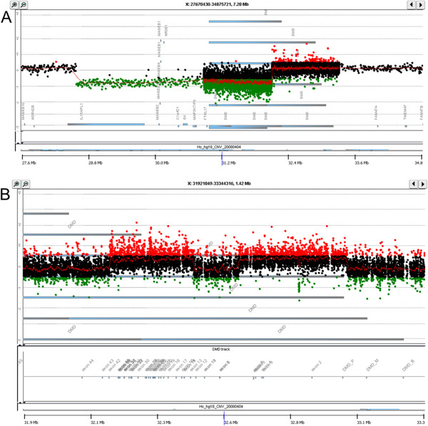
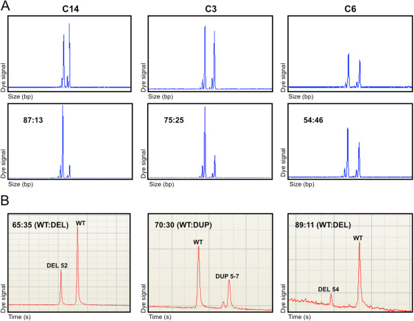
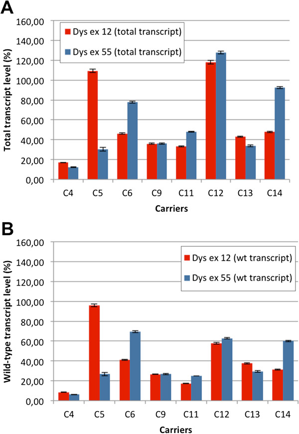
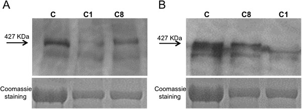
Methods: Eighteen females showing a mosaic pattern of dystrophin expression on muscle biopsy were recruited and classified as symptomatic (7) or asymptomatic (11), based on the presence or absence of muscle weakness. The causative DMD gene mutations were identified in all cases, and the X-inactivation pattern was assessed in muscle DNA. Transcriptional analysis in muscles was performed in all females, and relative quantification of wild-type and mutated transcripts was also performed in 9 carriers. Dystrophin protein was quantified by immunoblotting in 2 females.

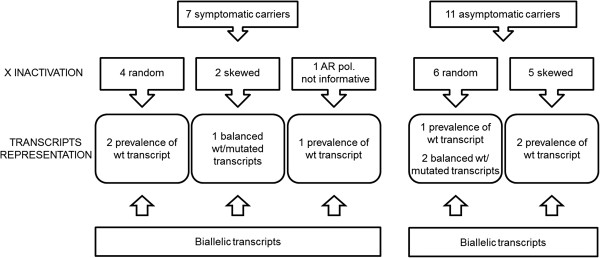
Results: The study highlighted a lack of relationship between dystrophic phenotype and X-inactivation pattern in females; skewed X-inactivation was found in 2 out of 6 symptomatic carriers and in 5 out of 11 asymptomatic carriers. All females were characterized by biallelic transcription, but no association was found between X-inactivation pattern and allele transcriptional balancing. Either a prevalence of wild-type transcript or equal proportions of wild-type and mutated RNAs was observed in both symptomatic and asymptomatic females. Moreover, very similar levels of total and wild-type transcripts were identified in the two groups of carriers.

Conclusions: This is the first study deeply exploring the DMD transcriptional behaviour in a cohort of female carriers. Notably, no relationship between X-inactivation pattern and transcriptional behaviour of DMD gene was observed, suggesting that the two mechanisms are regulated independently. Moreover, neither the total DMD transcript level, nor the relative proportion of the wild-type transcript do correlate with the symptomatic phenotype.


Figures

Figure 1
Figure 1
Immunohistochemical staining of muscle sections from carriers 1 and 2. A) Immunolabeling in symptomatic carrier 1 with NCL-DYS3 antibody (NH2-terminal) shows absence of staining in the majority of muscle fibers (approximately 80% in the whole section). B) Immunolabeling in symptomatic carrier 2 with DYS2 antibody (COOH-terminal) shows a mosaic pattern of dystrophin expression with coexistence of positive fibers, fibers with reduced or discontinuous staining and fibers with absence of labeling.
Figure 2
Figure 2
DMD-CGH array profile in carriers 16 and 8. A) In carrier 16, a deletion of 3.5 Mb was detected (chrX:g.(28.671.682_28.671.742)_(32.170.481_32.170.541)del). The 5’ breakpoint is located within intron 43 of the DMD gene and deletion covers the following downstream genes: FTHL17, MAP3K7IP3, GK, CXorf21, NR0B1, MAGEB4, MAGEB3, MAGEB2, MAGEB1, IL1RAPL1. B) In carrier 8, CGH analysis confirmed a non-contiguous duplication involving exons 1P-7 (chrX:g.(33.068.711_33.068.771)_(32.684.693_32.684.750)dup) and exons 13–42 (chrX:g.(32.523.766_32.523.826)_(32.228.415_32.228.475)dup). A polymorphic CNV is also visible within intron 2.
Figure 3
Figure 3
Comparison between X-inactivation pattern and transcript quantification. A) X-inactivation assay based on AR gene methylation. The upper line corresponds to PCR products of undigested muscle DNA, showing the two alleles at AR locus; the lower shows the products of DNA amplification after digestion with the methylation-sensitive enzymes HpaII and CfoI. B) RT-PCR analysis with a High Sensitivity DNA chip (Agilent). The ratios between wild-type and mutated transcripts are indicated. Carrier 14 is an asymptomatic female with a moderately skewed X-inactivation pattern of 87:13 and a transcript ratio of 65:35 (wt:deleted); symptomatic carrier 3 presented X-inactivation of 75:25 and a transcript ratio of 70:30 (wt:duplicated); carrier 6 is a symptomatic female featuring random X-inactivation of 54:46 and transcript ratio of 89:11 (wt:deleted).
Figure 4
Figure 4
Schematic representation of results from X-inactivation and transcriptional analysis. This schematic summarizes the results of X-inactivation studies and transcript quantification in the two groups of symptomatic and asymptomatic carriers, and highlights the absence of a relationship between X-inactivation pattern, transcriptional behaviour and dystrophic phenotype in female carriers.
Figure 5
Figure 5
Relative quantification of dystrophin transcript by real-time PCR.A) Histograms represent total transcript level of dystrophin gene in 8 female carriers, calculated on dystrophin exon 12 and exon 55, in respect to two control females (their mean value referred as 100%). Error bars represent mean ± SD. B) Transcriptional level of wild-type dystrophin allele, calculated from the relative ratio of wild-type and mutated transcripts.
Figure 6
Figure 6
Immunoblotting in carriers 1 and 8 and Coomassie staining for protein quantification by densitometric analysis. A) Immunoblot with DYS2 antibody (directed against carboxy terminal region of dystrophin) shows protein levels of 15% for carrier 1 and 70% for carrier 8, with respect to control. B) Immunoblot with DYS1 antibody (directed against rod domain) reveals 11% and 87% of normal protein levels for carriers 1 and 8, respectively.

Similar articles

Cited by

References

    1. Moser H, Emery AE. The manifesting carrier in Duchenne muscular dystrophy. Clin Genet. 1974;5(4):271–284. - PubMed
    1. Norman A, Harper P. A survey of manifesting carriers of Duchenne and Becker muscular dystrophy in Wales. Clin Genet. 1989;36(1):31–37. - PubMed
    1. Comi LI, Nigro G, Politano L, Petretta VR. The cardiomyopathy of Duchenne/Becker consultands. Int J Cardiol. 1992;34(3):297–305. doi: 10.1016/0167-5273(92)90028-2. - DOI - PubMed
    1. Kinoshita H, Goto Y, Ishikawa M, Uemura T, Matsumoto K, Hayashi YK, Arahata K, Nonaka I. A carrier of Duchenne muscular dystrophy with dilated cardiomyopathy but no skeletal muscle symptom. Brain Dev. 1995;17(3):202–205. doi: 10.1016/0387-7604(95)00018-7. - DOI - PubMed
    1. Hoogerwaard EM, Bakker E, Ippel PF, Oosterwijk JC, Majoor-Krakauer DF, Leschot NJ, Van Essen AJ, Brunner HG, van der Wouw PA, Wilde AA, de Visser M. Signs and symptoms of Duchenne muscular dystrophy and Becker muscular dystrophy among carriers in the Netherlands: a cohort study. Lancet. 1999;353:2116–2119. doi: 10.1016/S0140-6736(98)10028-4. - DOI - PubMed

Publication types

MeSH terms