

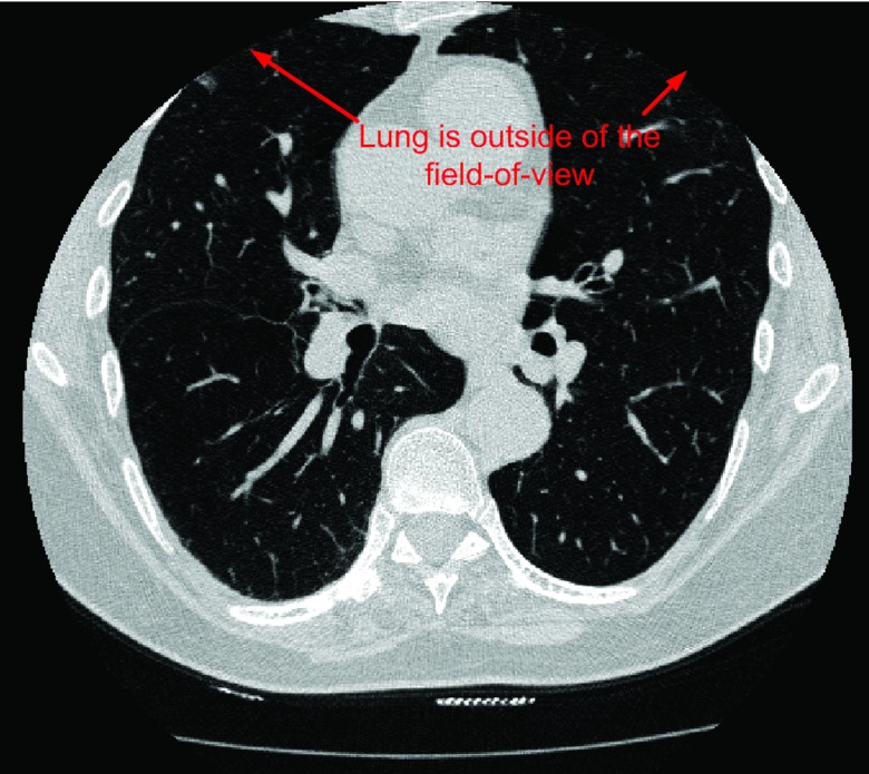
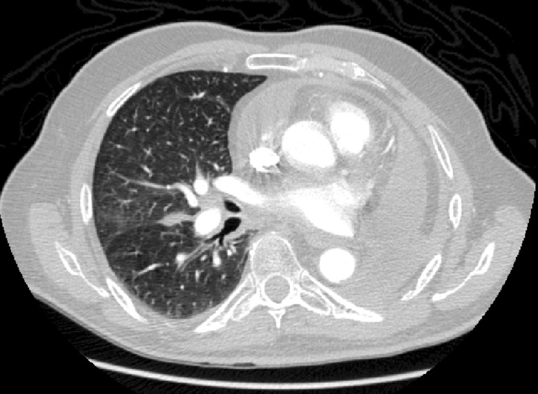
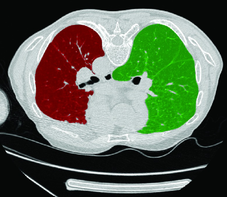
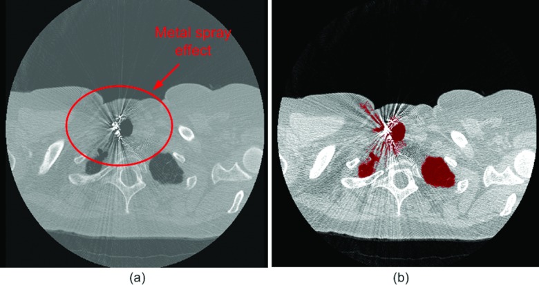
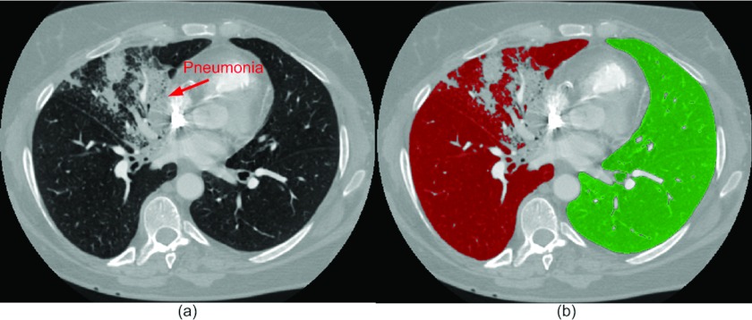
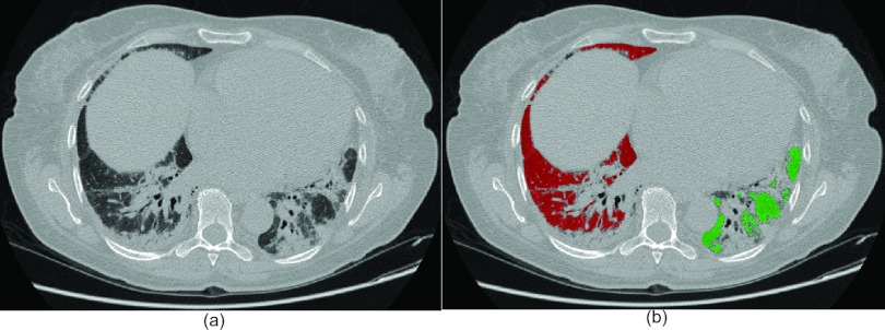
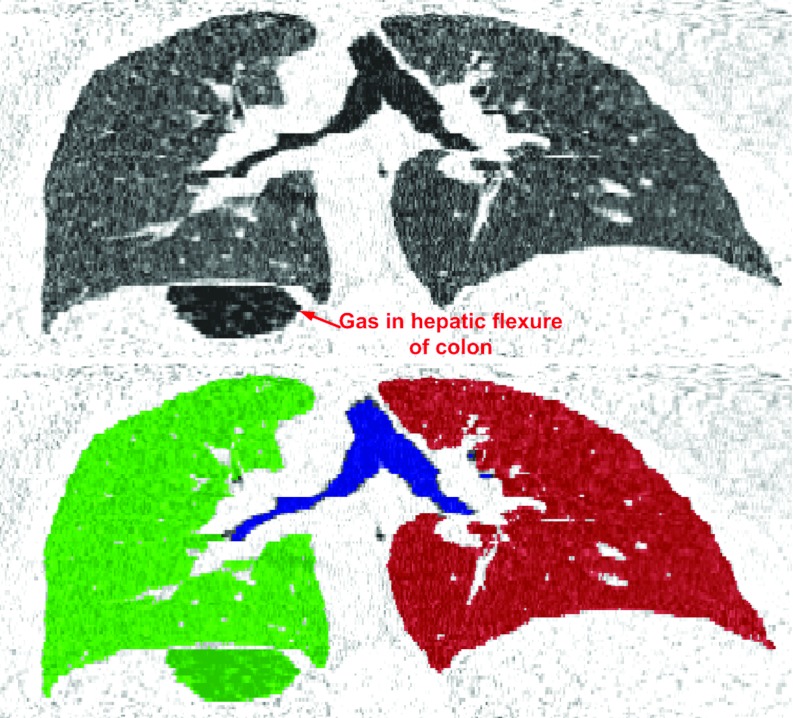
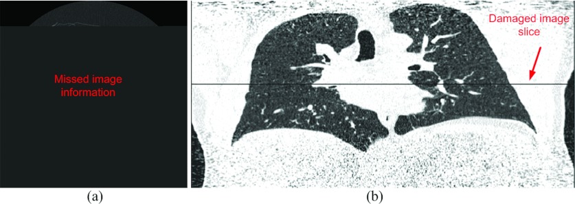
Illustration of the obstacles in computerized lung segmentation using examples
- PMID: 22894423
- PMCID: PMC3416879
- DOI: 10.1118/1.4737023
Illustration of the obstacles in computerized lung segmentation using examples
Abstract
Purpose: Automated lung volume segmentation is often a preprocessing step in quantitative lung computed tomography (CT) image analysis. The objective of this study is to identify the obstacles in computerized lung volume segmentation and illustrate those explicitly using real examples. Awareness of these "difficult" cases may be helpful for the development of a robust and consistent lung segmentation algorithm.
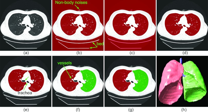
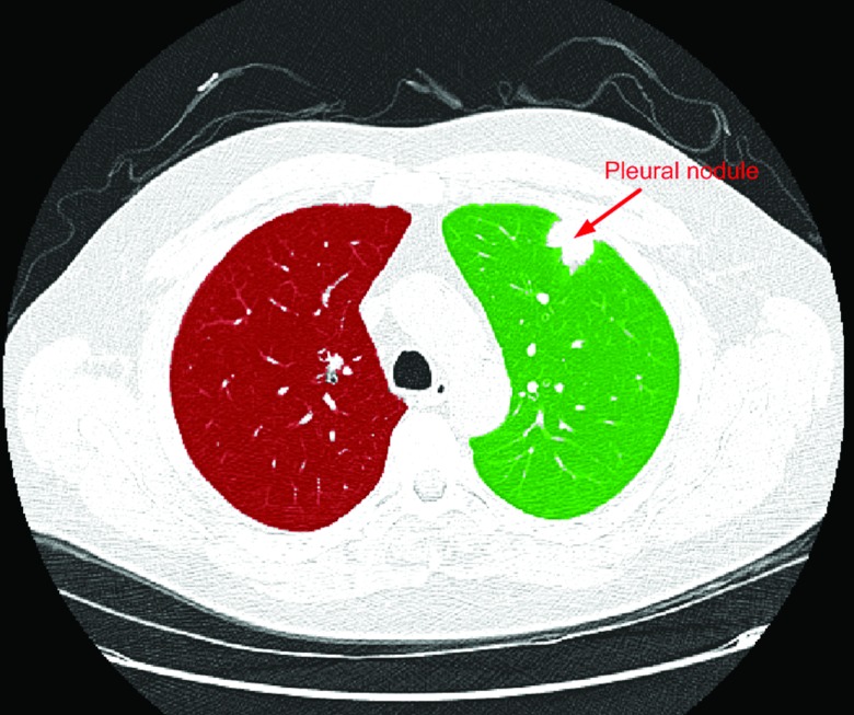
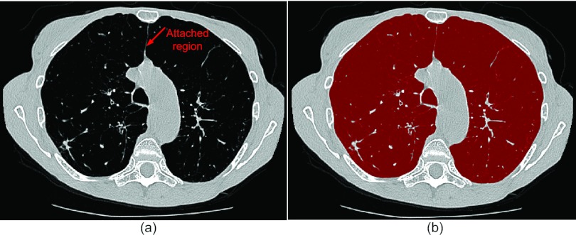
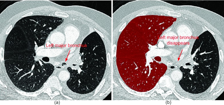
Methods: We collected a large diverse dataset consisting of 2768 chest CT examinations acquired on 2292 subjects from various sources. These examinations cover a wide range of diseases, including lung cancer, chronic obstructive pulmonary disease, human immunodeficiency virus, pulmonary embolism, pneumonia, asthma, and interstitial lung disease (ILD). The CT acquisition protocols, including dose, scanners, and reconstruction kernels, vary significantly. After the application of a "neutral" thresholding-based approach to the collected CT examinations in a batch manner, the failed cases were subjectively identified and classified into different subgroups.
Results: Totally, 121 failed examinations are identified, corresponding to a failure ratio of 4.4%. These failed cases are summarized as 11 different subgroups, which is further classified into 3 broad categories: (1) failure caused by diseases, (2) failure caused by anatomy variability, and (3) failure caused by external factors. The failure percentages in these categories are 62.0%, 32.2%, and 5.8%, respectively.
Conclusions: The presence of specific lung diseases (e.g., pulmonary nodules, ILD, and pneumonia) is the primary issue in computerized lung segmentation. The segmentation failures caused by external factors and anatomy variety are relatively low but unavoidable in practice. It is desirable to develop robust schemes to handle these issues in a single pass when a large number of CT examinations need to be analyzed.
Figures
References
-
- Ochsmann E., Carl T., Brand P., Raithel H. J., and Kraus T., “Inter-reader variability in chest radiography and HRCT for the early detection of asbestos-related lung and pleural abnormalities in a cohort of 636 asbestos-exposed subjects,” Int. Arch. Occup. Environ. Health 83(1), 39–46 (2010). 10.1007/s00420-009-0443-4 - DOI - PubMed
