Review
. 2012 Jul;17(7):071311.
doi: 10.1117/1.JBO.17.7.071311.

Diffuse optical imaging using spatially and temporally modulated light

Affiliations
Review

Diffuse optical imaging using spatially and temporally modulated light

Thomas D O'Sullivan et al. J Biomed Opt. 2012 Jul.

Abstract

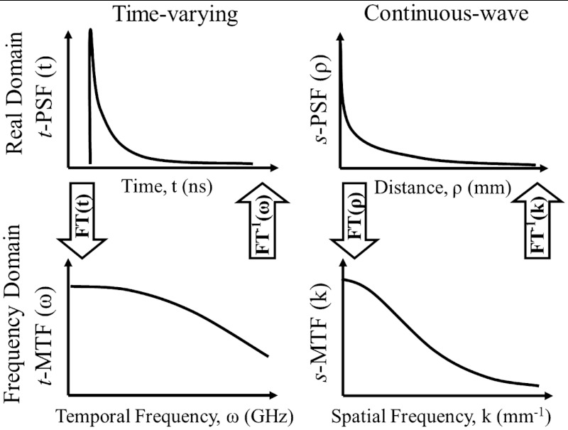
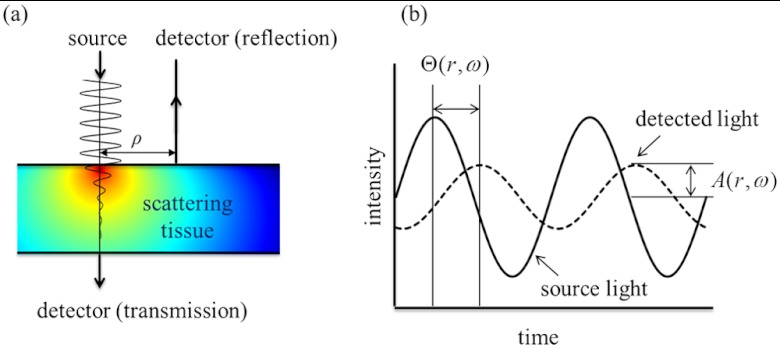
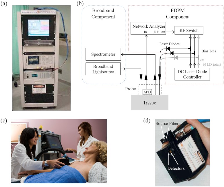
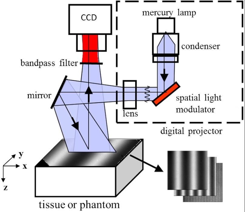
The authors describe the development of diffuse optical imaging (DOI) technologies, specifically the use of spatial and temporal modulation to control near infrared light propagation in thick tissues. We present theory and methods of DOI focusing on model-based techniques for quantitative, in vivo measurements of endogenous tissue absorption and scattering properties. We specifically emphasize the common conceptual framework of the scalar photon density wave for both temporal and spatial frequency-domain approaches. After presenting the history, theoretical foundation, and instrumentation related to these methods, we provide a brief review of clinical and preclinical applications from our research as well as our outlook on the future of DOI technology.


Figures

Fig. 1
Fig. 1
Four measurement domains of DOI: time domain (top left), temporal frequency domain (bottom left), real spatial domain (top right), and spatial frequency domain (bottom right). Figure reproduced with permission from Ref. .
Fig. 2
Fig. 2
Measurement of optical properties with FDPM; (a) A temporally modulated light source creates PDWs, which are attenuated in the tissue. The attenuated light can be detected in either reflection or transmission geometries. (b) The detected signal is diminished in amplitude and delayed in phase with respect to the source.
Fig. 3
Fig. 3
Frequency domain photon migration instrumentation; (a) A clinical DOSI instrument, (b) a block diagram of the broadband DOSI instrument, (c) a subject measurement with the DOSI instrument, and (d) the handpiece placed in contact with the tissue.
Fig. 4
Fig. 4
An SFDI Instrument, reproduced with permission from Ref. .
Fig. 5
Fig. 5
Data flow and processing steps for SFDI image creation. (a) Multiple frequencies (fx) are projected onto the sample, and remitted intensity I is captured. (b) Each illumination frequency is imaged at three phases, then demodulated and calibrated to yield Rd. (c) The Rd at each pixel is fit for and using a Monte Carlo light-transport model and lookup table to yield optical property maps. Reproduced with permission from Ref. .
Fig. 6
Fig. 6
Illustration of how tissue acts like a low-pass filter to spatially modulated illumination. Patterns with lower spatial frequencies propagate deeper into tissue.
Fig. 7
Fig. 7
DOSI images of chromophore values of a 65-year-old subject with carcinoma of the right breast, measuring 19×20×32  mm by ultrasound. The approximate tumor location is indicated by the black circle, confirmed by palpation. Measurements are taken in a grid pattern every 10 mm and interpolated to create these image maps.
Fig. 8
Fig. 8
Longitudinal DOSI measurements of a breast carcinoma during neoadjuvant chemotherapy (NAC) treatment. After surgery, this patient was confirmed to have no evidence of residual cancer and achieved a pathologic complete response (pCR). The inset value indicates the tumor to normal optical contrast in TOI.
Fig. 9
Fig. 9
Representative SFDI images (6×6  mm) of the barrel cortex of a rat undergoing a middle cerebral artery occlusion (MCAo), a model of ischemic stroke. (a) Chromophore maps pre and postMCAo. (b) Time course of cerebral hemodynamics from the barrel cortex as analyzed from these images. The error bars give the standard error of all pixels in a region of interest. Reproduced with permission from Ref. .
Fig. 10
Fig. 10
Representative optical property maps of the brain in a mouse model of Alzheimer’s disease taken with SFDI in vivo through intact skull. Pixel values in the ROI were averaged for each mouse and wavelength-dependent reduced scattering and absorption coefficient spectra were plotted for control and transgenic mice. Reproduced with permission from Ref. .
Fig. 11
Fig. 11
SFDI images of StO2 and corresponding digital images of a rat model of flap healing. The baseline image is shown at T=0, which was followed by 2 h of ischemia and two additional hours of treatment. The flap on the left in each image represents the control flap that was treated with normal saline after 2 h of ischemia and ultimately survived. The right flap in each image represents the experimental flap that was treated with hypertonic-hyperoncotic saline after 2 h of ischemia and eventually became necrotic within 24 to 48 h. This figure was reproduced with permission from data originally published in Ref. .

Similar articles

Cited by

References

    1. Jacques S. L., Pogue B. W., “Tutorial on diffuse light transport,” J. Biomed. Opt. 13(4), 041302 (2008).JBOPFO10.1117/1.2967535 - DOI - PubMed
    1. Elwell C. E., Cooper C. E., “Making light work: illuminating the future of biomedical optics,” Philos. T. Roy. Soc. A 369(1955), 4358–4379 (2011).PTRMAD10.1098/rsta.2011.0302 - DOI - PubMed
    1. Patterson M. S., Chance B., Wilson B. C., “Time resolved reflectance and transmittance for the non-invasive measurement of tissue optical properties,” Appl. Opt. 28(12), 2331–2336 (1989).APOPAI10.1364/AO.28.002331 - DOI - PubMed
    1. Cuccia D. J., et al. , “Quantitation and mapping of tissue optical properties using modulated imaging,” J. Biomed. Opt. 14(2), 024012 (2009).JBOPFO10.1117/1.3088140 - DOI - PMC - PubMed
    1. Arridge S. R., “Optical tomography in medical imaging,” Inverse Probl. 15(2), R41–R93 (1999).INPEEY10.1088/0266-5611/15/2/022 - DOI

Publication types

MeSH terms