

Analysis of second-harmonic-generation microscopy in a mouse model of ovarian carcinoma
- PMID: 22894485
- PMCID: PMC3389559
- DOI: 10.1117/1.JBO.17.7.076002
Analysis of second-harmonic-generation microscopy in a mouse model of ovarian carcinoma
Abstract
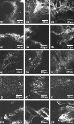
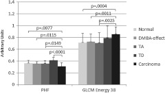
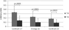
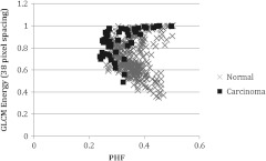
Second-harmonic-generation (SHG) imaging of mouse ovaries ex vivo was used to detect collagen structure changes accompanying ovarian cancer development. Dosing with 4-vinylcyclohexene diepoxide and 7,12-dimethylbenz[a]anthracene resulted in histologically confirmed cases of normal, benign abnormality, dysplasia, and carcinoma. Parameters for each SHG image were calculated using the Fourier transform matrix and gray-level co-occurrence matrix (GLCM). Cancer versus normal and cancer versus all other diagnoses showed the greatest separation using the parameters derived from power in the highest-frequency region and GLCM energy. Mixed effects models showed that these parameters were significantly different between cancer and normal (P<0.008). Images were classified with a support vector machine, using 25% of the data for training and 75% for testing. Utilizing all images with signal greater than the noise level, cancer versus not-cancer specimens were classified with 81.2% sensitivity and 80.0% specificity, and cancer versus normal specimens were classified with 77.8% sensitivity and 79.3% specificity. Utilizing only images with greater than of 75% of the field of view containing signal improved sensitivity and specificity for cancer versus normal to 81.5% and 81.1%. These results suggest that using SHG to visualize collagen structure in ovaries could help with early cancer detection.
Figures
Similar articles
-
Characterization of collagen fibers by means of texture analysis of second harmonic generation images using orientation-dependent gray level co-occurrence matrix method.J Biomed Opt. 2012 Feb;17(2):026007. doi: 10.1117/1.JBO.17.2.026007. J Biomed Opt. 2012. PMID: 22463039
-
Nonlinear optical microscopy: use of second harmonic generation and two-photon microscopy for automated quantitative liver fibrosis studies.J Biomed Opt. 2008 Nov-Dec;13(6):064010. doi: 10.1117/1.3041159. J Biomed Opt. 2008. PMID: 19123657
-
Mouse colorectal cancer an early detection approach using nonlinear microscopy.Biomed Mater Eng. 2014;24(6):3419-26. doi: 10.3233/BME-141166. Biomed Mater Eng. 2014. PMID: 25227053
-
Extracting diagnostic stromal organization features based on intrinsic two-photon excited fluorescence and second-harmonic generation signals.J Biomed Opt. 2009 Mar-Apr;14(2):020503. doi: 10.1117/1.3088029. J Biomed Opt. 2009. PMID: 19405709
-
Nonlinear optical microscopy signal processing strategies in cancer.Cancer Inform. 2014 Apr 2;13:67-76. doi: 10.4137/CIN.S12419. eCollection 2014. Cancer Inform. 2014. PMID: 24737930 Free PMC article. Review.
Cited by
-
Diagnostic potential of multimodal imaging of ovarian tissue using optical coherence tomography and second-harmonic generation microscopy.J Med Imaging (Bellingham). 2014 Jul 18;1(2):025501. doi: 10.1117/1.JMI.1.2.025501. J Med Imaging (Bellingham). 2014. PMID: 25798444 Free PMC article.
-
Multiscale anisotropy analysis of second-harmonic generation collagen imaging of mouse skin.J Biomed Opt. 2021 Jun;26(6):065002. doi: 10.1117/1.JBO.26.6.065002. J Biomed Opt. 2021. PMID: 34159763 Free PMC article.
-
On the quantitative analysis of lamellar collagen arrangement with second-harmonic generation imaging.Biomed Opt Express. 2024 Mar 29;15(4):2666-2680. doi: 10.1364/BOE.516817. eCollection 2024 Apr 1. Biomed Opt Express. 2024. PMID: 38633085 Free PMC article.
-
Squeezing the eggs to grow: The mechanobiology of mammalian folliculogenesis.Front Cell Dev Biol. 2022 Dec 2;10:1038107. doi: 10.3389/fcell.2022.1038107. eCollection 2022. Front Cell Dev Biol. 2022. PMID: 36531957 Free PMC article. Review.
-
Simultaneous multiplane imaging of human ovarian cancer by volume holographic imaging.J Biomed Opt. 2014 Mar;19(3):36020. doi: 10.1117/1.JBO.19.3.036020. J Biomed Opt. 2014. PMID: 24676382 Free PMC article.
References
-
- Cancer Facts and Figures 2011, American Cancer Society www.cancer.org (2011).
-
- Menon U., et al. , “Sensitivity and specificity of multimodal and ultrasound screening for ovarian cancer, and stage distribution of detected cancers: results of the prevalence screen of the UK Collaborative Trial of Ovarian Cancer Screening (UKCTOCS),” Lancet Oncol. 10(4), 327–340 (2009).LOANBN10.1016/S1470-2045(09)70026-9 - DOI - PubMed
Publication types
MeSH terms
Grants and funding
LinkOut - more resources
Full Text Sources
Medical
