. 2013 Feb;19(3-4):350-9.
doi: 10.1089/ten.TEA.2012.0274. Epub 2012 Nov 7.

Enhanced control of in vivo bone formation with surface functionalized alginate microbeads incorporating heparin and human bone morphogenetic protein-2

Affiliations

Enhanced control of in vivo bone formation with surface functionalized alginate microbeads incorporating heparin and human bone morphogenetic protein-2

Sunny Akogwu Abbah et al. Tissue Eng Part A. 2013 Feb.

Abstract

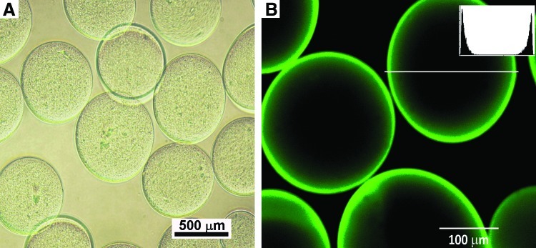
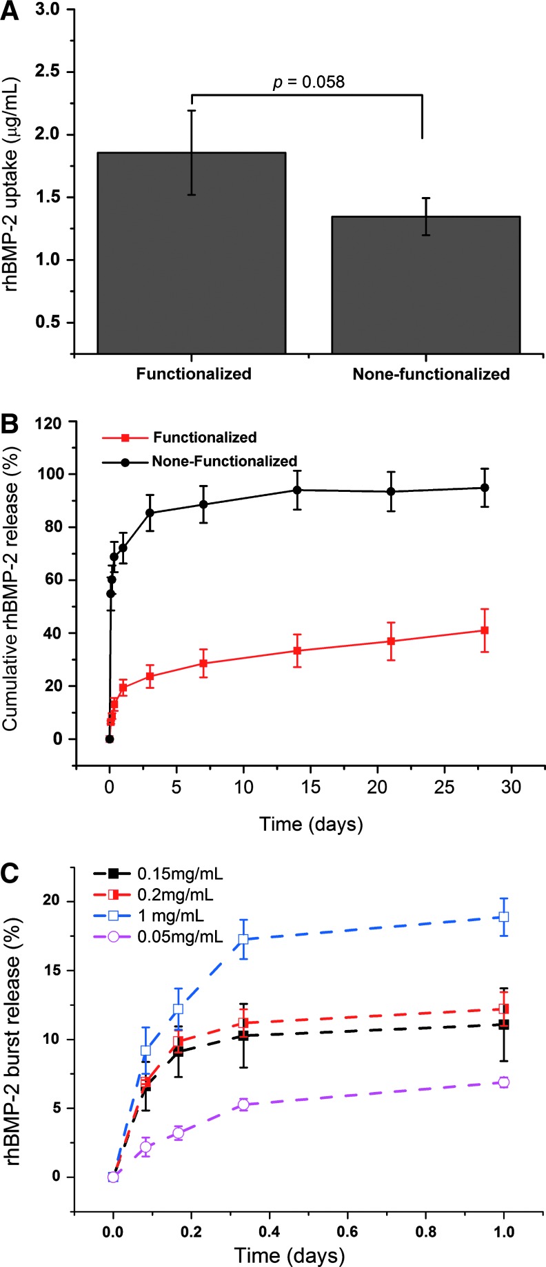
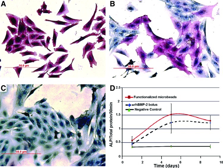
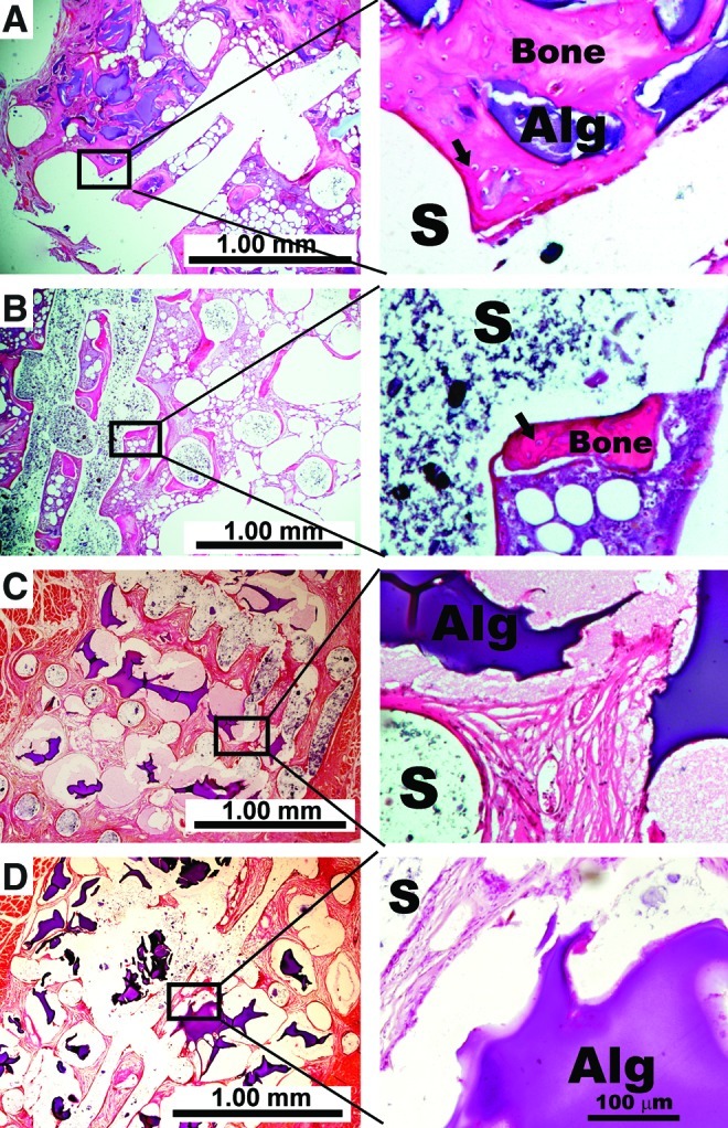
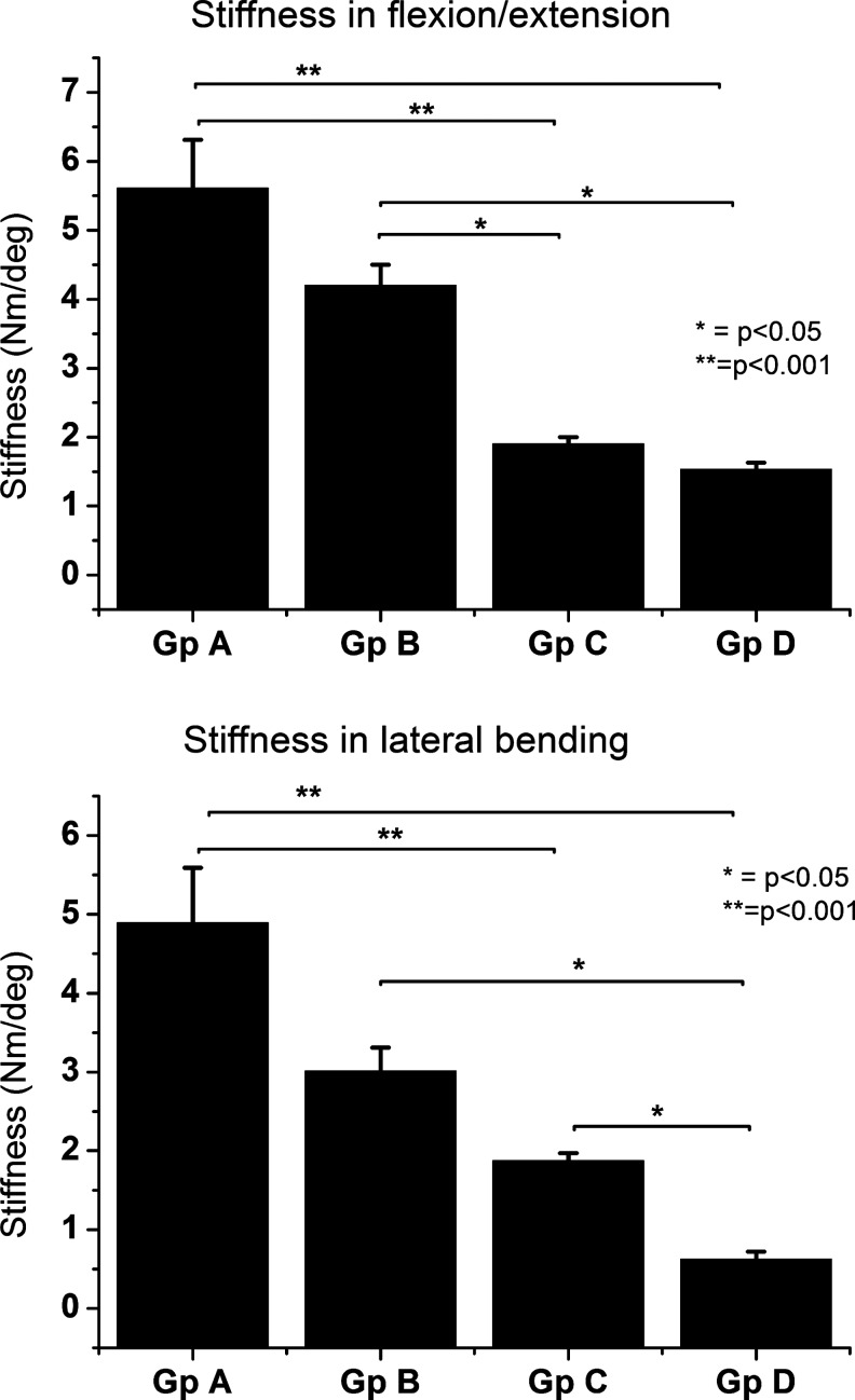
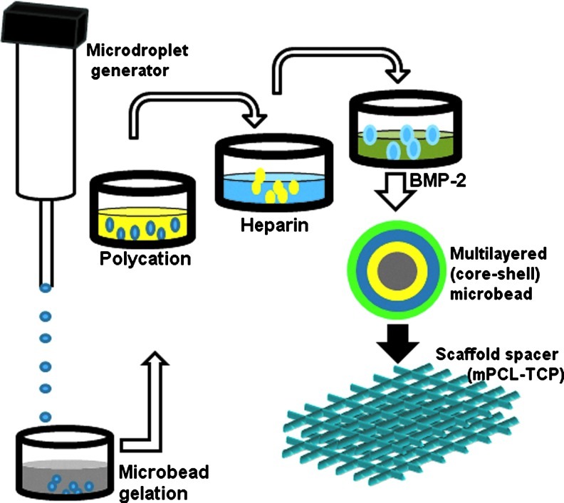
In this study, we tested the hypothesis that a surface functionalization delivery platform incorporating heparin onto strontium alginate microbeads surfaces would convert this "naive carriers" into "mini-reservoirs" for localized in vivo delivery of recombinant human bone morphogenetic protein-2 (rhBMP-2) that will induce functional bone regeneration. In vitro evaluation confirmed that (1) heparin incorporation could immobilize and prolong rhBMP-2 release for approximately 3 weeks; (2) a significant decrease (p<0.01) in rhBMP-2 burst release is attainable depending on initial protein load; and (3) rhBMP-2 released from surface functionalized microbeads retained bioactivity and stimulated higher alkaline phosphatase activity in cultured C(2)C(12) cells when compared with daily administration of fresh bolus rhBMP-2. Subsequently, surface functionalized microbeads were used for in vivo delivery of rhBMP-2 at local sites of posterolateral spinal fusion surgery in rats. The microbeads were loaded into the pores of medical-grade polyepsilone caprolactone-tricalcium phosphate scaffolds before implantation. Results revealed robust bone formation and a biomechanically solid fusion after 6 weeks. When compared with a control group consisting of an equivalent amount of rhBMP-2 that was directly adsorbed onto bare-surfaced microbeads with no heparin, a 5.3-fold increase in bone volume fraction and a 2.6-fold increase in bending stiffness (flexion/extension) were observed. When compared with collagen sponge carriers of rhBMP-2, a 1.5-fold and a 1.3-fold increase in bone volume fraction and bending stiffness were observed, respectively. More importantly, 3D micro-computed tomography images enabled the visualization of a well-contained newly formed bone at ipsilateral implant sites with surface functionalized rhBMP-2 delivery. This was absent with collagen sponge carriers where newly formed bone tissue was poorly contained and crossed over the posterior midline to contralateral implants. These findings are important because of complications with current rhBMP-2 delivery method, including excessive, uncontrolled bone formation.


Figures

FIG. 1.
FIG. 1.
Bright-field microscopy revealed microbeads of uniform morphology (A); Microbeads retained their morphological uniformity even after surface functionalization with heparin and heparin-binding proteins as observed on confocal laser scanning microscopy (B); this also confirms the uptake of fluorescein isothiocyanate-labeled heparin. Inset is histograph demonstrating the circumferential localization of green fluorescent dye intensity. Color images available online at www.liebertpub.com/tea
FIG. 2.
FIG. 2.
Monitoring the uptake (A) and release patterns (B and C) of recombinant human bone morphogenetic protein-2 (rhBMP-2) embedded within the surfaces of strontium crosslinked alginate microbeads using enzyme-linked immunosorbent assay methods. Color images available online at www.liebertpub.com/tea
FIG. 3.
FIG. 3.
Alkaline phosphatase enzyme activity of cultured C2C12 cells exposed to rhBMP-2 from surface functionalized microbeads (A) compared with bolus daily addition of equivalent rhBMP-2 doses (B) and negative control (beads with neither heparin nor rhBMP-2) (C). Measurements of alkaline phosphatase activity showed a similar trend between surface functionalized microbeads and the positive control group with a moderate increase in the surface functionalized group (D). Color images available online at www.liebertpub.com/tea
FIG. 4.
FIG. 4.
Reconstructed micro-CT images (A–D) and bone volumetric data (E) of harvested spinal segments after 6 weeks of implantation. New bone formation was observed in animals implanted with surface functionalized microbeads (A) and collagen sponge rhBMP-2 carriers (B). Peri-implant tissue mineralization and failure of bone formation were observed, respectively, in animals implanted with rhBMP-2 adsorbed directly onto alginate microbeads with no heparin (C) and rhBMP-2 microencapsulated in alginate core (D). Quantitative estimation of bone volume fraction (bone volume/total volume) showed significantly increased bone volume among surface functionalized alginate microbeads treatment group compared with the three control groups (E). *p<0.05 and **p<0.01.
FIG. 5.
FIG. 5.
Representative photomicrographs of histology sections stained with eosin and hematoxylin. New bone formation within medical grade polyepsilone caprolactone-tricalcium phosphate scaffold (S) pore spaces was confirmed among surface functionalized microbeads (A) and collagen sponge implants groups (B) at 6 weeks. Among animals implanted with rhBMP-2 adsorbed directly to microbeads without heparin (C) and animals implanted with microencapsulated rhBMP-2 (D), there was no evidence of new bone formation within the scaffold pore spaces. Bone formation was by direct ossification in the two groups with new bone formation. High magnification images show osteocytes in their lacunae (arrows) as well as fragmented remnants of alginate (Alg) matrix with no evidence of fibrous tissue encapsulation or massive cell infiltration. Color images available online at www.liebertpub.com/tea
FIG. 6.
FIG. 6.
Stiffness analysis (mean±standard deviation) of rat spinal segments evaluated in flexion/extension and lateral bending after 6 weeks of posterolateral fusion surgery. Gp A=surface functionalized alginate microbeads with heparin and rhBMP-2, Gp B=rhBMP-2 loaded unto collagen sponges, Gp C=rhBMP-2 adsorbed directly to microbeads without heparin, and Gp D=microencapsulated rhBMP-2.
FIG. 7.
FIG. 7.
Schematic of the surface functionalization procedure depicting layer-by-layer polyelectrolyte deposition for condensing heparin onto the surface of alginate microbeads and further entrapping rhBMP-2. Color images available online at www.liebertpub.com/tea

References

    1. Reddi A.H. Morphogenesis and tissue engineering of bone and cartilage: inductive signals, stem cells, and biomimetic biomaterials. Tissue Eng. 2000;6:351. - PubMed
    1. Carragee E.J. Hurwitz E.L. Weiner B.K. A critical review of recombinant human bone morphogenetic protein-2 trials in spinal surgery: emerging safety concerns and lessons learned. Spine J. 2011;11:471. - PubMed
    1. Lee J.W. Lee S. Lee S.H. Yang H.S. Im G.I. Kim C.S., et al. Improved spinal fusion efficacy by long-term delivery of bone morphogenetic protein-2 in a rabbit model. Acta Orthop. 2011;82:756. - PMC - PubMed
    1. Khan S.A. Nelson M.S. Pan C. Gaffney P.M. Gupta P. Endogenous heparan sulfate and heparin modulate bone morphogenetic protein-4 signaling and activity. Am J Physiol Cell Physiol. 2008;294:1387C. - PubMed
    1. Dejima K. Kanai M.I. Akiyama T. Levings D.C. Nakato H. Novel contact-dependent bone morphogenetic protein (BMP) signaling mediated by heparan sulfate proteoglycans. J Biol Chem. 2011;286:17103. - PMC - PubMed

Publication types

MeSH terms

LinkOut - more resources