. 2012 Aug 16:9:198.
doi: 10.1186/1742-2094-9-198.

Adenosine A2B receptor-mediated leukemia inhibitory factor release from astrocytes protects cortical neurons against excitotoxicity

Affiliations

Adenosine A2B receptor-mediated leukemia inhibitory factor release from astrocytes protects cortical neurons against excitotoxicity

Shamsudheen Moidunny et al. J Neuroinflammation. .

Abstract

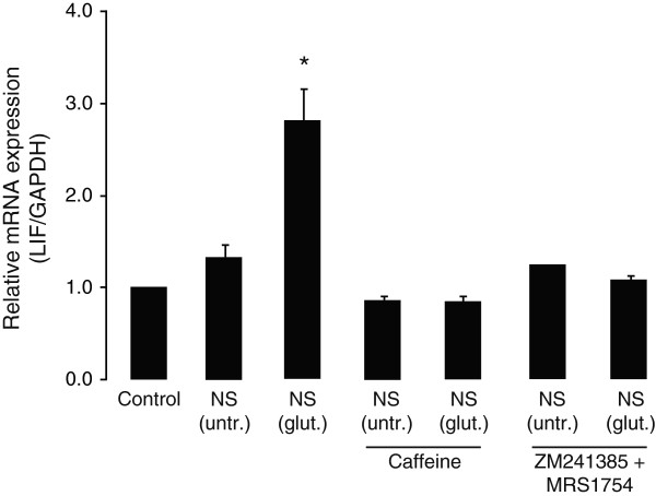
Background: Neuroprotective and neurotrophic properties of leukemia inhibitory factor (LIF) have been widely reported. In the central nervous system (CNS), astrocytes are the major source for LIF, expression of which is enhanced following disturbances leading to neuronal damage. How astrocytic LIF expression is regulated, however, has remained an unanswered question. Since neuronal stress is associated with production of extracellular adenosine, we investigated whether LIF expression in astrocytes was mediated through adenosine receptor signaling.

Methods: Mouse cortical neuronal and astrocyte cultures from wild-type and adenosine A(2B) receptor knock-out animals, as well as adenosine receptor agonists/antagonists and various enzymatic inhibitors, were used to study LIF expression and release in astrocytes. When needed, a one-way analysis of variance (ANOVA) followed by Bonferroni post-hoc test was used for statistical analysis.

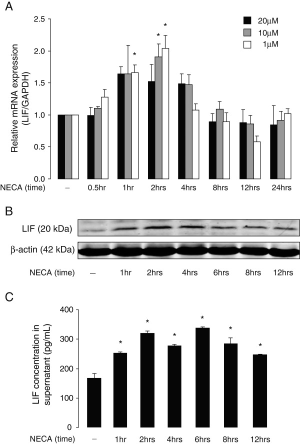
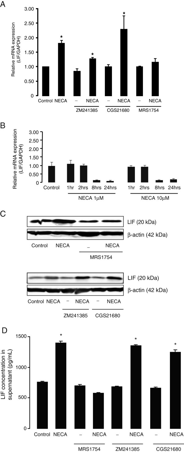
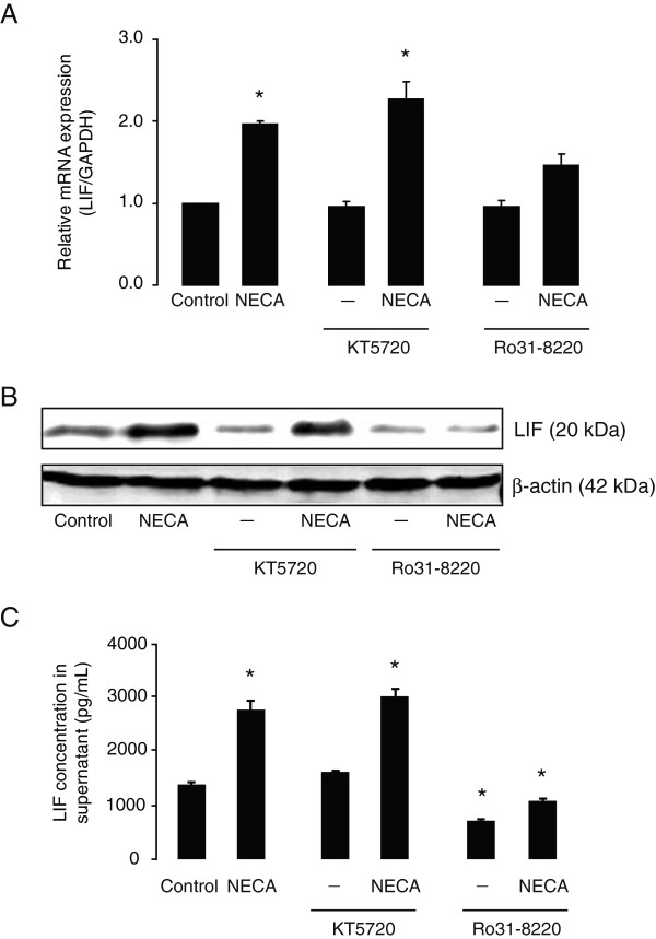
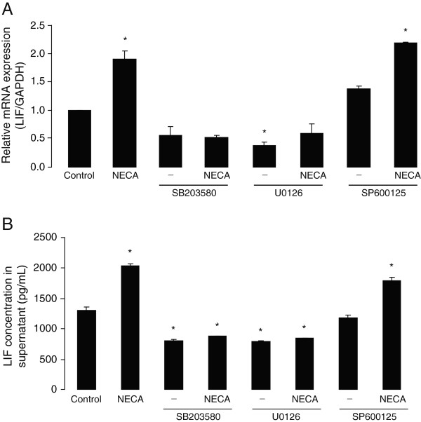
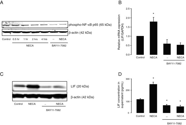
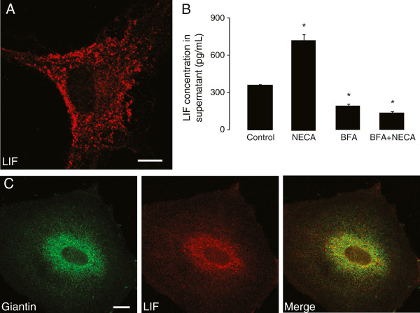
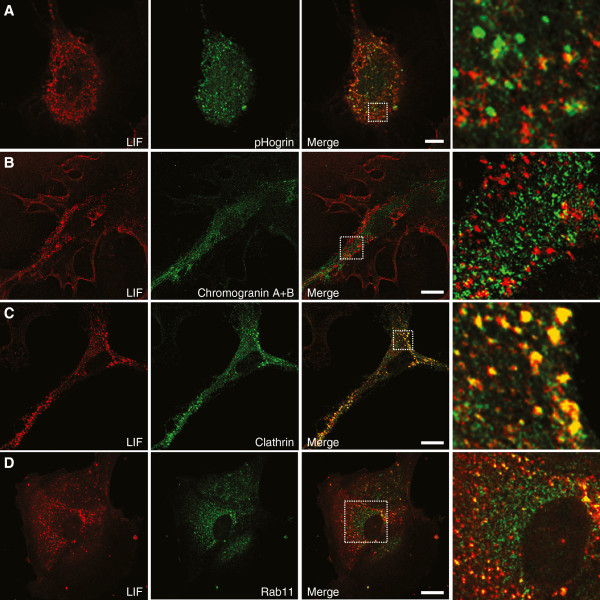
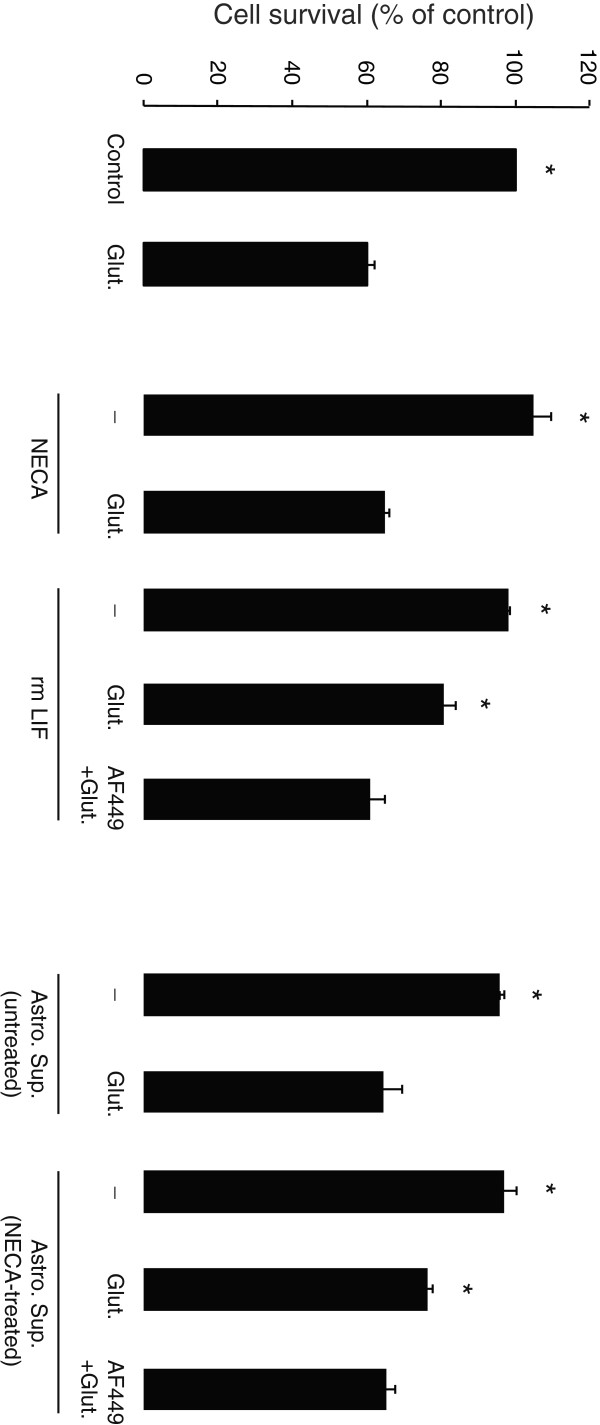
Results: We show here that glutamate-stressed cortical neurons induce LIF expression through activation of adenosine A(2B) receptor subtype in cultured astrocytes and require signaling of protein kinase C (PKC), mitogen-activated protein kinases (MAPKs: p38 and ERK1/2), and the nuclear transcription factor (NF)-κB. Moreover, LIF concentration in the supernatant in response to 5'-N-ethylcarboxamide (NECA) stimulation was directly correlated to de novo protein synthesis, suggesting that LIF release did not occur through a regulated release pathway. Immunocytochemistry experiments show that LIF-containing vesicles co-localize with clathrin and Rab11, but not with pHogrin, Chromogranin (Cg)A and CgB, suggesting that LIF might be secreted through recycling endosomes. We further show that pre-treatment with supernatants from NECA-treated astrocytes increased survival of cultured cortical neurons against glutamate, which was absent when the supernatants were pre-treated with an anti-LIF neutralizing antibody.

Conclusions: Adenosine from glutamate-stressed neurons induces rapid LIF release in astrocytes. This rapid release of LIF promotes the survival of cortical neurons against excitotoxicity.


Figures

Figure 1
Figure 1
Glutamate-stressed cortical neurons induce leukemia inhibitory factor (LIF) gene expression in primary cortical astrocytes, by a mechanism dependent on astrocytic adenosine receptor activation. Supernatants of primary mouse cortical neurons (NS) collected 18 hours following treatment without (untr.) or with glutamate (glut.; 50 μM, for 1 hour) were applied (1:1 dilution by volume) to the primary cultured cortical astrocytes for 2 hours. Where indicated, astrocytes were pre-treated with caffeine (50 μM) or a cocktail of adenosine A2A and A2B receptor antagonists (A2A antagonist: ZM 241385, 250 nM; A2B antagonist: MRS 1754, 250 nM), for 30 minutes before incubation with neuronal supernatants and were analyzed for LIF mRNA expression (relative to GAPDH) using real-time PCR. Data are normalized to the control and presented as Mean ± SEM of three independent experiments. P < 0.05.
Figure 2
Figure 2
NECA increases leukemia inhibitory factor (LIF) expression and secretion levels in primary mouse astrocytes. (A) Primary cortical astrocytes were treated without or with NECA (1, 10 or 20 μM) for 0.5, 1, 2, 4, 8, 12 and 24 hours. Cells were then analyzed for LIF mRNA expression (relative to GAPDH) by real-time PCR. Data are normalized to the control and presented as Mean ± SEM of three independent experiments. P < 0.05. (B) shows Western blot analysis of cultured astrocytes treated without or with NECA (1 μM) for 1, 2, 4, 6, 8 and 12 hours to determine LIF protein levels. β-actin served as a loading control. (C) shows a representation of sandwich ELISA experiment performed to detect LIF content in supernatants of cultured astrocytes that were treated without or with NECA (1 μM) for 1, 2, 4, 6, 8 and 12 hours. Each bar corresponds to the mean concentration of LIF in triplicate samples; error bars indicate SEM. Observations were confirmed by repeating the experiments two additional times. P < 0.05. NECA, 5′-N-ethylcarboxamide.
Figure 3
Figure 3
NECA-induced leukemia inhibitory factor (LIF) expression and secretion levels in primary astrocytes are dependent on adenosine A2Breceptor activation. Primary cortical astrocytes were treated with the adenosine analog, NECA (1 μM) or a selective adenosine A2A receptor agonist (CGS 21680, 1 μM) for 2 hours (for real-time PCR) or 4 hours (for Western blot and ELISA). Where indicated, astrocytes were pre-treated for 30 minutes with selective adenosine A2A receptor antagonist (ZM 241385, 250 nM) or adenosine A2B receptor antagonist (MRS 1754, 250 nM), prior to NECA stimulation. (A and B) show real-time PCR analyses of LIF gene expression (relative to GAPDH) in wild-type and A2B knock-out astrocytes, respectively. Data are normalized to the control and presented as Mean ± SEM of three independent experiments. P < 0.05. (C) shows Western blot analyses to detect LIF protein levels in wild-type astrocytes. β-actin served as a loading control. (D) shows a representation of sandwich ELISA experiment performed to detect LIF content in supernatants of cultured wild-type astrocytes. Each bar corresponds to the mean concentration of LIF in triplicate samples; error bars indicate SEM. Observations were confirmed by repeating the experiments two additional times. P < 0.05. NECA, 5′-N-ethylcarboxamide.
Figure 4
Figure 4
Effect of signaling pathway inhibitors on survival of cultured astrocytes. Primary cortical astrocytes were treated without or with different signaling pathway inhibitors used in this study (U 0126, 5 to 10 μM; SB 203580, 10 to 20 μM; BAY 11–7082, 10 to 20 μM; Ro 31–8220, 250 to 500nM; KT 5720, 250 to 500nM; SP 600125, 10 to 20 μM) for 24 hours and cell survival assessed by a colorimetric MTT assay. The optical densities were measured at 570 nm, with a 630 nm and blank correction. Data are normalized to percent of control (vehicle) and presented as Mean ± S.E.M of three independent experiments. P < 0.05, when compared to control.
Figure 5
Figure 5
NECA-induced leukemia inhibitory factor (LIF) expression and secretion levels in primary astrocytes are blocked by protein kinase C (PKC) inhibitor, but not by protein kinase A (PKA) inhibitor. Primary cortical astrocytes were treated without or with NECA (1 μM) for 2 hours (for real-time PCR) or 4 hours (for Western blot and ELISA). Where indicated, astrocytes were pre-treated for 30 minutes with specific inhibitors of PKA (KT 5720, 250 nM) or PKC (Ro 31–8220, 250 nM), prior to NECA stimulation. (A) shows real-time PCR analysis of LIF gene expression (relative to GAPDH). Data are normalized to the control and presented as Mean ± SEM of three independent experiments. P < 0.05. (B) shows Western blot analysis to detect LIF protein levels. β-actin served as a loading control. (C) shows a representation of sandwich ELISA experiment performed to detect LIF content in supernatants of cultured astrocytes. Each bar corresponds to the mean concentration of LIF in triplicate samples; error bars indicate SEM. Observations were confirmed by repeating the experiments two additional times. P < 0.05. NECA, 5′-N-ethylcarboxamide.
Figure 6
Figure 6
Basal and NECA-induced leukemia inhibitory factor (LIF) expression and secretion levels in primary astrocytes are dependent on ERK1/2- and p38- but not on JNK-MAPK activation. Primary cortical astrocytes were treated without or with NECA (1 μM) for 2 hours (for real-time PCR) or 4 hours (for ELISA). Where indicated, astrocytes were pre-treated for 2 hours with specific inhibitors of ERK1/2 (U 0126, 5 μM), p38 (SB 203580, 10 μM) or JNK (SP 600125, 10 μM) pathways, prior to NECA stimulation. (A) shows real-time PCR analysis of LIF gene expression (relative to GAPDH). Data are normalized to the control and presented as Mean ± SEM of three independent experiments. P < 0.05. (B) shows a representation of sandwich ELISA experiment performed to detect LIF content in supernatants of cultured astrocytes. Each bar corresponds to the mean concentration of LIF in triplicate samples; error bars indicate SEM. Observations were confirmed by repeating the experiments two additional times. P < 0.05. ERK, extracellular signal-regulated kinase; JNK, c-Jun N-terminal kinase; MAPK, mitogen-activated protein kinase; NECA, 5′-N-ethylcarboxamide.
Figure 7
Figure 7
Basal and NECA-induced leukemia inhibitory factor (LIF) expression and secretion levels in primary astrocytes are dependent on NF-κB activation. (A) shows Western blot analysis of primary cortical astrocytes treated without or with NECA (1 μM; for 0.5, 1, 2 and 4 hours) to detect phosphorylation at Ser536 of NF-κB p65 (RelA) proteins. Where indicated, cells were pre-treated with selective inhibitor of NF-κB (BAY 11–7082, 10 μM) for 2 hours prior to NECA stimulation. β-actin served as a loading control. Subsequently, astrocytes were treated without or with NECA (1 μM) for 2 hours (for real-time PCR) and 4 hours (for Western blot and ELISA), in presence or absence of BAY 11–7082 (10 μM, added 2 hours prior to NECA stimulation). (B) shows real-time PCR analysis of LIF gene expression (relative to GAPDH). Data are normalized to the control and presented as Mean ± SEM of three independent experiments. P < 0.05. (C) shows Western blot analysis to detect LIF protein levels. β-actin served as a loading control. (D) shows a representation of sandwich ELISA experiment performed to detect LIF content in supernatants of cultured astrocytes. Each bar corresponds to the mean concentration of LIF in triplicate samples; error bars indicate SEM. Observations were confirmed by repeating the experiments two additional times. P < 0.05. NECA, 5′-N-ethylcarboxamide.
Figure 8
Figure 8
Leukemia inhibitory factor (LIF) secretion in primary astrocytes is constitutive and independent of NECA stimulation. (A) illustrates LIF immunocytochemistry in cultured cortical astrocytes, where LIF is found in vesicle-like structures throughout the cytoplasm. Scale bar corresponds to 10 μm. (B) shows complete co-localization of LIF with Giantin, a marker of Golgi apparatus, when cultured astrocytes were treated with Brefeldin A (BFA, 5 μg/mL for 1 hour). Scale bar corresponds to 10 μm. (C) shows a representation of sandwich ELISA experiment performed to detect LIF content in supernatants of cultured astrocytes that were treated without or with NECA (1 μM) for 4 hours. Where indicated, cells were pre-treated with BFA (5 μg/mL) for 1 hour prior to NECA stimulation. Each bar corresponds to the mean concentration of LIF in triplicate samples; error bars indicate SEM. Observations were confirmed by repeating the experiments two additional times. P < 0.05. NECA, 5′-N-ethylcarboxamide.
Figure 9
Figure 9
Leukemia inhibitory factor (LIF) co-localizes with clathrin and Rab11 but not with large dense-core vesicle markers. Immunostaining performed in cultured astrocytes revealed that LIF does not co-localize with pHogrin (A) and Chromogranin A & B (B), which are both markers for large dense-core vesicles. However, we observed a massive co-localization of LIF with clathrin (C) and a partial co-localization with Rab11 (D). Scale bars correspond to 10 μm.
Figure 10
Figure 10
Effect of untreated and NECA-treated astrocyte supernatants on survival of cultured cortical neurons against excitotoxicity. Primary cortical neurons were treated without or with recombinant mouse Leukemia inhibitory factor (rmLIF, 0.1 ng/mL), NECA (1 μM) or astrocyte supernatants (untreated or treated with 1 μM NECA for 4 hours; diluted 1:1 in neuronal culture medium) for 24 hours. Where indicated, rmLIF and NECA-treated astrocyte supernatants were treated with an anti-LIF neutralizing antibody (AF449; 100 ng/mL for 1.5 hours) before applying them to cultured neurons. Subsequently, the neurons were treated without or with glutamate (Glut.; 50 μM for 1 hour) and cell survival assessed 24 hours after glutamate treatment by a colorimetric MTT assay. The optical densities were measured at 570 nm, with a 630 nm and blank correction. Data are normalized to percent of control and presented as Mean ± S.E.M of four independent experiments. P < 0.05, when compared to glutamate-treated condition. NECA, 5′-N-ethylcarboxamide.

Similar articles

Cited by

References

    1. Heinrich PC, Behrmann I, Haan S, Hermanns HM, Muller-Newen G, Schaper F. Principles of interleukin (IL)-6-type cytokine signalling and its regulation. Biochem J. 2003;374:1–20. doi: 10.1042/BJ20030407. - DOI - PMC - PubMed
    1. Patterson PH. The emerging neuropoietic cytokine family: first CDF/LIF, CNTF and IL-6; next ONC, MGF, GCSF? Curr Opin Neurobiol. 1992;2:94–97. doi: 10.1016/0959-4388(92)90169-L. - DOI - PubMed
    1. Patterson PH. Leukemia inhibitory factor, a cytokine at the interface between neurobiology and immunology. Proc Natl Acad Sci USA. 1994;91:7833–7835. doi: 10.1073/pnas.91.17.7833. - DOI - PMC - PubMed
    1. Kurek JB, Bower JJ, Romanella M, Koentgen F, Murphy M, Austin L. The role of leukemia inhibitory factor in skeletal muscle regeneration. Muscle Nerve. 1997;20:815–822. doi: 10.1002/(SICI)1097-4598(199707)20:7<815::AID-MUS5>3.0.CO;2-A. - DOI - PubMed
    1. Kwon YW, Abbondanzo SJ, Stewart CL, Gurney ME. Leukemia inhibitory factor influences the timing of programmed synapses withdrawal from neonatal muscles. J Neurobiol. 1995;28:35–50. doi: 10.1002/neu.480280105. - DOI - PubMed

Publication types