

Polymorphism-specific PCR enhances the diagnostic performance of American tegumentary leishmaniasis and allows the rapid identification of Leishmania species from Argentina
- PMID: 22894734
- PMCID: PMC3449195
- DOI: 10.1186/1471-2334-12-191
Polymorphism-specific PCR enhances the diagnostic performance of American tegumentary leishmaniasis and allows the rapid identification of Leishmania species from Argentina
Abstract
Background: The diagnosis of the leishmaniases poses enormous challenges in Argentina. The Polymorphism-Specific PCR (PS-PCR) designed and validated in our laboratories has been proven effective for typifying the Leishmania genus from cultured material. Here we evaluated the performance of this method in the diagnosis of American tegumentary leishmaniasis (ATL) and the rapid identification of Leishmania spp. directly from clinical specimens.
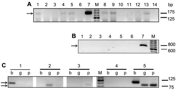
Methods: A total of 63 patients from northwestern Argentina, with cutaneous or mucocutaneous lesions, underwent an ATL diagnosis protocol which included clinical examination, Leishmanin skin test, and microscopic examination of dermal smears. In addition, we performed PS-PCR on DNA directly extracted from the specimens scraped from the lesions.
Results: Out of the 63 patients, 44 were classified as ATL cases and 19 as non-ATL cases. The diagnostic sensitivity of the microscopic analysis of dermal smears and PS-PCR individually were 70.5% and 81%, respectively. When performing both tests in parallel, this parameter increased significantly to 97.6% (p = 0.0018). The specificities, on the other hand, were 100%, 84.2%, and 83.3% for the combination, respectively (p > 0.05). Using the PS-PCR analysis we successfully identified the Leishmania spp. in 31 out of the 44 ATL cases. Twenty-eight (90.3%) cases were caused by L. (V.) braziliensis, two (6.5%) by L. (V.) guyanensis, and one (3.2%) by L. (V.) panamensis.
Conclusions: The efficacy of the ATL diagnosis was significantly improved by combining the dermal smear examination with a PS-PCR analysis. Our strategy allowed us to reach the diagnosis of ATL with high accuracy regarding the species of the etiological agent in 70.5% of the cases. Moreover, we diagnosed two cases of the disseminated cutaneous form caused by L. (V.) braziliensis and a cutaneous case due to L. (V.) panamensis infection, both findings reported for the first time in Argentina.
Figures

References
-
- Report of the Scientific Workving Group meeting on Leishmaniasis. World Health Organization, Geneva; 2004. pp. 5–16.
-
- Sosa Estani S, Campanini A, Sinagra A, Luna C, Peralta M, Coutada V, Medina L, Riarte A, Salomon D, Gomez A. et al. Clinical features and diagnosis of mucocutaneous leishmaniasis in patients of an endemic area in Salta. Medicina (B Aires) 1998;58(6):685–691. - PubMed
-
- Frank FM, Fernandez MM, Taranto NJ, Cajal SP, Margni RA, Castro E, Thomaz-Soccol V, Malchiodi EL. Characterization of human infection by Leishmania spp. in the Northwest of Argentina: immune response, double infection with Trypanosoma cruzi and species of Leishmania involved. Parasitology. 2003;126(Pt 1):31–39. - PubMed
-
- Marco JD, Barroso PA, Calvopina M, Kumazawa H, Furuya M, Korenaga M, Cajal SP, Mora MC, Rea MM, Borda CE. et al. Species assignation of Leishmania from human and canine American tegumentary leishmaniasis cases by multilocus enzyme electrophoresis in North Argentina. Am J Trop Med Hyg. 2005;72(5):606–611. - PubMed
Publication types
MeSH terms
LinkOut - more resources
Full Text Sources
Miscellaneous
