. 2012 Aug 15:11:99.
doi: 10.1186/1475-2840-11-99.

Far infra-red therapy promotes ischemia-induced angiogenesis in diabetic mice and restores high glucose-suppressed endothelial progenitor cell functions

Affiliations

Far infra-red therapy promotes ischemia-induced angiogenesis in diabetic mice and restores high glucose-suppressed endothelial progenitor cell functions

Po-Hsun Huang et al. Cardiovasc Diabetol. .

Abstract

Background: Far infra-red (IFR) therapy was shown to exert beneficial effects in cardiovascular system, but effects of IFR on endothelial progenitor cell (EPC) and EPC-related vasculogenesis remain unclear. We hypothesized that IFR radiation can restore blood flow recovery in ischemic hindlimb in diabetic mice by enhancement of EPCs functions and homing process.

Materials and methods: Starting at 4 weeks after the onset of diabetes, unilateral hindlimb ischemia was induced in streptozotocin (STZ)-induced diabetic mice, which were divided into control and IFR therapy groups (n = 6 per group). The latter mice were placed in an IFR dry sauna at 34°C for 30 min once per day for 5 weeks.

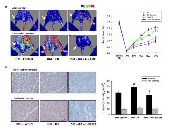
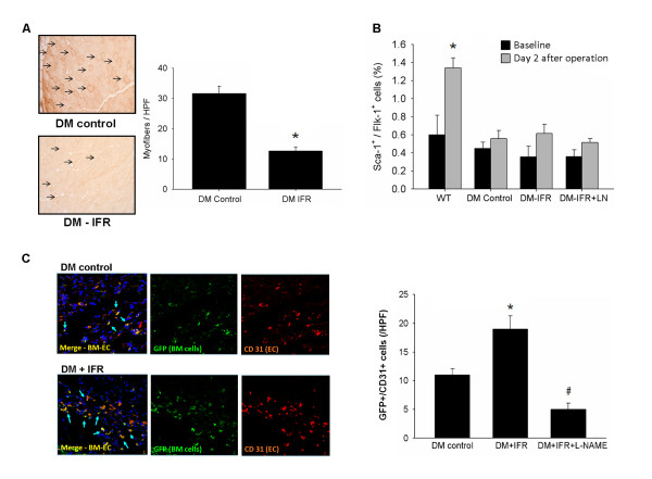
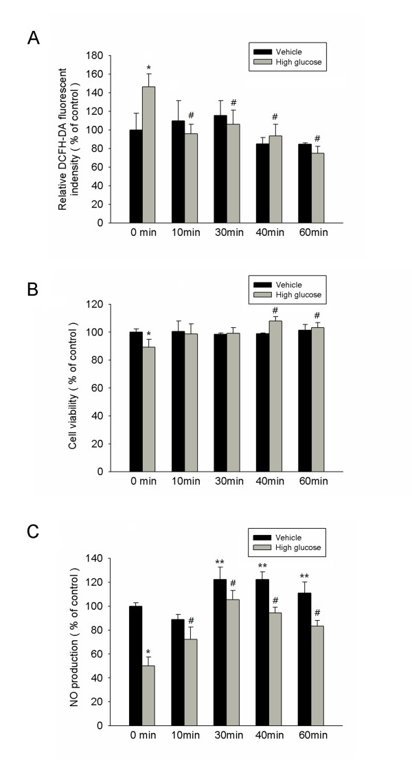
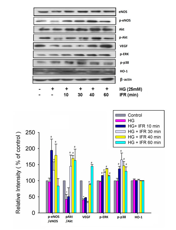
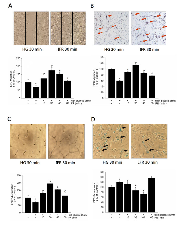
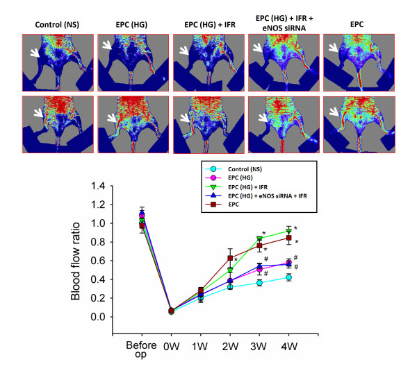
Results: Doppler perfusion imaging demonstrated that the ischemic limb/normal side blood perfusion ratio in the thermal therapy group was significantly increased beyond that in controls, and significantly greater capillary density was seen in the IFR therapy group. Flow cytometry analysis showed impaired EPCs (Sca-1(+)/Flk-1(+)) mobilization after ischemia surgery in diabetic mice with or without IFR therapy (n = 6 per group). However, as compared to those in the control group, bone marrow-derived EPCs differentiated into endothelial cells defined as GFP(+)/CD31(+) double-positive cells were significantly increased in ischemic tissue around the vessels in diabetic mice that received IFR radiation. In in-vitro studies, cultured EPCs treated with IFR radiation markedly augmented high glucose-impaired EPC functions, inhibited high glucose-induced EPC senescence and reduced H(2)O(2) production. Nude mice received human EPCs treated with IFR in high glucose medium showed a significant improvement in blood flow recovery in ischemic limb compared to those without IFR therapy. IFR therapy promoted blood flow recovery and new vessel formation in STZ-induced diabetic mice.

Conclusions: Administration of IFR therapy promoted collateral flow recovery and new vessel formation in STZ-induced diabetic mice, and these beneficial effects may derive from enhancement of EPC functions and homing process.


Figures

Figure 1
Figure 1
Effects of far infrared (IFR) therapy on blood flow recovery and new vessels formation in STZ-induced diabetic mice. (A) Representative results of laser Doppler measurements before operation and 4 weeks after hindlimb ischemia surgery in wild-type mice, control (vehicle), IFR therapy, and IFR + NG-nitro-L-arginine methyl ester (L-NAME) mice. Color scale illustrates blood flow variations from minimal (dark blue) to maximal (red) values. Arrows indicate ischemic (right) limb after hindlimb ischemia surgery. Doppler perfusion ratio (ischemic/non-ischemic hind limb) over time in the different groups. Administration of L-NAME in drinking water abolished the beneficial effect of IFR therapy in diabetic mice. (*p < 0.05 compared with DM-control; #p < 0.05 compared with DM-FIR; n = 6) (B) Mice were sacrificed 3 weeks after surgery and capillaries in the ischemic muscles were visualized by anti-CD31 immunostaining. Results are mean ± standard error of mean (SEM). (*p < 0.05 compared with DM-control; #p < 0.05 compared with DM-IFR; n = 6).
Figure 2
Figure 2
Effects of IFR radiation on oxidative stress, EPC mobilization after hindlimb ischemia and tissue homing in STZ-induced diabetic mice. (A) Effect of IFR on oxidative stress in ischemic muscles of STZ-induced diabetic mice. Nitrotyrosine (n = 4 per group) immunostaining of ischemic muscles extracted on day 21 in control (vehicle), and in mice that had received IFR radiation. (*p < 0.05 compared with DM-control) (B) EPCs (defined as Sca-1+/Flk-1+ cells) mobilization after tissue ischemia was determined by flow cytometry in STZ-induced diabetic mice given the vehicle, IFR or IFR + L-NAME. (*p < 0.05 compared with WT-baseline; n = 6 per group) (C) STZ-induced diabetes was created in FVB mice that received eGFP mouse bone marrow cells. By immunofluorescence staining, STZ-induced diabetic mice in IFR group had more GFP+/CD31+ double-positive cells in ischemic muscle than those in the vehicle group. (*p < 0.05 compared with DM-control; #p < 0.05 compared with DM-FIR; n = 6).
Figure 3
Figure 3
Morphology and characterization of human endothelial progenitor cells (EPCs) from peripheral blood. (A) Peripheral blood mononuclear cells (MNCs) were plated on a fibronectin-coated culture dish on the first day. (B) Four days after plating, adherent early EPCs with a spindle shape were shown. (C) Three weeks after plating, ECFCs with a cobblestone-like morphology were selected, reseeded, and grown to confluence. (D-I) ECFC characterization was performed by immunohistochemical staining. Most of the EPC expressed endothelial and hematopoietic stem cell markers, VE-cadherin, PECAM-1 (CD31), CD34, KDR, AC133, and eNOS, which are considered critical markers of EPCs. Cells were counterstained with 4',6-diamidino-2-phenylindole (DAPI) for the nuclei (blue).
Figure 4
Figure 4
IFR therapy decreased reactive oxidative stress, recovered EPC proliferation, and increased NO production in high-glucose conditions. (A) High glucose markedly increased H2O2 production determined by the relative DCFH-DA fluorescent intensity, and the administration of IFR therapy suppressed high glucose-induced reactive oxidative stress (ROS) index in EPCs culture. (*p < 0.05 compared with control - 0 min; #p < 0.05 compared with high glucose - 0 min) (B) The effect of IFR radiation on EPCs proliferation was analyzed by MTT assay. (*p < 0.05 compared with control - 0 min; #p < 0.05 compared with high glucose - 0 min) (C) Nitrate production (as NO content) in culture medium was measured by Griess reagent. High glucose-suppressed NO production in cultured late EPC s. After 4 days of incubation, IFR radiation increased NO production with or with high glucose conditions. (*p < 0.05 compared with control - 0 min; #p < 0.05 compared with high glucose - 0 min; **p < 0.05 compared with control – 0 min; n = 4 for each experiment).
Figure 5
Figure 5
Effects of IFR radiation on eNOS, Akt, p-ERK, p-38 MAPK, VEGF and HO-1 production in cultured EPCs. Administration of IFR radiation on cultured ECFCs for 10, 30, and 40 min followed by treatment of EPCs in high-glucose conditions significantly upregulated high glucose-impaired eNOS production and eNOS activity (p-eNOS/total eNOS). Treatment of IFR therapy for 30, 40 and 60 min also upregulated Akt activation (p-Akt/total Akt). In addition, administration IFR radiation also promoted VEGF production, p-ERK, and p-38 MAPK in high glucose conditions, but not HO-1. Each bar graph shows the summarized data from four separate experiments by densitometry after normalization. Data are means ± SEM; n = 4 in each experiment. (*p < 0.05 compared with control; #p < 0.05 compared with HG group.).
Figure 6
Figure 6
Effects of IFR therapy on EPC migration, tube formation, and senescence in vitro. (A,B) Scratch test and modified Boyden chamber assay were used to assess the migratory function of ECFCs in high glucose conditions. Boyden chamber assay using VEGF as a chemoattracting factor was used to evaluate the effects of IFR radiation on EPC migration. (C) An in vitro angiogenesis assay for late ECFCs used ECMatrix gel. Representative photos for in vitro angiogenesis are shown. Cells were stained with crystal violet, and the averages of the total area of complete tubes formed by cells were compared by using computer software. (D) To determine the onset of cellular aging, acidic ß-galactosidase was used as a biochemical marker for acidification, typical for ECFCs senescence. Data are means ± SEM; n = 4 in each experiment.
Figure 7
Figure 7
IFR treated-EPC transplantation improved blood perfusion in the ischemic hindlimb. (A) Representative images of hindlimb blood flow measured by laser Doppler and quantitative analysis of blood flow expressed as perfusion ratio of the ischemic to the contralateral (non-operated) hindlimb immediately after hindlimb ischemia surgery and 3 weeks after intramuscular injection of normal saline, EPC-treated with high glucose (EPC-HG), EPC-treated with high glucose and FIR therapy (EPC-HG + IFR), or EPC treated with high glucose and IFR and eNOS siRNA (EPC-HG + eNOS siRNA + IFR). (*p < 0.05 compared with control; #p < 0.05 compared with EPC-HG + IFR; n = 6).

Similar articles

Cited by

References

    1. Folkman J. Seminars in Medicine of the Beth Israel Hospital, Boston. Clinical applications of research on angiogenesis. N Engl J Med. 1995;333(26):1757–1763. doi: 10.1056/NEJM199512283332608. - DOI - PubMed
    1. Freedman S, Isner JM. Therapeutic angiogenesis for ischemic cardiovascular disease. J Mol Cell Cardiol. 2001;33(3):379–393. doi: 10.1006/jmcc.2000.1329. - DOI - PubMed
    1. Rosengart TK, Lee LY, Patel SR, Sanborn TA, Parikh M, Bergman GW, Hachamovitch R, Szulc M, Kligfield PD, Okin PM, Hahn RT, Devereux RB, Post MR, Hackett NR, Foster T, Grasso TM, Lesser ML, Isom OW, Crystal RG. Angiogenesis gene therapy: phase I assessment of direct intramyocardial administration of an adenovirus vector expressing VEGF121 cDNA to individuals with clinically significant severe coronary artery disease. Circulation. 1999;100(5):468–474. doi: 10.1161/01.CIR.100.5.468. - DOI - PubMed
    1. Nathan DM, Cleary PA, Backlund JY, Genuth SM, Lachin JM, Orchard TJ, Raskin P, Zinman B. Diabetes Control and Complications Trial/Epidemiology of Diabetes Interventions and Complications (DCCT/EDIC) Study Research Group: Intensive diabetes treatment and cardiovascular disease in patients with type 1 diabetes. N Engl J Med. 2005;353(25):2643–2653. - PMC - PubMed
    1. Adeghate E. Molecular and cellular basis of the etiology and management of diabetic cardiomyopathy: a short review. Mol Cell Biochem. 2004;261(1–2):187–191. - PubMed

Publication types

MeSH terms