

Review
doi: 10.2174/157340312801784952.
Point of care cardiac ultrasound applications in the emergency department and intensive care unit--a review
Affiliations
- PMID: 22894759
- PMCID: PMC3406278
- DOI: 10.2174/157340312801784952
Item in Clipboard
Review
Point of care cardiac ultrasound applications in the emergency department and intensive care unit--a review
Curr Cardiol Rev.
2012 May.
Abstract
The use of point of care echocardiography by non-cardiologist in acute care settings such as the emergency department (ED) or the intensive care unit (ICU) is very common. Unlike diagnostic echocardiography, the scope of such point of care exams is often restricted to address the clinical questions raised by the patient's differential diagnosis or chief complaint in order to inform immediate management decisions. In this article, an overview of the most common applications of this focused echocardiography in the ED and ICU is provided. This includes but is not limited to the evaluation of patients experiencing hypotension, cardiac arrest, cardiac trauma, chest pain and patients after cardiac surgery.
Figures
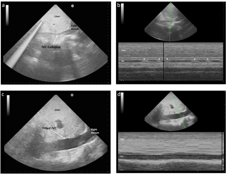
IVC images taken in the same spontaneously breathing patient with sepsis at the bedside in the ED. 1a 2D image shows a small
calibre IVC collapsing during spontaneous breathing. 1b M-mode image of the same patient shows collapse with each breath (denoted by
'*'). 1c 2D image of the same patient after 90 minutes and 3L of crystalloid shows larger calibre IVC and, as is seen in 1d An M-mode demonstrates
the absence of size variation with respiration, suggestive of adequate fluid resuscitation.
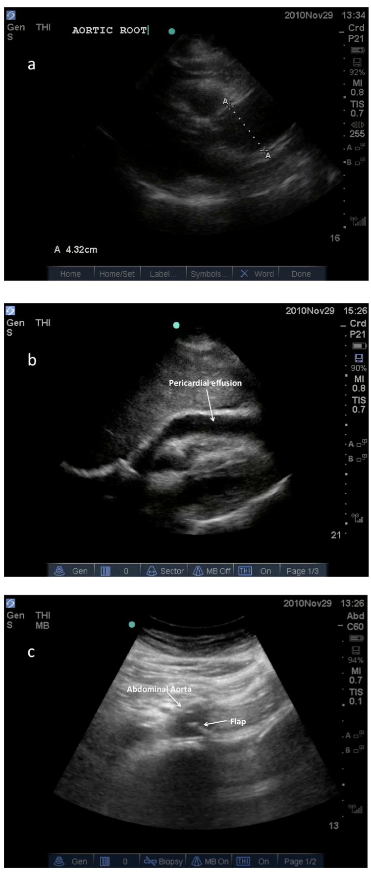
Images from a hemodynamically unstable patient who
presented to the ED with undifferentiated chest pain and abdominal
pain. Immediate point of care ultrasound was performed revealing
the following images: 2a: Parasternal long axis view demonstrating
enlarged ascending aorta and aortic root. 2b: Sub-xiphoid view
demonstrating pericardial effusion. 2c: Transabdominal view of the
proximal abdominal aorta where a mobile, intraluminal, echogenic
line was seen, suggestive of an intimal flap. After the patient was
stabilized, CT scan confirmed a Stanford type A aortic dissection
extending from the aortic root to the iliac bifurcation.
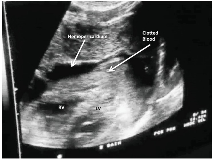
Hemopericardium as seen from a sub-xiphoid view after a
penetrating injury to right ventricle (RV). Note clotted blood in
pericardium.
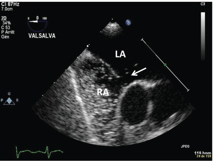
Right-to-left shunt. On this modified mid-esophageal bicaval
view from a bedside, focused TEE in the ICU, agitated saline
bubbles (arrow) can be seen passing from the Right Atrium (RA) to
the Left Atrium (LA) through a defect in the intra-atrial septum.
References
-
- Plummer D, Brunette D, Asinger R, Ruiz E. Emergency department echocardiography improves outcome in penetrating cardiac injury. Ann Emerg Med. 1992;21(6):709–12. - PubMed
-
- Labovitz A, Noble V Bierig M, et al. Focused cardiac ultrasound in the emergent setting: a consensus statement of the American Society of Echocardiography and American College of Emergency Physicians. J Am Soc Echocardiogr. 2010;23(12):1225–30. - PubMed
-
- Mayo P, Beaulieu Y, Doelken P, et al. American College of Chest Physicians/La Société de Réanimation de Langue Française statement on competence in critical care ultrasonography. Chest. 2009;135(4):1050–60. - PubMed
-
- Stewart W, Douglas P, Sagar K, et al. Echocardiography in emergency medicine: a policy statement by the American Society of Echocardiography and the American College of Cardiology. The Task Force on Echocardiography in Emergency Medicine of the American Society of Echocardiography and the Echocardiography TPEC Committees of the American College of Cardiology. J Am Soc Echocardiogr. 1999;12(1):82–4. - PubMed
-
- Moore C, Copel J. Point-of-care ultrasonography. N Engl J Med. 2011;364(8):749–57. - PubMed
Publication types
MeSH terms
LinkOut - more resources
Full Text Sources
Medical
