Review
. 2012 Sep;7(9):987-93.
doi: 10.4161/epi.21567. Epub 2012 Aug 16.

Epigenetics within the matrix: a neo-regulator of fibrotic disease

Affiliations
Review

Epigenetics within the matrix: a neo-regulator of fibrotic disease

Claire M Robinson et al. Epigenetics. 2012 Sep.

Abstract

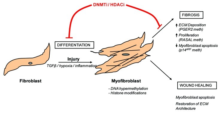
Fibrosis of any tissue is characterized by excessive extracellular matrix accumulation that ultimately destroys tissue architecture and eventually abolishes normal organ function. Although much research has focused on the mechanisms underlying disease pathogenesis, there are still no effective antifibrotic therapies that can reverse, stop or delay the formation of scar tissue in most fibrotic organs. As fibrosis can be described as an aberrant wound healing response, a recent hypothesis suggests that the cells involved in this process gain an altered heritable phenotype that promotes excessive fibrotic tissue accumulation. This article will review the most recent observations in a newly emerging field that links epigenetic modifications to the pathogenesis of fibrosis. Specifically, the roles of DNA methylation and histone modifications in fibrotic disease will be discussed.


Figures

None
Figure 1. Fibroblast to myofibroblast differentiation and the role of epigenetic modifications Following injury, fibroblasts differentiate into myofibroblasts. This process is encouraged by pro-fibrotic stimulants, such as transforming growth factor β (TGFβ), inflammatory cytokines and hypoxia. Under normal circumstances, wound-healing myofibroblasts contribute to tissue remodeling and repair and are eventually removed by apoptosis. In contrast, fibrotic myofibroblasts are hyperproliferative and continually secrete pro-fibrotic cytokines and collagens into their surrounding environment. These cells are resistant to apoptosis and are therefore continually present in the tissue. As this review highlights, DNA hypermethylation as well as histone modifications have been implicated in controlling this process. Hence, the use of DNA methyltransferase inhibitors (DNMTi) or histone deacetylase inhibitors (HDACi) may be beneficial in preventing myofibroblast differentiation.

Similar articles

Cited by

References

    1. Wynn TA. Cellular and molecular mechanisms of fibrosis. J Pathol. 2008;214:199–210. doi: 10.1002/path.2277. - DOI - PMC - PubMed
    1. Mehal WZ, Iredale J, Friedman SL. Scraping fibrosis: expressway to the core of fibrosis. Nat Med. 2011;17:552–3. doi: 10.1038/nm0511-552. - DOI - PMC - PubMed
    1. Phan SH. Biology of fibroblasts and myofibroblasts. Proc Am Thorac Soc. 2008;5:334–7. doi: 10.1513/pats.200708-146DR. - DOI - PMC - PubMed
    1. Darby IA, Hewitson TD. Fibroblast differentiation in wound healing and fibrosis. Int Rev Cytol. 2007;257:143–79. doi: 10.1016/S0074-7696(07)57004-X. - DOI - PubMed
    1. Grgic I, Duffield JS, Humphreys BD. The origin of interstitial myofibroblasts in chronic kidney disease. Pediatr Nephrol. 2012;27:183–93. doi: 10.1007/s00467-011-1772-6. - DOI - PMC - PubMed

Publication types