. 2012 Sep 15;11(18):3433-42.
doi: 10.4161/cc.21732. Epub 2012 Aug 16.

p53 negatively regulates Aurora A via both transcriptional and posttranslational regulation

Affiliations

p53 negatively regulates Aurora A via both transcriptional and posttranslational regulation

Chun-Chi Wu et al. Cell Cycle. .

Abstract

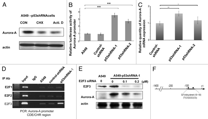
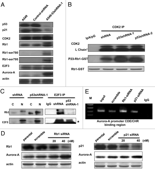
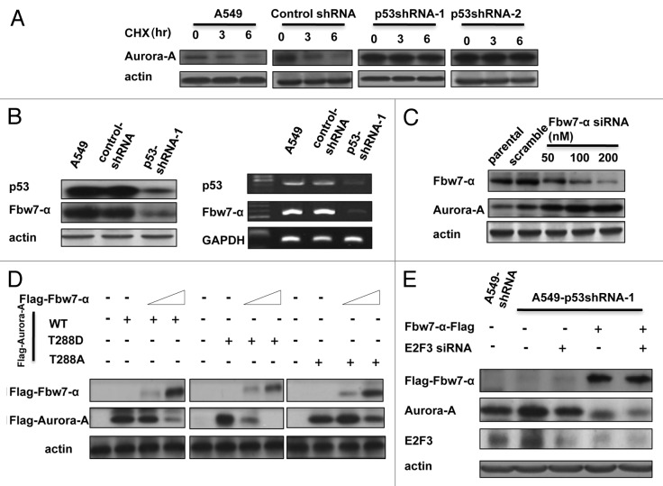
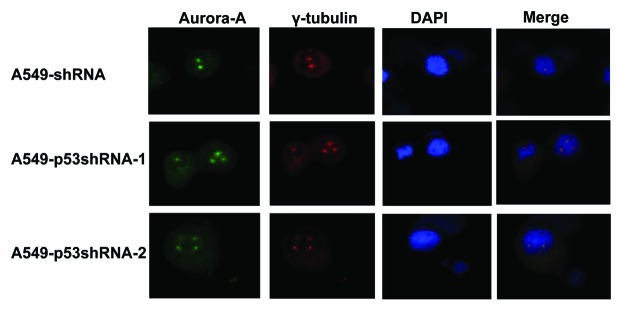
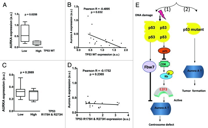
p53 plays an important role in mitotic checkpoint, but what its role is remains enigmatic. Aurora A is a Ser/Thr kinase involved in correcting progression of mitosis. Here, we show that p53 is a negative regulator for Aurora A. We found that p53 deficiency leads to Aurora A elevation. Ectopic expression of p53 or DNA damage-induced expression of p53 can suppress the expression of Aurora A. Mechanistic studies show that p53 is a negative regulator for Aurora A expression through both transcriptional and posttranslational regulation. p53 knockdown in cancer cells reduces the level of p21, which, in turn, increases the activity of CDK2 followed by induction of Rb1 hyperphosphorylation and its dissociation with transcriptional factor E2F3. E2F3 can bind to Aurora A gene promoter, potentiating Aurora A gene expression and p53 deficiency, enhancing the binding of E2F3 on Aurora A promoter. Also, p53 deficiency leads to decelerating Aurora A's turnover rate, due to the fact that p53 deficiency causes the downregulation of Fbw7α, a component of E3 ligase of Aurora A. Consistently, p53 knockdown-mediated Aurora A elevation is mitigated when Fbw7α is ectopically expressed. Thus, p53-mediated Aurora A degradation requires Fbw7α expression. Significantly, inverse correlation between p53 and Aurora A elevation is translated into the deregulation of centrosome amplification. p53 knockdown leads to high percentages of cells with abnormal amplification of centrosome. These data suggest that p53 is an important negative regulator of Aurora A, and that loss of p53 in many types of cancer could lead to abnormal elevation of Aurora A and dysregulated mitosis, which provides a growth advantage for cancer cells.


Figures

None
Figure 1. p53 is a negative regulator of Aurora A. (A) The parental and p53-knockdown A549 stable clones were harvested and subjected to western blotting using the indicated antibodies. (B) A549 cells were transfecetd with various dosages of p53 siRNA for 48 h, the cells were then collected and subjected to western blotting using the indicated antibodies. (C) Various dosages of p53 inhibitor PFT were added into A549 cells, 48 h later, the cells were collected and subjected to western blot. (D) The adenovirus particles containing p53-wild type or p53-mut (DNA binding mutant) gene were infected into A549-p53shRNA stable cells. The cells were then harvested and subjected to western blotting using the indicated antibodies. (E) p53-null H1299 cells were infected with adenovirus particles described in (D). (F) A549-control shRNA, A549-p53 shRNA and H1299 cells were treated with indicated doses of cisplatin for 48 h, the cells were then collected, lysed and subjected to western blotting using the indicated antibodies.
None
Figure 2. p53 affects the binding ability of E2F3 on Aurora A promoter region. (A) A549-p53shRNA cells were treated with actinomycin D (500 ng/ml) or cyclohexmide (50 μg/ml), respectively, for 9 h, the cells were then collected and subjected to western blot using the indicated antibodies. (B) Aurora A promoter (+15 to -1400 bps)-luciferase vector was cotransfected with internal control luciferase vector into wild type or p53-knockdown A549 cells for 48 h, the cells were then harvested and subjected to luciferase assay. (C) The expression levels of Aurora A of A549-shRNA, A549 p53-shRNA-1 and A549-p53shRNA-2 cells were determined by qPCR. (D) A549, A549-shRNA and A549-p53shRNA-1 cells were analyzed for chromatin immunoprecipitation (ChIP) using IgG, E2F1, E2F2, E2F3 antibody, respectively. The immunoprecipitated chromatin fragments were then amplified by PCR reaction using primer pairs that corresponded to E2F binding element (CDE/CHR) in Aurora A promoter region. (E) A549-p53shRNA-1 cells were transfected with various doses of E2F3 siRNA for 48 h followed by western blot using anti-E2F3 and Aurora A antibodies, respectively. (F) A diagram of the E2F3 binding region in Aurora A promoter.
None
Figure 3. The p21-cdk-Rb1-E2F3 pathway is involved in the upregulation of Aurora A. (A) The western blot of p21, Rb1, Rb1-Ser 780, Rb1-Ser795, E2F3, p53 and Aurora A of parental-, vehicle- and A549 p53-shRNA-1 cells were performed. (B) The endogenous CDK2 of A549-shRNA, p53-shRNA-1 and p53shRNA-2 cells were immunoprecipitated this was then followed by a kinase reaction using Rb1-GST as a substrate in the presence of γ-p32 [ATP]. Asterisk indicates IgG light chain. (C) Cytoplasmic and nuclear fractions were prepared from the A549-shRNA and A549-p53shRNA-1 cells and were then immunoprecipitated with anti-E2F3 antibody or with IgG alone. The immunoprecipitation complex was then immunoblotted with the indicated antibodies. Asterisk indicates IgG light chain. (D) A549-shRNA cells were transfected with different doses of Rb1 siRNA (left panel), p21 siRNA (right panel) or scrambled siRNA, respectively. The cell lysates were subjected to western blot using the indicated antibodies. (E) Similar transfection experiments described above were performed, and the cells were collected 60 h after transfection followed by chromatin-immunoprecipitation using anti E2F3 antibody. The CDE/CHR region of the Aurora A promoter was then amplified by PCR using the indicated primer set.
None
Figure 4. Fbw-7a acts as a regulator in p53-mediated downregulation of Aurora A. (A) A549 cells were transfected with indicated shRNA-vector or p53-knockdown RNA and treated with cyclohexamide (CHX) for the indicated hours; the cells were then lysed and subjected to western blot using anti-Aurora A antibody. (B) The proteins or total RNAs of indicated cells were extracted, and subjected to western blot, or RT-PCR analysis was performed to examine the protein and RNA levels of Fbw7-α. (C) Various doses of Fbw7-α siRNA were transfected into A549 cells, 48 h later, the cells were collected and subjected to western blot using anti-Fbw7 or Aurora A antibody, respectively. (D) Indicated Aurora A constructs were transfected into 293T cells, followed by transfection with various dosages of Fbw7-α. The cell lysates were subjected to western blot using anti-Flag antibody. (E) The indicated plasmids or siRNA were transfected into A549-p53shRNA-1 cells. The cell lysates were subjected to western blot using anti-Flag, E2F3 and Aurora A antibodies, respectively.
None
Figure 5. The knockdown of p53 induces the amplification of multiple centrosomes in cells. A549-shRNA, A549-p53shRNA-1 and A549-p53shRNA-2 cells were immunostained with indicated antibodies for fluorescent microscopy. Cell nuclei were stained with DAPI.
None
Figure 6. wt p53 expression level inversely correlates with Aurora A expression in human ovarian cancer samples. (A) Aurora A expression is suppressed in ovarian cancer patients with high levels of TP53. Gene expression profiles of p53 and Aurora A were retrieved from The Cancer Genome Atlas’ ovarian cancer patients microarray data sets and analyzed with Nexus Expression 2.0 (BioDiscovery). Unpaired Student’s t-test was done using Prism 5.0d. (B) Aurora A expression is inversely correlated with wt TP53 level in ovarian cancer patients. Pearson Correlation Test was done using Prism 5.0d (C) Aurora A expression levels are not affected by mutated p53 in ovarian cancer patients. Gene expression profiles were retrieved from The Cancer Genome Atlas’ ovarian cancer patient microarray data sets and analyzed with Nexus Expression 2.0 (BioDiscovery). TP53 mutations R175H and R273H were determined by sequencing analysis. Unpaired Student t-test was done using Prism 5.0d. (D) There is no inverse correlation between Aurora A expression and TP53 mutants R175H and R273H levels in ovarian cancer patients. Pearson Correlation Test was done using Prism 5.0d. (E) Conclusion model.

Comment in

Similar articles

Cited by

References

    1. Toledo F, Wahl GM. Regulating the p53 pathway: in vitro hypotheses, in vivo veritas. Nat Rev Cancer. 2006;6:909–23. doi: 10.1038/nrc2012. - DOI - PubMed
    1. Soussi T, Béroud C. Assessing TP53 status in human tumours to evaluate clinical outcome. Nat Rev Cancer. 2001;1:233–40. doi: 10.1038/35106009. - DOI - PubMed
    1. Brown JM, Attardi LD. The role of apoptosis in cancer development and treatment response. Nat Rev Cancer. 2005;5:231–7. doi: 10.1038/nrc1560. - DOI - PubMed
    1. Koumenis C, Alarcon R, Hammond E, Sutphin P, Hoffman W, Murphy M, et al. Regulation of p53 by hypoxia: dissociation of transcriptional repression and apoptosis from p53-dependent transactivation. Mol Cell Biol. 2001;21:1297–310. doi: 10.1128/MCB.21.4.1297-1310.2001. - DOI - PMC - PubMed
    1. Lakin ND, Jackson SP. Regulation of p53 in response to DNA damage. Oncogene. 1999;18:7644–55. doi: 10.1038/sj.onc.1203015. - DOI - PubMed

Publication types

MeSH terms

Substances