. 2012 Nov;13(13):1244-54.
doi: 10.4161/cbt.21460. Epub 2012 Aug 16.

Oridonin in combination with imatinib exerts synergetic anti-leukemia effect in Ph+ acute lymphoblastic leukemia cells in vitro by inhibiting activation of LYN/mTOR signaling pathway

Affiliations

Oridonin in combination with imatinib exerts synergetic anti-leukemia effect in Ph+ acute lymphoblastic leukemia cells in vitro by inhibiting activation of LYN/mTOR signaling pathway

Yong Guo et al. Cancer Biol Ther. 2012 Nov.

Abstract

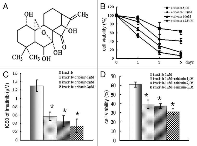
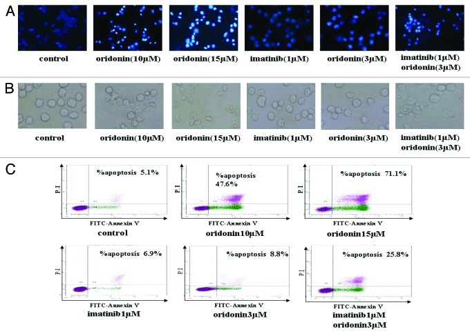
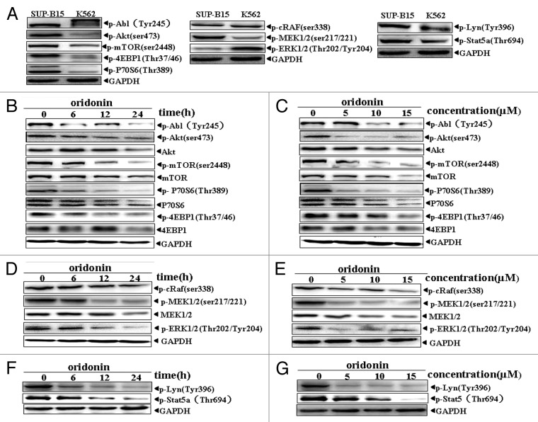
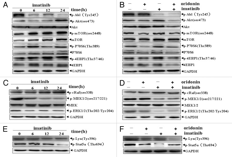
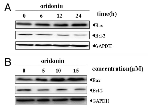
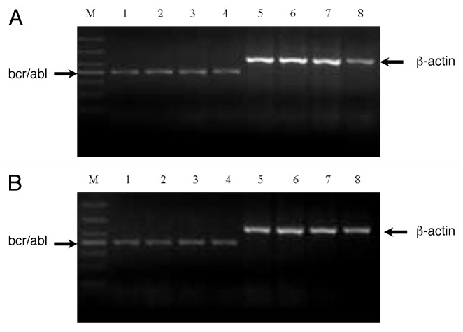
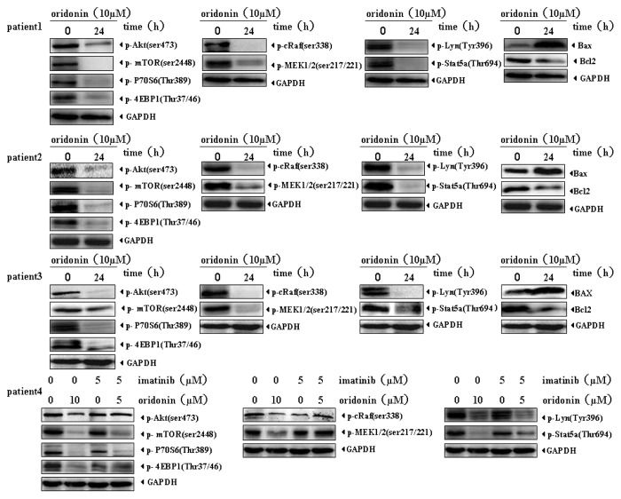
Philadelphia chromosome-positive acute lymphoblastic leukemia (Ph+ ALL) is triggered by constitutively activated BCR-ABL and SRC family tyrosine kinases.They account for the activations of multiple growth-signaling pathways, including Raf/MEK/ERK, Akt/mTOR and STAT5 pathways. The BCR-ABL tyrosine kinase inhibitor imatinib is the standard treatment for Ph+ leukemia and plays efficacious role in CML. However, imatinib has few inhibitory effects on SRC tyrosine kinase with response rate of Ph+ ALL lower and relapse more frequent and quicker compared with CML. Previous studies showed that oridonin inhibits proliferation and induces apoptosis in many tumor cells. However, the anticancer activity and mechanism of oridonin in Ph+ ALL is unknown. To investigate the anticancer activity of oridonin, we examined its role in constitutively activated Akt/mTOR, Raf/MEK/ERK, STAT5 and SRC pathway, mRNA level of bcr/abl gene, cell viability and apoptosis in Ph+ ALL SUP-B15 cells. Furthermore, we detected synergetic effect of oridonin plus imatinib. Our results showed that oridonin inhibiting activations of LYN (one of SRC family kinases) and ABL and their downstream Akt/mTOR, Raf/MEK/ERK and STAT5 pathways, downregulated Bcl-2 but upregulated Bax protein and then induced apoptosis in Ph+ ALL cells. Oridonin plus imatinib exerted synergetic effects by overcoming imatinib defect of upregulating Akt/mTOR and LYN signaling. Additionally, we examined the effect of oridonin on the signaling pathways in the primary specimens from Ph+ ALL patients. Our data showed that oridonin remarkably suppressed activations of Akt/mTOR, Raf/MEK and STAT5 pathway in these primary specimens and oridonin with imatinib exerted synergetic suppressive effects on mTOR, STAT5 and LYN signaling in one imatinib resistant patient specimen. Additional evaluation of oridonin as a potential therapeutic agent for Ph+ ALL seems warranted.


Figures

None
Figure 1. Chemical structure of oridonin and cytotoxic effect of oridonin alone or in combination with imatinib on SUP-B15 cells. (A) chemical structure of oridonin.B, SUP-B15 cells were incubated with 0, 5, 7.5, 10, 12.5 μM oridonin for 1 d, 3 d and 5 d. At the end of incubation, MTT methods determined the cell survival rates. Cell viability is expressed as the percentage of cell survival compared with the control. (B) SUP-B15 cells were incubated with imatinib alone or in combination with 1 μM, 2 μM and 3 μM oridonin for 72 h. At the end of incubation, MTT methods were used to determine the cell survival rates and the IC50 of imatinib. (C) The cell viability treated with imatinib alone or in combination with indicated concentration of oridonin for 72 h. D,The IC50 of imatinib alone or in combination with indicated concentration of oridonin for 72 h. Data were from three independent experiments. *p < 0.05 was used to compare the effects of compounds.
None
Figure 2. Oridonin and imatinib induced apoptosis in SUP-B15 cells. (A) The SUP-B15 cells were treated with indicated oridonin, imatinib, or combination for 24 h, changes in cellular morphology were examined by a light microscope. The membrane of control cells was lubricious and integrity, treatment of 3 μM oridonin, 1 μM imatinib showed little morphologic changes, but oridonin plus imatinib-treated cells showed bluffing and lobulate appearances of membrane ( × 400). (B) The cells were treated with indicated oridonin, imatinib or in combination for 24 h, then stained with hoechst 33258 fluorescent. Changes in cellular nuclei were observed under a fluorescence microscope. The nuclei of control cells were round and were homogeneously stained. Treatment of 3 μM oridonin, 1 μM imatinib showed little morphologic changes, similar to control cells, while oridonin plus imatinib-treated cells showed marked blebbing of the nuclei and apoptotic bodies ( × 200). (C) The cells were treated with indicated oridonin, imatinib or in combination for 24 h and apoptosis was examined using the AnnexinV-FITC /PI apoptosis detection kit.
None
Figure 3. Oridonin inhibited constitutively activation of growth-signaling pathway in SUP-B15 cells. Western blot analysis with indicated anti-phosphotyrosine, phosphoserine and phosphothreonine antibodies showed Akt/mTOR and RAF/MEK/ERK signaling pathway, Abl, STAT5 and LYN protein kinase were constitutively active in SUP-B15 cells, compared with the expression in K562 cells (A). The cells were incubated with 7.5 μM oridonin for 6, 12, 24 h or with 5, 10, 15 μM doses of oridonin for 24 h. Cells were then harvested and total proteins were extracted. Equal amounts of protein from each sample were separated on SDS-PAGE and immunoblotted with indicated antibodies, GAPDH was used as a loading control. Oridonin inhibited constitutively activation of Abl, Akt/mTOR (B and C), Raf/MEK/ERK (D and E) and STAT5,LYN (F and G) signaling pathway in time- and dose-dependent manner. The results shown were representative of two independent experiments.
None
Figure 4. Effects of imatinib alone or in combination with oridonin on growth-signaling pathways in SUP-B15 cells. The SUP-B15 cells were incubated with 1μM imtinib for 6, 12, 24 h, or with 1 μM imatinib plus 3 μM oridonin for 24 h. They were then harvested and total proteins were extracted. Equal amounts of protein from each sample were separated on SDS-PAGE and immunoblotted with indicated antibodies, GAPDH was used as a loading control. Imatinib upregulated Akt/mTOR signaling pathway (A), inhibited constitutively activation of MEK, ERK signaling proteins (C) and STAT5 signaling (E) and had no significant effect on RAF and LYN signaling proteins (C,E). Oridonin overcame the upregulation of Akt/mTOR signaling pathway by imatinib. (B), Oridonin plus imatinib had no synergetic inhibitory effects on activation of MEK, ERK (D) and STAT5 (F) signaling proteins. The combination of oridonin and imatinib exerted significant inhibitory effect on activation of LYN tyrosine kinase (F). The results shown were representative of two independent experiments.
None
Figure 5. Oridonin modulated the expression of pro-apoptotic protein Bax and anti-apoptotic protein Bcl-2 in SUP-B15 cells. The cells were incubated with 7.5 μM oridonin for 6, 12, 24 h (A), or with 5, 10, 15 μM doses of oridonin for 24 h (B). Equal amounts of protein from each sample were separated on SDS-PAGE and immunoblotted with antibodies against Bcl2 and Bax. GAPDH was used as a loading control. The results shown were representative of two independent experiments. Oridonin upregulated the expression of pro-apoptotic protein Bax and downregulated the expression of anti-apoptotic protein Bcl-2 in SUP-B15 cells.
None
Figure 6. Analysis of mRNA expression of bcr-abl gene in SUP-B15 cells. The cells were treated with indicated oridonin, imatinib, or in combination for 24 h. Total RNA was isolated and RT-PCR was performed with β-actin primers as a control with the same amount of RNA. Oridonin, imatinib alone, or thier combination had no effects on the mRNA expression of bcr-abl gene in SUP-B15 cells. (A) Lane 1: control; Lane 2: oridonin 5 μM; Lane 3: oridonin 10 μM; Lane 4: oridonin 15μM; Lane 5: β-actin of conrtol; Lane 6: β-actin of oridonin 5μM; Lane 7: β-actin of oridonin 10μM. Lane 8: β-actin of oridonin 15 μM. (B) Lane 1: control; Lane 2: oridonin 3μM; Lane 3: imatinib 1μM; Lane 4: oridonin 3μM and imtinib 1μM; Lane 5: β-actin of control; Lane 6: β-actin of oridonin 3 μM; Lane 7: β-actin of imatinib 1μM; Lane 5: β-actin of oridonin 3 μM and imatinib 1 μM.
None
Figure 7. Oridonin downregulates signaling pathways in Ph+ ALL patient primary cells. The mononuclear cells from Ph+ ALL patients were treated with 5 μM oridonin,10 μM oridonin, 5 μM imatinib, or 5 μM oridonin with 5 μM imatinib for 24 h. Total proteins were extracted and equal amounts of protein from each sample were separated on SDS-PAGE and immunoblotted with indicated antibodies. GAPDH was used as a loading control. Oridonin significantly suppressed Akt/mTOR, Raf/MEK, STAT5 and LYN signaling pathways in all four clinical specimens. In patient 4 who progressed under imatinib therapy, 5 μM imatinib had little inhibitory effects on Akt/mTOR, Raf/MEK, STAT5 and LYN signaling while, 5 μM imatinib with 5 μM oridonin significantly inhibited the activation of mTOR, LYN and STAT5 signalings, but had no synergetic inhibitory effects on Raf/MEK.

Similar articles

Cited by

References

    1. Laurent E, Talpaz M, Kantarjian H, Kurzrock R. The BCR gene and philadelphia chromosome-positive leukemogenesis. Cancer Res. 2001;61:2343–55. - PubMed
    1. Kurzrock R, Kantarjian HM, Druker BJ, Talpaz M. Philadelphia chromosome-positive leukemias: from basic mechanisms to molecular therapeutics. Ann Intern Med. 2003;138:819–30. - PubMed
    1. Daley GQ, Van Etten RA, Baltimore D. Induction of chronic myelogenous leukemia in mice by the P210bcr/abl gene of the Philadelphia chromosome. Science. 1990;247:824–30. doi: 10.1126/science.2406902. - DOI - PubMed
    1. Lugo TG, Pendergast AM, Muller AJ, Witte ON. Tyrosine kinase activity and transformation potency of bcr-abl oncogene products. Science. 1990;247:1079–82. doi: 10.1126/science.2408149. - DOI - PubMed
    1. Cobaleda C, Gutiérrez-Cianca N, Pérez-Losada J, Flores T, García-Sanz R, González M, et al. A primitive hematopoietic cell is the target for the leukemic transformation in human philadelphia-positive acute lymphoblastic leukemia. Blood. 2000;95:1007–13. - PubMed

Publication types

MeSH terms