

Case Reports
doi: 10.1097/JTO.0b013e3182614ab5.
Next-generation sequencing identifies and immunohistochemistry confirms a novel crizotinib-sensitive ALK rearrangement in a patient with metastatic non-small-cell lung cancer
Affiliations
- PMID: 22895149
- PMCID: PMC3645938
- DOI: 10.1097/JTO.0b013e3182614ab5
Item in Clipboard
Case Reports
Next-generation sequencing identifies and immunohistochemistry confirms a novel crizotinib-sensitive ALK rearrangement in a patient with metastatic non-small-cell lung cancer
J Thorac Oncol.
2012 Sep.
No abstract available
Conflict of interest statement
NP, MI – nothing to declare.
GAO, GP, VM, DL, PJS, and MTC are employees and equity holders of Foundation Medicine
FRH: Consultant (advisory board) for Pfizer (compensated) and research agreement through University of Colorado with Ventana/Roche.
LSG: Employee of Oncotest-Teva Pharmaceutical Industries, Petach Tikva, Israel
Figures

Panel A Using a manual FISH technique, paraffin sections were hybridized with fluorescent ALK Break-Apart probe (Abbott Molecular). The specimen displayed an atypical pattern of double 3′ALK signals (red) fused with the 5′ALK signal (green), which was classified as negative for ALK gene rearrangement (< 15% of cells with split signals, 3′ ALK and 5′ ALK signals apart by >2 times signal size or single 3′ ALK signal, 50 cells scored) Panels B, C, and D.
ALK IHC was performed using a primary antibody (clone D5F3, Cell Signaling Technology, Inc). All paraffin tissue sections were stained with ALK and were reviewed by two pathologists. The tumor cells have predominantly strong staining (3+) in 50% of the tumor cells (lower right). The remaining tumor cells showed (2+) in 30% (lower left) and (1+) in 20% of the tumor cells thus resulting in a histologic (H) score of 230 on a 0–300 scale. The top right is 200X and bottom two panels are 400X.
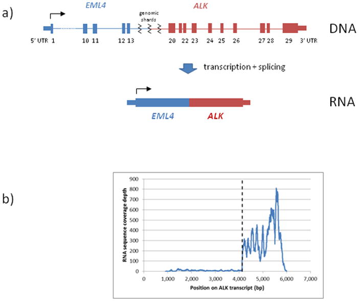
a) Hypothetical structure of rearrangement in genomic DNA (top) – complex rearrangement placing EML4 exons 1–13 upstream of ALK exons 20–29, separated by small genomic shards; RNA (bottom) – transcription and splicing remove connective region and generate a canonical EML4-ALK fusion transcript; b) Expression of ALK as measured by RNA-seq coverage, dashed line denotes boundary between exons 19 and 20.

Chest (A) and Pelvic (B) PET CT scans before, after 4 weeks and after 4 months of crizotinib.
References
-
- Rikova K, Guo A, Zeng Q, et al. Global survey of phosphotyrosine signaling identifies oncogenic kinases in lung cancer. Cell. 2007;131:1190–203. - PubMed
Publication types
MeSH terms
Substances
Grants and funding
LinkOut - more resources
Full Text Sources
Other Literature Sources
Medical
