. 2012 Dec;132(12):2791-9.
doi: 10.1038/jid.2012.261. Epub 2012 Aug 16.

Diacylglycerol kinase regulates tyrosinase expression and function in human melanocytes

Affiliations

Diacylglycerol kinase regulates tyrosinase expression and function in human melanocytes

Masakazu Kawaguchi et al. J Invest Dermatol. 2012 Dec.

Abstract

Diacylglycerol (DAG) increases the melanin content of human melanocytes in vitro and increases the pigmentation of guinea pig skin in vivo, but the mechanism(s) underlying those effects remain unknown. In this study, we characterized the role of diacylglycerol kinase (DGK), which phosphorylates DAG to generate phosphatidic acid, in the regulation of pigmentation. Ten isoforms of DGK have been identified, and we show that DGKζ is the most abundant isoform expressed by human melanocytic cells. Melanin content, tyrosinase activity, and tyrosinase protein levels were significantly reduced by a DGK inhibitor, but tyrosinase and microphthalmia-associated transcription factor messenger RNA (mRNA) levels were not changed by that inhibition, and there were no effects on the expression of other melanogenesis-related proteins. Isoform-specific small interfering RNAs showed that knockdown of DGKζ decreased melanin content and tyrosinase expression in melanocytic cells. Overexpression of DGKζ increased tyrosinase protein levels, but did not increase tyrosinase mRNA levels. Glycosidase digestion revealed that inhibition of DGK reduced only the mature form of tyrosinase, and the decrease of tyrosinase resulting from DGK inhibition could be blocked partially by protease inhibitors. These results suggest that DGK regulates melanogenesis via modulation of the posttranslational processing of tyrosinase, which may be related with the protein degradation machinery.


Figures
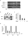
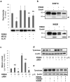
Figure 1
Figure 1. Expression of DGK isoform mRNAs and proteins in melanocytic cells and effects of levels of tyrosinase and melanin
(a) Expression levels of DGK isoform mRNAs examined by RT-PCR. (b) Expression of DGKη and DGKζ examined by Western blot analysis using anti-DGK antibodies. Each lane represents lysates from NHEMs, SK-Mel-23 cells, B16F10 cells and A293 cells; arrowheads indicate the bands of interest. (c) Melanin content in NHEMs treated with the DGK inhibitor. Results are averages of 3 independent experiments ± S.D. *P<0.05 compared to the control. (d) NHEMs were cultured with 1-2.5 μM DGK inhibitor and tyrosinase activity was examined; results are averages of 3 independent experiments ± S.D. **P<0.01 compared to the control at each time point.
Figure 2
Figure 2. Effect of the DGK inhibitor on melanogenesis and on the expression of melanocyte-specific factors in NHEMs
melanogenesis-related proteins analyzed by Western blot (a) and real-time RT-PCR analysis (b) to characterize tyrosinase and MITF mRNA levels in DGK inhibitor-treated cells. Results are averages of 3 independent experiments ± S.D.
Figure 3
Figure 3. Effect of the DGK inhibitor on the post-translational processing of tyrosinase and proteasomal degradation
(a) Levels of tyrosinase in B16F10 cells after treatment with the DGK inhibitor (20 μM) for 48 h in the presence or absence of proteasome or lysosomal protease inhibitors (120 nM MG132 or 3 μM ALLN); results are expressed as the % of the untreated control in each experiment and are means ± S.D. for triplicate determinations. **P<0.01 compared to the relevant control without the inhibitor; NS = not significant. (b) B16F10 cells or NHEMs (5 μg protein/lane) were treated with the DGK inhibitor and extracts of those cells were then digested with or without EndoH as noted, after which immunoreactive tyrosinase bands were detected by Western blotting. Arrowheads indicate the position of the unglycosylated tyrosinase band. Each experiment was performed 3 times with similar results. (c) Melanin content in B16F10 cells treated with αMSH in the presence or absence of the DGK inhibitor (10 to 30 μM) for 48 h; αMSH was added to the culture medium 1 h after treatment with the DGK inhibitor. Results are averages of 3 independent experiments ± S.D. **P<0.01 compared to the relevant control with or without αMSH; NS = not significant. (d) Effect of αMSH and R59949 on tyrosinase protein levels; experiments were carried out as detailed for 3c above, and were repeated 3 times with similar results. (e) NHEMs were treated with the DGK inhibitor (1 μM) for 24 h in the absence or presence of PD98059 (20 μM) or Gö6983 (1 μM), and tyrosinase protein levels were examined. This experiment was performed 3 times with similar results.
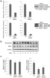
Figure 4
Figure 4. Effect of the DGK inhibition on melanogenesis in SK-Mel-23 cells
(a) Efficiency of siRNAs on the expression of DGKζ or DGKζ isoforms in SK-Mel-23 cells. The expression level of each DGK isoform was determined by real-time RT-PCR. Western blots on the right show protein levels of DGKζ or DGKζ isoforms in the siRNA-treated cells as noted. (b) Effect of DGK isoform silencing on tyrosinase and MITF protein levels. (c) Effect of DGKη or DGKζ silencing on melanin content. Results are averages of 3 independent experiments ± S.D. *P< 0.05 compared to the control (SC).
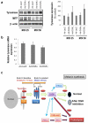
Figure 5
Figure 5. Effect of over-expression of DGKζ on tyrosinase levels
(a) NHEMs were transduced with 25 or 50 MOI of adenovirus expressing DGKα or DGKζ for 24 h, and 48 h after transduction, tyrosinase and MITF levels were examined by Western blotting; experiments were performed 3 times with similar results. (b) Real-time RT-PCR analysis of tyrosinase mRNA levels; results are averages of 3 independent experiments ± S.D. no significant differences were noted. (c) Scheme summarizing the action of DGK to regulate the function of tyrosinase. Tyrosinase enters the cis-Golgi as an EndoH-sensitive (~70 kDa) protein and exits the trans-Golgi network as an EndoH-resistant (~80 kDa) protein. After maturation in the Golgi, tyrosinase is trafficked either to melanosomes for melanin synthesis or to the proteolytic degradation machinery. The proteolysis of tyrosinase is divided into two pathways, one that is integrated into the ER associated degradation in the ubiquitin proteasome system, while the other is integrated into the endosomal/lysosomal degradation system. Inhibition of DGK activity disrupts the post-translational processing of tyrosinase from the Golgi, which leads to a dramatic decrease of functional tyrosinase. Dotted arrows represent processes reported in other studies as discussed in the text

Comment in

References

    1. Allan AE, Archambault M, Messana E, et al. Topically applied diacylglycerols increase pigmentation in guinea pig skin. J Invest Dermatol. 1995;105:687–692. - PubMed
    1. Ando H, Kondoh H, Ichihashi M, et al. Approaches to identify inhibitors of melanin biosynthesis via the quality control of tyrosinase. J Invest Dermatol. 2007;127:751–761. - PubMed
    1. Ando H, Watabe H, Valencia JC, et al. Fatty acids regulate pigmentation via proteasomal degradation of tyrosinase - a new aspect of ubiquitin-proteasome function. J Biol Chem. 2004;279:15427–15433. - PubMed
    1. Ando H, Wen Z-M, Kim H-Y, et al. Intracellular composition of fatty acid affects the processing and function of tyrosinase through the ubiquitin-proteasome pathway. Biochem J. 2006;394:43–50. - PMC - PubMed
    1. Avila-Flores A, Santos T, Rincon E, et al. Modulation of the mammalian target of rapamycin pathway by diacylglycerol kinase-produced phosphatidic acid. J Biol Chem. 2005;280:10091–10099. - PubMed

Publication types

MeSH terms

LinkOut - more resources