. 2012 Aug 15;32(33):11187-200.
doi: 10.1523/JNEUROSCI.0684-12.2012.

Opioid activation of toll-like receptor 4 contributes to drug reinforcement

Affiliations

Opioid activation of toll-like receptor 4 contributes to drug reinforcement

M R Hutchinson et al. J Neurosci. .

Abstract

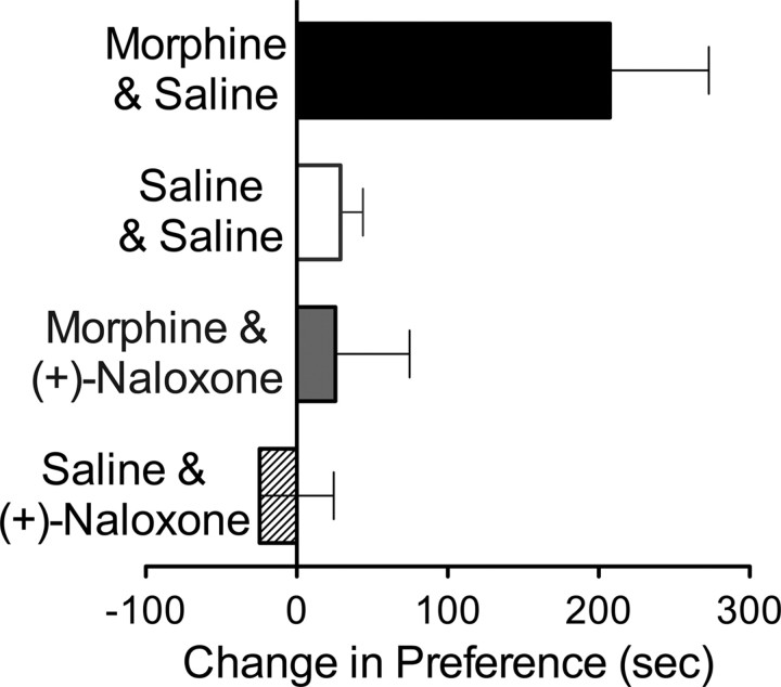
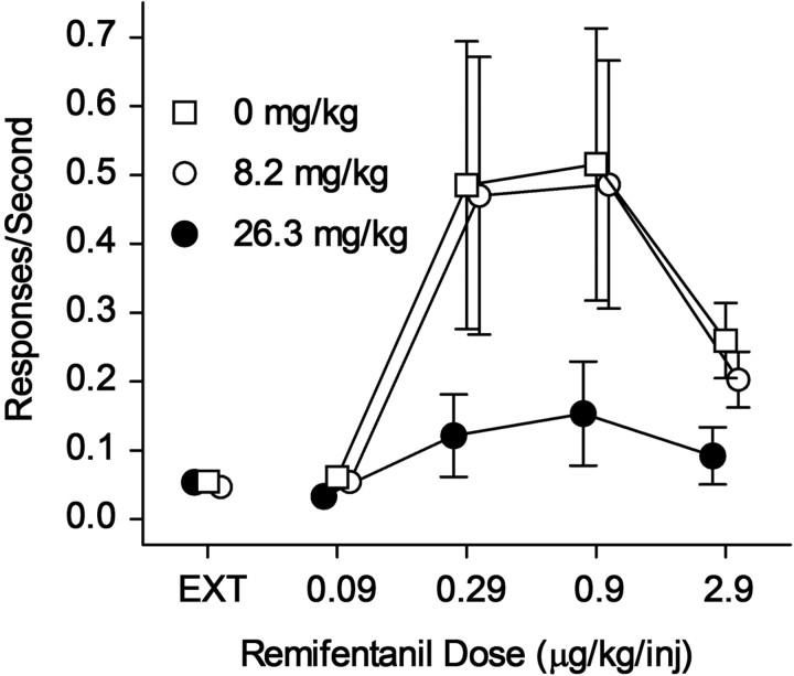
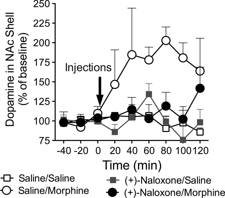
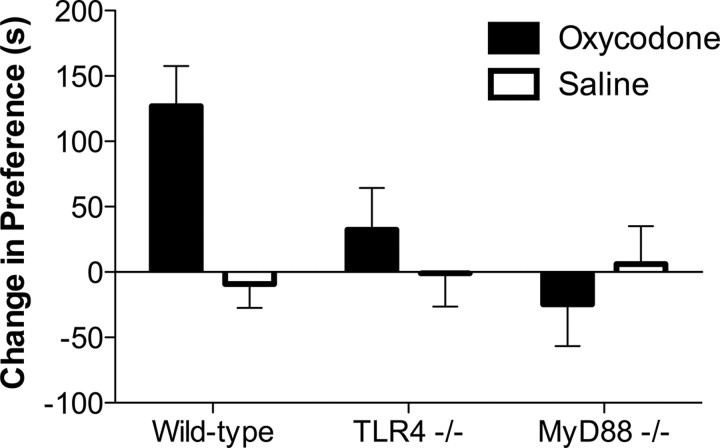
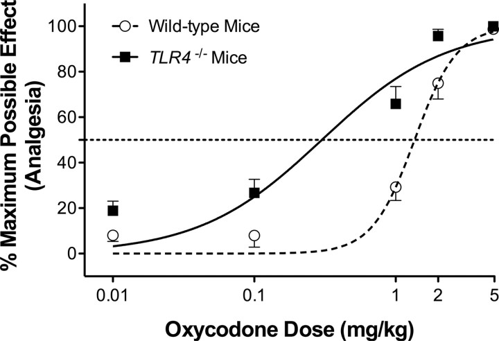
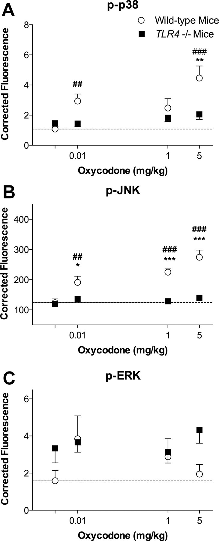
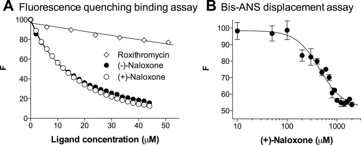
Opioid action was thought to exert reinforcing effects solely via the initial agonism of opioid receptors. Here, we present evidence for an additional novel contributor to opioid reward: the innate immune pattern-recognition receptor, toll-like receptor 4 (TLR4), and its MyD88-dependent signaling. Blockade of TLR4/MD2 by administration of the nonopioid, unnatural isomer of naloxone, (+)-naloxone (rats), or two independent genetic knock-outs of MyD88-TLR4-dependent signaling (mice), suppressed opioid-induced conditioned place preference. (+)-Naloxone also reduced opioid (remifentanil) self-administration (rats), another commonly used behavioral measure of drug reward. Moreover, pharmacological blockade of morphine-TLR4/MD2 activity potently reduced morphine-induced elevations of extracellular dopamine in rat nucleus accumbens, a region critical for opioid reinforcement. Importantly, opioid-TLR4 actions are not a unidirectional influence on opioid pharmacodynamics, since TLR4(-/-) mice had reduced oxycodone-induced p38 and JNK phosphorylation, while displaying potentiated analgesia. Similar to our recent reports of morphine-TLR4/MD2 binding, here we provide a combination of in silico and biophysical data to support (+)-naloxone and remifentanil binding to TLR4/MD2. Collectively, these data indicate that the actions of opioids at classical opioid receptors, together with their newly identified TLR4/MD2 actions, affect the mesolimbic dopamine system that amplifies opioid-induced elevations in extracellular dopamine levels, therefore possibly explaining altered opioid reward behaviors. Thus, the discovery of TLR4/MD2 recognition of opioids as foreign xenobiotic substances adds to the existing hypothesized neuronal reinforcement mechanisms, identifies a new drug target in TLR4/MD2 for the treatment of addictions, and provides further evidence supporting a role for central proinflammatory immune signaling in drug reward.


Figures

Figure 1.
Figure 1.
(+)-Naloxone blocks the place conditioning effects of morphine. Morphine (5 mg/kg, i.p.; 4 pairings) injected with saline (black bar) produced significant place preference (two-way ANOVA with Bonferroni post hoc test, p < 0.01). (+)-Naloxone (1 mg/kg, s.c.) injected just before morphine blocked morphine-induced preferences (gray bar). In the absence of morphine, (+)-naloxone and saline were without effect on place conditioning. Data are means per group ± SEMs, p = 0.01, n = 6–11/group.
Figure 2.
Figure 2.
(+)-Naloxone suppresses remifentanil self-administration. Ordinates, responses per second; abscissae, remifentanil injection dose in ug/kg/injection. Ext; extinction. 0 mg/kg (+)-naloxone (open squares), 8.2 mg/kg (+)-naloxone (open circles), and 26.3 mg/kg (+)-naloxone (filled circles). (+)-Naloxone was administered intraperitoneally at 5 min before sessions. Note that (+)-naloxone dose dependently decreased remifentanil self-administration. Data are means ± SEMs; n = 6/group.
Figure 3.
Figure 3.
Probe placements in the nucleus accumbens shell in the in vivo microdialysis experiment. Probe placements are depicted using plates adapted from the atlas of Paxinos and Watson (1998). Not all probes can be seen due to overlapping placements.
Figure 4.
Figure 4.
(+)-Naloxone suppresses morphine-induced dopamine release in the nucleus accumbens shell. In vivo microdialysis was used to test the effect of (+)-naloxone-(1 mg/kg, s.c.) on morphine- (6 mg/kg, s.c.) induced increases in extracellular dopamine in the nucleus accumbens shell. After three baseline samples, rats received saline plus saline, saline plus morphine, (+)-naloxone plus saline, or (+)-naloxone plus morphine. Repeated-measures ANOVA revealed a significant effect of treatment condition (p < 0.01). Data are means per group ± SEMs; n = 4/group.
Figure 5.
Figure 5.
Oxycodone conditioned place preference is significantly reduced in TLR4 or MyD88−/− compared with wild-type mice. Deficiencies in TLR4 or MyD88 render mice with significantly reduced oxycodone conditioned place preference; main strain (F(2,72) = 3.21, p < 0.046) and drug effects (F(1,72) = 3.00, p = 0.049) with a significant interaction (F(2,72) = 4.76, p = 0.01). Post hoc analysis revealed a significant place preference induced by oxycodone in wild-type mice (t = 3.8, p < 0.001), but no significant effect of oxycodone in the TLR4−/− or MyD88−/− animals (t < 0.79, p > 0.05). n = 11–17 per group. Data are means ± SEMs.
Figure 6.
Figure 6.
Remifentanil analgesia is potentiated by coadministration of (+)-naloxone, as predicted if remifentanil acts as a TLR4 agonist. Following baseline (BL) latency assessments with radiant heat to the tail and hindpaws, (+)-naloxone (75 mg/kg) or vehicle (saline) was subcutaneously administered, 10 min before the first intrathecal dosing. A total of three intrathecal injections of remifentanil or vehicle (glycine) were administered 15 min apart, given the brief half-life of remifentanil. Remifentanil produced a significant and brief analgesia in both the tail (top; F = 44.8, p < 0.05) and hindpaws (bottom; F = 23.29, p < 0.05) when compared with glycine vehicle. Remifentanil analgesia was robustly potentiated by (+)-naloxone (F = 44.8; p < 0.05). Arrows indicate timing of the remifentanil or glycine vehicle intrathecal injections. Data are means ± SEMs; n = 6/group.
Figure 7.
Figure 7.
Oxycodone is a more potent analgesic in TLR4−/− mice compared with wild-type mice. TLR4−/− mice demonstrate significantly longer hotplate latencies when administered with oxycodone compared with wild-type controls. ED50 wild-type 1.36 mg/kg versus TLR4−/− 0.26 mg/kg; F(2,71) = 24.1; p < 0.0001. n = 7–8 animals/dose.
Figure 8.
Figure 8.
(+)-Naloxone does not alter morphine levels in brain (hippocampus). To define whether the results from the conditioned place preference or in vivo microdialysis studies may be attributed to blockade by (+)-naloxone of morphine reaching the CNS, morphine levels were measured in hippocampus, where this brain structure was chosen simply due to its size and ease of isolation. Rats were injected subcutaneously with 1 mg/kg (+)-naloxone or saline 5 min before intraperitoneal 6 mg/kg morphine; naive rats served as negative controls. Either 5 or 30 min after injection, rats were decapitated, hippocampi were harvested, and morphine levels quantified by HPLC (Van Crugten et al., 1997; Doverty et al., 2001). No differences in brain levels of morphine were detected, comparing (+)-naloxone and saline groups administered morphine (two-way ANOVA; p > 0.05). Data are means ± SEMs; n = 4/group.
Figure 9.
Figure 9.
Oxycodone causes TLR4-dependent increases in p38 and JNK phosphorylation. TLR4−/− mice had significantly reduced oxycodone-induced MAP kinase signaling. Phosphorylation of MAPK proteins (p38, JNK, and ERK) were measured from wild-type and TLR4−/− spinal cords following acute oxycodone administration. Oxycodone caused significant dose dependent elevations in the phosphorylation of p38 (A) and JNK (B) in wild-type but not TLR4−/− mice. An oxycodone effect was not observed in ERK phosphorylation (C). n = 7–8 animals per dose. Post hoc WT versus TLR4−/−, *p < 0.05, **p < 0.01, ***p < 0.001. Post hoc vehicle versus dose, ##p < 0.01, ###p < 0.001.
Figure 10.
Figure 10.
Remifentanil, its major metabolite remifentanil acid, and morphine each dock to TLR4 and MD2 complex. Morphine, remifentanil, and its opioid inactive metabolite, remifentanil acid, were determined to dock in silico to the lipopolysaccharide-binding domain of MD2. Comparisons of the docking energy estimates (y-axis) for remifentanil and remifentanil acid across the multiple conformational states (x-axis) suggest that such docking conformations preferred the formation of the active signaling heterodimer TLR4/MD2 complex rather than the MD2 alone conformation. In contrast, morphine failed to display any such docking energy preference to the TLR4/MD2 complex states, instead demonstrating high affinity for all states.
Figure 11.
Figure 11.
Biophysical determination of remifentanil binding to MD2 in vitro. A, Titration curves of fluorescence quenching assay with the increasing drug concentrations. Remifentanil binds to MD2 and causes the quenching of expressed MD2 intrinsic fluorescence in vitro, while roxithromycin, a compound used as a negative control, shows no MD2 binding activity. KD = 6.0 ± 1.1 μm for the remifentanil-MD2 interaction. B, Replotting the data from A according to the equation: lg (F0/F − 1) = −lgKD + n × lg ([remifentanil]), revealing a slope of 1.06 ± 0.07 and a KD = 8.2 ± 1.1 μm for the remifentanil-MD2 interaction. C, Specificity of remifentanil binding is revealed by its failure to bind protein A. Remifentanil demonstrates negligible binding to a negative control protein, protein A. As the excitation, 280 nm was used, and emission at 308 nm (peak position) was plotted against the titrated remifentanil concentration. Note: the binding constants derived here are likely underestimating the affinity constants of remifentanil to MD2 due to the lack of cofactors found in vivo. Data are means ± SEMs; n = 2 replications per group.
Figure 12.
Figure 12.
In silico docking of (+)-naloxone in the lipopolysaccharide-binding pocket of MD2, an essential coreceptor of TLR4. Morphine, remifentanil, and its opioid inactive metabolite, remifentanil acid (displayed as the overlapping stick symbol structures), were determined to dock in silico to the lipopolysaccharide-binding domain of MD2 (represented as a ribbon peptide), in a conformation that spatially overlap with (+)-naloxone (spatial cloud representing preferred docking conformation) and morphine in silico docking.
Figure 13.
Figure 13.
(+)-Naloxone binds to MD2 in vitro. A, Both (−)-naloxone and (+)-naloxone bind to MD2 and cause the quenching of expressed MD2 intrinsic fluorescence in vitro, while roxithromycin, a compound used as a negative control, shows no MD2 binding activity. B, (+)-Naloxone caused the decrease of Bis-ANS fluorescence from the Bis-ANS-MD2 complex, suggesting that (+)-naloxone replaces Bis-ANS binding to MD2. Note: the binding constants derived here are likely underestimating the affinity constants of (+)-naloxone to MD2 due to the lack of cofactors found in vitro. Data are means ± SEMs; n = 2 replications/group.

References

    1. Agrawal RG, Hewetson A, George CM, Syapin PJ, Bergeson SE. Minocycline reduces ethanol drinking. Brain Behav Immun. 2011;25(Suppl 1):S165–S169. - PMC - PubMed
    1. Beardsley PM, Shelton KL, Hendrick E, Johnson KW. The glial cell modulator and phosphodiesterase inhibitor, AV411 (ibudilast), attenuates prime- and stress-induced methamphetamine relapse. Eur J Pharmacol. 2010;637:102–108. - PMC - PubMed
    1. Bland ST, Hutchinson MR, Maier SF, Watkins LR, Johnson KW. The glial activation inhibitor AV411 reduces morphine-induced nucleus accumbens dopamine release. Brain Behav Immun. 2009;23:492–497. - PMC - PubMed
    1. Bowers MS, Kalivas PW. Forebrain astroglial plasticity is induced following withdrawal from repeated cocaine administration. Eur J Neurosci. 2003;17:1273–1278. - PubMed
    1. Bsibsi M, Ravid R, Gveric D, van Noort JM. Broad expression of Toll-like receptors in the human central nervous system. J Neuropathol Exp Neurol. 2002;61:1013–1021. - PubMed

Publication types

MeSH terms

LinkOut - more resources