. 2012 Aug 15;32(33):11259-70.
doi: 10.1523/JNEUROSCI.0948-12.2012.

State-dependent modulation of breathing in urethane-anesthetized rats

Affiliations

State-dependent modulation of breathing in urethane-anesthetized rats

Silvia Pagliardini et al. J Neurosci. .

Abstract

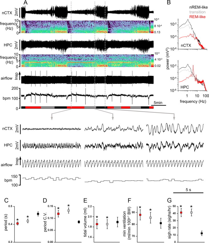
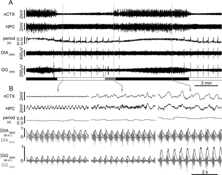
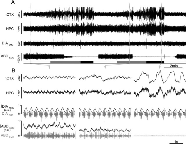
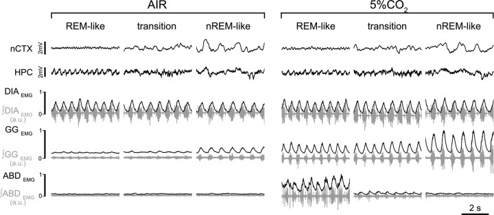
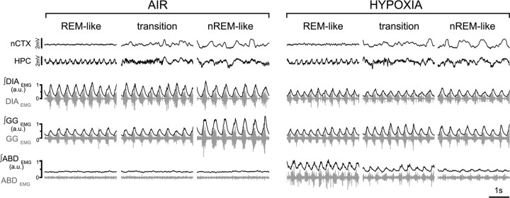
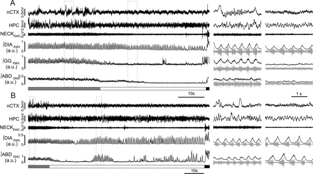
Respiratory activity is most fragile during sleep, in particular during paradoxical [or rapid eye movement (REM)] sleep and sleep state transitions. Rats are commonly used to study respiratory neuromodulation, but rodent sleep is characterized by a highly fragmented sleep pattern, thus making it very challenging to examine different sleep states and potential pharmacological manipulations within them. Sleep-like brain-state alternations occur in rats under urethane anesthesia and may be an effective and efficient model for sleep itself. The present study assessed state-dependent changes in breathing and respiratory muscle modulation under urethane anesthesia to determine their similarity to those occurring during natural sleep. Rats were anesthetized with urethane and respiratory airflow, as well as electromyographic activity in respiratory muscles were recorded in combination with local field potentials in neocortex and hippocampus to determine how breathing pattern and muscle activity are modulated with brain state. Measurements were made in normoxic, hypoxic, and hypercapnic conditions. Results were compared with recordings made from rats during natural sleep. Brain-state alternations under urethane anesthesia were closely correlated with changes in breathing rate and variability and with modulation of respiratory muscle tone. These changes closely mimicked those observed in natural sleep. Of great interest was that, during both REM and REM-like states, genioglossus muscle activity was strongly depressed and abdominal muscle activity showed potent expiratory modulation. We demonstrate that, in urethane-anesthetized rats, respiratory airflow and muscle activity are closely correlated with brain-state transitions and parallel those shown in natural sleep, providing a useful model to systematically study sleep-related changes in respiratory control.


Figures

Figure 1.
Figure 1.
State-dependent modulation of breathing in urethane-anesthetized rats. A, Long-term EEG recordings at nCTX and HPC sites and corresponding power spectrograms, respiratory airflow, and breathing rate (bpm) show the rhythmic alternation of brain activity and the associated changes in respiratory flow and breathing frequency across brain states. Spectrograms indicate the prevalence of power at ∼1 Hz in both nCTX and HPC during nREM-like state and the prevalence of electrical power at ∼4 Hz in the HPC during REM-like states. The transition state is characterized by weak nCTX (1 Hz) and HPC (3–4 Hz) power. Schematic blocks at the bottom of the plot indicate time spent in REM-like (red), transition (gray), and nREM-like (black) epochs. Dashed vertical gray lines indicate the time point from which the traces displayed below are taken. Magnification of traces in REM-like (left), transition (middle), and nREM-like (right) states. B, Power spectral analysis for nCTX (top) and HPC (bottom) during nREM-like, REM, and transition states further indicate the prevalence of ∼1 Hz power in nCTX and HPC during nREM-like state and the prevalence of ∼4 Hz power in HPC during REM-like events. C–G, Average pooled period, CV of the period, tidal volume, minute ventilation, and sigh rate in REM-like (red), transition (gray), and nREM-like (black) epochs. Asterisks indicate statistical significance (p < 0.05) of states (REM-like or transition) relative to nREM-like epochs. C, D, n = 23; E, F, n = 15; G, n = 18.
Figure 2.
Figure 2.
State-dependent modulation of GG muscle activity in urethane-anesthetized rats. A, Simultaneous EEG (nCTX and HPC) and EMG (DIA and GG) with ongoing respiratory period across brain-state alternations. Schematic blocks at the bottom of the plot indicate time spent in REM-like (white), transition (gray), and nREM-like (black) epochs. B, Details of EEG traces, period, raw EMG (gray), and rectified and integrated EMG traces of DIA and GG (black). Note the lack of state-dependent modulation of DIA and the strong activation of GGEMG during nREM-like state (n = 14).
Figure 3.
Figure 3.
State-dependent modulation of abdominal muscle activity in urethane-anesthetized rats. A, Simultaneous EEG (nCTX and HPC) and EMG (DIA and ABD) across brain-state alternations. Schematic blocks at the bottom of the plot indicate time spent in REM-like (white), transition (gray), and nREM-like (black) epochs. B, Details of EEG traces, raw EMG (gray), and rectified and integrated EMG traces of DIA and ABD (black). Note the expiratory-modulated activation of ABDEMG activity during REM-like and transition states (n = 7).
Figure 4.
Figure 4.
Hypercapnic response across brain states. EEG (nCTX and HPC) and EMG (DIA, GG, and ABD; raw traces in gray; rectified and integrated EMG traces in black) in control (left) and during hypercapnia (5% CO2 in air; right) during REM-like, transition, and nREM-like epochs. Note the state-dependent potentiation of GGEMG and ABDEMG activity in hypercapnia (n = 8).
Figure 5.
Figure 5.
Hypoxic response across brain states. EEG (nCTX and HPC) and EMG (DIA, GG, and ABD; raw traces in gray; rectified and integrated EMG traces in black) in control (left) and during hypoxia (13.3% O2; right) in REM-like, transition, and nREM-like epochs. Note the state-dependent potentiation of ABDEMG activity and the lack of state-dependent modulation of GGEMG during nREM-like epochs under hypoxia (n = 8).
Figure 6.
Figure 6.
State-dependent modulation of breathing in natural sleep. A, EEG (nCTX and HPC) and EMG (raw neck, and rectified and integrated traces of DIA, GG, and ABD) activity during transition from nREM into REM, followed by a sudden awakening. Note the reduction of both tonic (baseline activity) and respiratory-modulated EMG activity in GG. Similarly, ABDEMG activity decreases at the beginning of the REM epoch. Details of nREM and REM epochs (boxes on long trace recordings) are shown on the right of the panel. B, Long trace and details from a different nREM–REM–wake epoch recorded in the same rat show the occurrence of recurrent activation of expiratory ABDEMG activity during REM (n = 7; 42 of 103 REM epochs longer than 10 s).

Similar articles

Cited by

References

    1. Abbott SB, Stornetta RL, Coates MB, Guyenet PG. Phox2b-expressing neurons of the parafacial region regulate breathing rate, inspiration, and expiration in conscious rats. J Neurosci. 2011;31:16410–16422. - PMC - PubMed
    1. Antkowiak B. How do general anaesthetics work? Naturwissenschaften. 2001;88:201–213. - PubMed
    1. Aserinsky E. Periodic respiratory pattern occurring in conjunction with eye movements during sleep. Science. 1965;150:763–766. - PubMed
    1. Aserinsky E, Kleitman N. Regularly occurring periods of eye motility, and concomitant phenomena, during sleep. Science. 1953;118:273–274. - PubMed
    1. Baldwin DN, Suki B, Pillow JJ, Roiha HL, Minocchieri S, Frey U. Effect of sighs on breathing memory and dynamics in healthy infants. J Appl Physiol. 2004;97:1830–1839. - PubMed

Publication types

MeSH terms