. 2012 Aug 15;32(33):11343-55.
doi: 10.1523/JNEUROSCI.1436-12.2012.

Gβ3 is required for normal light ON responses and synaptic maintenance

Affiliations

Gβ3 is required for normal light ON responses and synaptic maintenance

Anuradha Dhingra et al. J Neurosci. .

Abstract

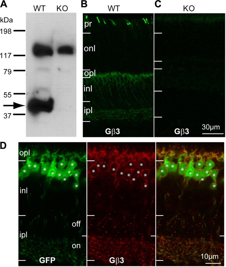
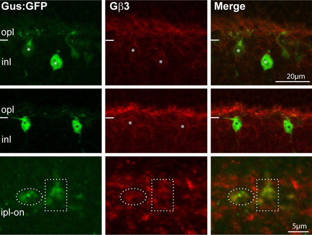
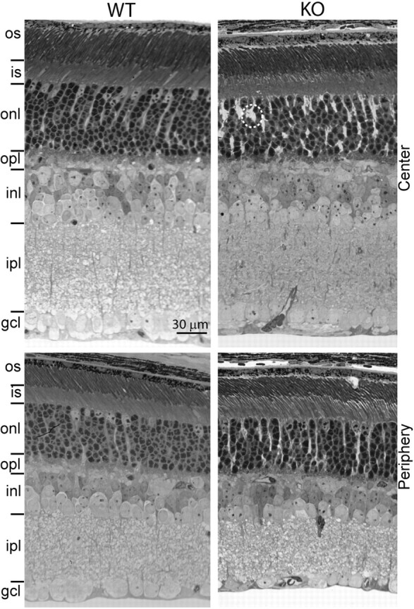
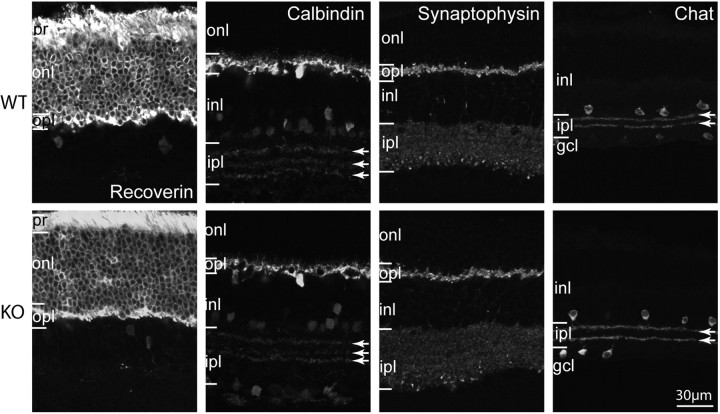
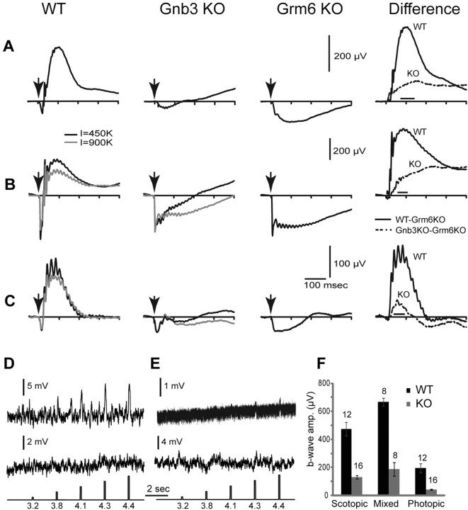
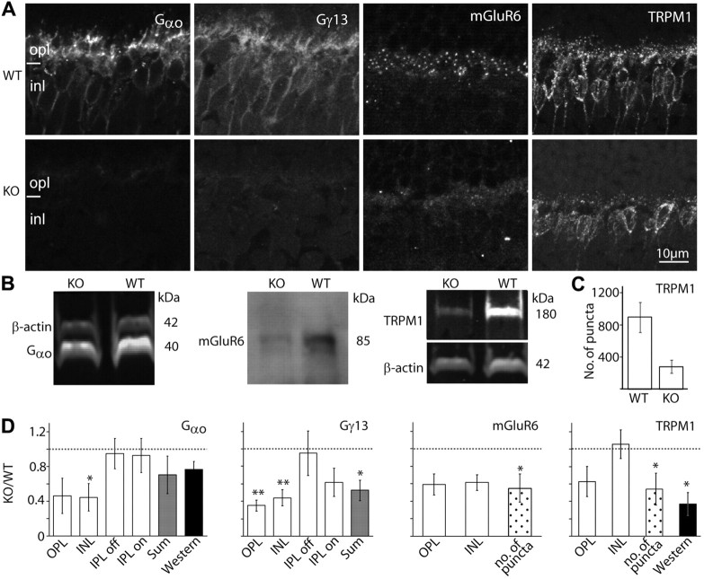
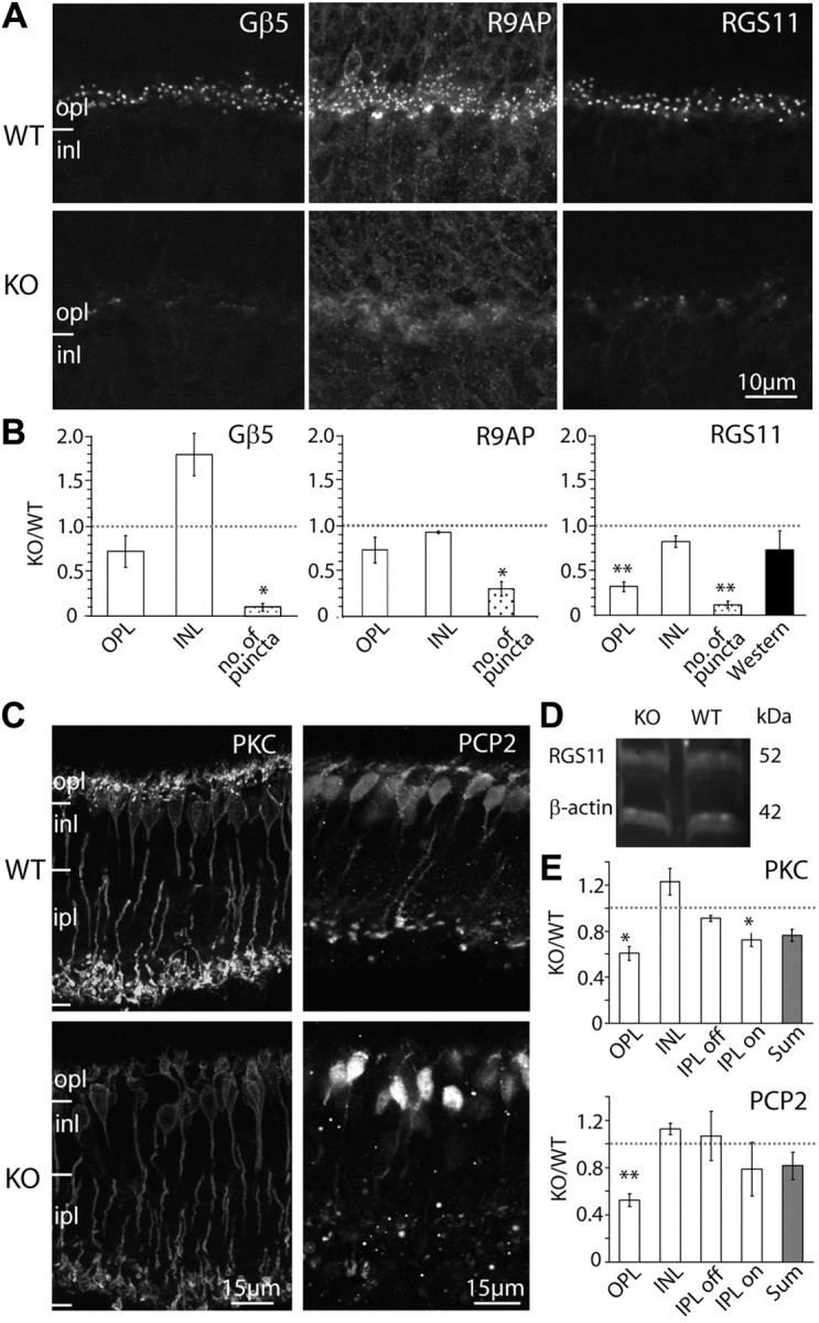
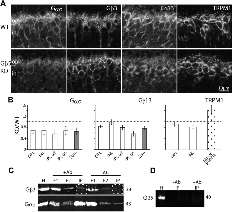
Heterotrimeric G-proteins, comprising Gα and Gβγ subunits, couple metabotropic receptors to various downstream effectors and contribute to assembling and trafficking receptor-based signaling complexes. A G-protein β subunit, Gβ(3), plays a critical role in several physiological processes, as a polymorphism in its gene is associated with a risk factor for several disorders. Retinal ON bipolar cells express Gβ(3), and they provide an excellent system to study its role. In the ON bipolar cells, mGluR6 inverts the photoreceptor's signal via a cascade in which glutamate released from photoreceptors closes the TRPM1 channel. This cascade is essential for vision since deficiencies in its proteins lead to complete congenital stationary night blindness. Here we report that Gβ(3) participates in the G-protein heterotrimer that couples mGluR6 to TRPM1. Gβ(3) deletion in mouse greatly reduces the light response under both scotopic and photopic conditions, but it does not eliminate it. In addition, Gβ(3) deletion causes mislocalization and downregulation of most cascade elements and modulators. Furthermore, Gβ(3) may play a role in synaptic maintenance since in its absence, the number of invaginating rod bipolar dendrites is greatly reduced, a deficit that was not observed at 3 weeks, the end of the developmental period.


Figures

Figure 1.
Figure 1.
3 is expressed by cones and all types of ON bipolar cells. A, Western blots for Gβ3 in WT and Gnb3-null (KO) mice show a specific band at the expected molecular weight of 38 kDa (arrow) and a higher nonspecific band at ∼130 kDa. B, Immunostaining for Gβ3 in wild-type shows strong staining of cone outer segments (in the photoreceptor layer) and weaker staining in the outer plexiform layer and the inner nuclear layer. C, All staining is absent in the null mouse. D, Higher magnification of the stained inner retina of the Grm6-GFP mouse that expresses GFP in all and only ON bipolar cells. Strong Gβ3 staining appears in the dendrites and somas, and weak staining is present in the ON sublamina of the inner plexiform layer (ipl). Note that all GFP-expressing cells also express Gβ3 (asterisks) and vice versa. pr, Photoreceptor layer; onl, outer nuclear layer; opl, outer plexiform layer; inl, inner nuclear layer.
Figure 2.
Figure 2.
Type 7 cone bipolar cells express Gβ3. A retina of a transgenic mouse, Gus8.4GFP (which expresses GFP weakly in certain rod bipolar cells and strongly in type 7 cone bipolar cells), stained for Gβ3 shows that bright GFP-labeled somas are stained for Gβ3 (asterisks). Dotted ellipses encircle type 7 axon terminals in the ipl, and these terminals are weakly stained for Gβ3. Abbreviations are defined as in Figure 1.
Figure 3.
Figure 3.
Retinal layers of wild-type and Gnb3-null mice have similar appearance and thickness. Semithin (1 μm; radial view) epon radial sections of retinas (4 weeks old) stained with toluidine blue clearly show all the retinal layers. This picture is representative of three adult WTs and three adult KOs. The layers in general have a normal appearance, but in certain regions, Muller cells appear to occupy bigger spaces than normal (within the dotted circle). Top panels show the center of the retina, and bottom panels show the periphery. os, Outer segments; is, inner segments; gcl, ganglion cell layer. Other abbreviations are defined as in Figure 1.
Figure 4.
Figure 4.
Gross morphology of all cell classes is normal in the Gnb3-null retina. Immunostaining using several cell markers for WT (top) and Gnb3-null (KO; bottom) retinas. Recoverin labels all photoreceptors throughout the cell including outer and inner segments, somas, and photoreceptor terminals. All of these segments are stained in both wild-type and null retinas. Calbindin labels horizontal cells strongly and certain amacrine cells weakly. Horizontal cells and the three bands in the ipl (arrows) appear similar. Synaptophysin staining shows that the two synaptic layers are similar. Choline acetyl transferase (Chat) labels the cholinergic amacrine cells that stratify in the OFF (top band) and ON sublaminas (lower band) of the ipl; these bands are unaffected by the absence of Gβ3. Data are representative of three (synaptophysin) to eight (calbindin) images from three different animals for each genotype. Abbreviations are defined as in Figure 1.
Figure 5.
Figure 5.
Light responses in Gnb3-null ON bipolar cells are dramatically reduced, but not eliminated. A–C, Averaged ERGs of WT, Gnb3-null (KO), and Grm6-null (KO) mice under three different conditions. Arrows indicate a stimulus flash. In A, animals were dark adapted overnight and stimulated with a flash that produced 10 photoisomerizations per rod, which is below cone threshold. These represent scotopic conditions. The right panel represents the average Grm6 KO record subtracted from the average WT (solid lines) or Gnb3-KO record (dotted lines). This difference between the original ERG record and the Grm6 KO record is an estimate of the pure b-wave in these two mouse lines. In B, animals were dark adapted and stimulated with a saturated light flash of 450,000 (black traces) or 900,000 (gray) R* per rod. This flash stimulates both rods and cones, so the ERG represents the mixed rod- and cone-driven responses. Subtracted records on the right are for the lower intensity (black). In C, mice were adapted to a bright background (513 nm; 27,000 R*/rod/s) that completely suppressed the cGMP-activated current of the rods. They were then stimulated with an intense white flash that isomerizes 1% of the M-cone pigment and 0.1% of the UV-cone pigment in adult mice. ERG traces represent cone-driven responses, i.e., photopic conditions. For the mixed and photopic responses (B, C), only the records for 450,000 R* per rod were used for subtraction (because the Grm6-KO was stimulated only with this intensity). The number of records averaged for Grm6-null mice was eight for scotopic and mixed conditions and six for photopic conditions. D, E, Whole-cell recordings from wild-type (D) and Gnb3-KO (E) rod bipolar cells under current-clamp mode. Two examples are given for each genotype; the top one shows a light response and the bottom does not. The response of the Gnb3-KO cell in the top trace is very small, but clearly above the noise. Interestingly, the noise level in this cell was much lower than the rest (same recording procedures). Perhaps this is the reason the response can be extracted. The lowest traces show the timing and intensity of the light stimuli; these are given in log photons μm−2 per flash. F, A histogram quantifying the average b-wave amplitude for wild-type and Gnb3-null eyes under the three recording conditions. The time windows used to average these values for each condition are indicated by the horizontal line just above the x-axis in the difference ERGs of A–C. Numbers above the histogram bars indicate the numbers of averaged records. For this and subsequent figures, error bars indicate SEs.
Figure 6.
Figure 6.
The essential cascade elements are down regulated. A, Immunostaining of WT (top) and Gnb3-null (KO; bottom) retinas for Gαo, Gγ13, mGluR6, and TRPM1. Staining in the null mouse was greatly reduced. B, Western blots for Gαo and TRPM1 were done with the Odyssey reagents (bands are brighter than background), and for mGluR6 were performed with the ECL technique (bands are darker than background). Bands' identities and molecular weights are indicated on the sides of the blots. All three proteins are downregulated in the null mouse. C, Bar chart showing the average number of TRPM1-positive puncta in the outer plexiform layer (within a retinal area of 1680 μm2) in the WT and null retinas (n = 3 for each). D, Relative immunostaining intensity (i.e., staining intensity in null divided by staining intensity in wild type) for various proteins in the layers of the retina (white bars), sum intensity of all layers (gray bars), and relative intensity of the protein bands (Western blots, dark bars). For staining patterns that show puncta in the opl, the relative number of puncta is also shown (dotted bars). *p < 0.05; **p < 0.01. Western blot quantification for mGluR6 was not performed because of the need to use ECL; however, in every set, the mGluR6 band in the null retina is less intense than that in wild type. The dotted horizontal line shows unity, i.e., when wild-type and null retinas are the same. All experiments were performed on at least three sets, where a set included a null and wild-type retina from littermates; when a littermate was not available, an age-matched wild type was used. For this and subsequent figures, the same settings are used for each pair of pictures. Abbreviations are defined as in Figure 1.
Figure 7.
Figure 7.
3 deletion heavily affects modulators of the mGluR6 cascade that display a punctate appearance, but only mildly affects the diffusely distributed modulators. A, Immunostaining of WT (top) and Gnb3-null (KO; bottom) mice for Gβ5, R9AP, and RGS11. Punctate staining is greatly reduced in the null mouse. B, Relative immunostaining (as defined in Fig. 6C) for these proteins in the outer plexiform and inner nuclear layers of the retina (white bars), relative puncta count (dotted bars), and band intensity from Western blots (for RGS11 only; dark bar). Punctate staining is greatly reduced in the null mouse. C, Immunostaining of WT (top) and Gnb3-null (KO; bottom) mice for the diffuse modulators PKC and Ret-PCP2. D, Western blot for RGS11 shows that the band intensity is similar between wild-type and Gnb3-null mice. E, Relative immunostaining of various layers of the retina (white bars) and sum intensities of all layers (gray bars). *p < 0.05; **p < 0.01. Abbreviations are defined as in Figure 1.
Figure 8.
Figure 8.
Gnb3-null rod bipolar dendrites are not maintained in the invagination of the rod terminal. A, B, Electron micrographs of rod terminals (RT). In WT retina, numerous profiles of rod terminals (outlined by dotted lines) are seen with one or two rod bipolar dendrites (b) postsynaptic to the rod's synaptic ribbon (r). In Gnb3-null (KO) retina, bipolar dendrites are usually smaller and are rarely observed. Horizontal cell processes (h) are seen with the same frequency in WT and KO. C, Bar graph describes the percent of profiles in which a ribbon, at least one horizontal cell process (Horiz.), or at least one rod bipolar dendrite are observed. For each category, we show the percentage at 3 weeks (3w) and 4–7 weeks (A) for wild type (black) and Gnb3 KO (gray). The number of profiles analyzed for WT were 218 (3w; n = 2) and 395 (A; n = 3), and for Gnb3-null they were 351 (3w; n = 2) and 404 (A; n = 3). The difference between WTs and nulls at 4–7 weeks was significant (χ2 test, p < 0.005). D, E, Electron micrographs of cone terminals (CT). These terminals (outlined by dotted lines) are seen in both wild-type and null retinas, but in null retinas they appear darker with a density that resembles their neighboring rod terminals. mit, Mitochondrion. Scale bar: (in E) A, B, 0.455 μm; D, E, 1 μm. F–I, ERG records at different ages in wild-type (F, G) and null (H, I) mice. To compare shapes, we averaged traces from each age group and normalized this average trace to its minimum (peak of a-wave). The numbers of records averaged for each trace were 4 (3w), 6 (4–8w), and 2 (30w) for WT and 4 (3w), 6 (4–8w), and 4 (30w) for Gnb3-null. In G and I, the time scale is expanded to show the initial oscillations. Note that in wild-type mice (G), the oscillations at 3 weeks lag behind those in older adults (gray and black arrows, respectively), while in null mice (I), they appear simultaneously (gray arrows), although they all lag behind the wild-type oscillations. Note the different time scales in G and I.
Figure 9.
Figure 9.
3 rather than Gβ5 forms the heterotrimer Go. A, Immunostaining in wild-type (top) and Gβ5-null (bottom) retinas for Gαo, Gβ3, Gγ13, and TRPM1. Fixed retinas and sections were obtained from J. Chen. B, Relative staining intensity (as defined in Fig. 6C) for Gαo, Gγ13, and TRPM1 in several retinal layers and their sum. For TRPM1, the number of puncta was calculated as in previous figures. Gβ3 staining was not quantified because staining only worked on one set of tissues. However, results from the Chen lab showed similar staining intensity in wild-type and null tissues (personal communications). Average ratios represent results from at least three sets of experiments. C, Coimmunoprecipitation using a monoclonal antibody to pull down Gαo and its interacting proteins. The top blot was probed for Gβ3, and bottom one for Gαo. The relevant lanes, i.e., immunoprecipitated (IP) protein with and without the antibody, are boxed with dotted squares. H, Homogenate; F1, F2, flow through. D, Coimmunoprecipitation samples probed for Gβ5.

References

    1. Ball SL, Pardue MT, McCall MA, Gregg RG, Peachey NS. Immunohistochemical analysis of the outer plexiform layer in the nob mouse shows no abnormalities. Vis Neurosci. 2003;20:267–272. - PubMed
    1. Berrebi AS, Mugnaini E. Characteristics of labeling of the cerebellar Purkinje neuron by L7 antiserum. J Chem Neuroanat. 1992;5:235–243. - PubMed
    1. Blake BL, Wing MR, Zhou JY, Lei Q, Hillmann JR, Behe CI, Morris RA, Harden TK, Bayliss DA, Miller RJ, Siderovski DP. G beta association and effector interaction selectivities of the divergent G gamma subunit G gamma(13) J Biol Chem. 2001;276:49267–49274. - PubMed
    1. Blanks JC, Adinolfi AM, Lolley RN. Synaptogenesis in the photoreceptor terminal of the mouse retina. J Comp Neurol. 1974;156:81–93. - PubMed
    1. Bomsel M, Mostov K. Role of heterotrimeric G proteins in membrane traffic. Mol Biol Cell. 1992;3:1317–1328. - PMC - PubMed

Publication types

MeSH terms

LinkOut - more resources